

The area of children's drawing has been of interest in the literature for the past century, with the emotional and psychological aspects of children's art expression being of particular interest to psychologists, psychiatrists, and art therapists (Malchiodi, Reference Malchiodi1998). Cognitive uses of children's human figure drawings can be traced to Goodenough (Reference Goodenough1926), who developed the Draw-A-Man test based on the assumption that certain aspects of children's drawings correlated to a child's mental age, and could therefore be used as a measure of cognitive development.
Following on from Goodenough (Reference Goodenough1926), many studies (e.g., Barrett & Eames, Reference Barrett and Eames1996; Chappell & Steitz, Reference Chappell and Steitz1993) have been conducted to demonstrate the ages and stages of children's human figure drawings, with these studies consistently showing that, as children grow older, their drawings of the human figure include more parts and become more detailed, connected, and realistic. As proposed by Crider (Reference Crider, Bibace and Walsh1989), children's body knowledge development appears to progress from primitive, global concepts to a more complex, detailed and abstract understanding, with their depictions of the human body generally reflecting this.
Children's drawings of internal anatomy have tended to follow similar developmental trends, with earlier studies in this area finding that, with increasing age, children were able to depict more internal body parts (Amann-Gainotti & Antenore, Reference Amann-Gainotti and Antenore1990; Brumback, Reference Brumback1977; Glaun & Rosenthal, Reference Glaun and Rosenthal1987), and were more aware of the correct location of internal body organs (Brumback, Reference Brumback1977; McEwing, Reference McEwing1996). In addition, children appeared to develop a better understanding of the physiological functioning and significance of organs (Clarke & Newell, Reference Clarke and Newell1997; Eiser & Patterson, Reference Eiser and Patterson1983; McEwing, Reference McEwing1996), as well as developing an awareness of internal anatomy as being interconnected to make up bodily systems (Gellert, Reference Gellert1962; Glaun & Rosenthal, Reference Glaun and Rosenthal1987).
Consistent with a developing knowledge of internal body organs, researchers have found that young children (i.e., 5–7 years) often held misconceptions about the location and function of body parts (Glaun & Rosenthal, Reference Glaun and Rosenthal1987; McEwing, Reference McEwing1996). From about the age of 9 years, however, children were found to make considerable gains in knowledge of internal anatomy to obtain a growing awareness of the integration and function of body parts (Jones, Badger, & Moore, Reference Jones, Badger and Moore1992). At this stage of development, the majority of children knew of the brain, with some children also having a rudimentary knowledge of the liver, lungs, and of the interconnectedness and function of parts such as the stomach and intestines. It is thought that not until adolescence was there a real understanding of organisational and functional systems within the body (Amann-Gainotti & Antenore, Reference Amann-Gainotti and Antenore1990; Jones et al., Reference Jones, Badger and Moore1992). Even so, Youngblutt (Reference Youngblutt1994) emphasised that there was no strict age-to-stage correspondence in the acquisition of body knowledge, and that children could stay in some stages longer than others.
More recent research has tended to take an educational approach, with an interest in how children may come to acquire internal body knowledge. Dempster and Stears (Reference Dempster and Stears2014), for example, assessed internal body knowledge with a sample of 7-year olds – i.e., before formal instruction in human anatomy – and found that, whilst certain organs seemed to be well known (the stomach and bones were drawn by approximately 55% of the children, followed closely by the heart, drawn by approximately 50%), they were depicted individually. Very few children drew organ systems, i.e., two or more correctly connected organs in roughly the correct position in the body, however. It was hypothesised that children at this age drew their knowledge principally from personal experience of their own body's functioning gained in a social, rather than from a more formal, context.
Reiss and Tunnicliffe (Reference Reiss, Tunnicliffe and Behrendt2001) had previously looked at children's understanding of internal body structure, but with a sample of children ranging in age from 4 years to 14 years. Drawings were rated on a scale of 1 to 7, with level 1 being no representation of internal structure, through level 2 (one or more internal organs placed at random) to level 7 (comprehensive representation of four or more organ systems). These authors did not report findings by age group but reported more generally that, whilst there was a rapid increase in knowledge from the 4 to the 6 year olds, subsequent increases in internal body knowledge as depicted in drawings became successively smaller. The authors also reported, similar to the later Dempster and Stears (Reference Dempster and Stears2014) study, that most of the students lacked much understanding of organ systems, with their depictions of internal anatomy consisting of ‘a scattered assemblage of isolated organs and incomplete organ systems’ (p. 105). Also, similar to the Dempster and Stears study was the finding that individual organs seemed to be better known and more frequently depicted, with 93% of students drawing the heart, 87% of children drawing bones, and 66% of children drawing the lungs and stomach.
Even when more specific instructions about the body were requested, developmental trends were still apparent. Garcia-Barros and colleagues required children aged 4 to 7 years to depict where food, drink and air that entered the body went (Garcia-Barros, Martinez-Losada, & Garrido, Reference Garcia-Barros, Martinez-Losada and Garrido2011). These authors found that, in general, the children had a more accurate view of the digestive system than of the respiratory system with, in particular, children aged 6 and 7 years having an accurate idea of the digestive system as being a continuous tube with one or two main organs – stomach/intestine – going from the mouth to the bottom of the body. Children tended to have a less accurate conception of the respiratory system, however, with the youngest children not identifying any of the respiratory system organs. The older children generally recognised the lungs, although the authors reported that there were ‘a large number’ (p. 2118) of subjects who regarded the stomach as the only breathing-related organ, suggesting that these children saw air as reaching the only cavity in the body they could easily identify: the stomach.
Children's knowledge of the function of internal organs was explored by Jaakkola and Slaughter (Reference Jaakkola and Slaughter2002) through interviews with children aged 4 to 10 years of age. Consistent with results from previous studies, these authors found that children's general factual knowledge about the body increased between the ages of 4 and 8 years of age, and that children's knowledge of both organ location and organ function seemed to increase at approximately the same rate across this age range. More surprising was the finding that there was a ‘dramatic increase’ (p. 337) between the ages of 4 and 6 in children's understanding that organ function sustained life. The authors concluded that their results suggested a change in children's bodily understanding between the ages of 4 and 6 years, and proposed that this signified the point at which life became the core of children's understanding of bodily function, in the sense that the ultimate goal of bodily function is the biological goal of maintaining life. This latter finding, particularly in children so young, has not been noted in other studies, although the question of the purpose of organs may not have been specifically posed and, in any case, is unlikely to have arisen in the more open-ended requests to draw what is inside the body.
With respect to primary school-aged children at least, they have been increasingly exposed to school-based health education programmes as well as an increasing range of printed and audio-visual material about the human body (e.g., Stowell & Leake, Reference Stowell and Leake2012; Wynne & Silver, Reference Wynne and Silver2009). Whilst such material could be assumed to lead to a greater awareness and knowledge in children of their own bodies Schmidt (Reference Schmidt2001), however, noted from her own research that children still tended to draw incorrect representations of body contents. This author considered the possibility that primary school-age children, in particular, may have difficulty incorporating what they had learned from printed and audio-visual material because they cannot easily visualise the interior of their own bodies. Glaun and Rosenthal (Reference Glaun and Rosenthal1987) had previously commented upon the difficulties children experienced in trying to conceptualise that which is not directly observable, and believed that children seemed to remain relatively ignorant about the insides of their bodies until adolescence. More recently, however, Garcia-Barros et al. (Reference Garcia-Barros, Martinez-Losada and Garrido2011) hypothesised that knowledge of internal body organs may be influenced by empirical experience, noting that children aged 4 to 7 years had a more accurate view of the digestive system than that of the respiratory system. Whilst even 4-year olds could identify the stomach as the place where food goes, the authors hypothesised that these children may not have recognised any respiratory system organs possibly due to a belief that air is capable of spreading inside the body. Indeed, Schmidt (Reference Schmidt2001) had earlier reported that children under 9 years did not seem to have an understanding of lung function and the concepts of inspired and expired air.
As the only study in this area to have been conducted with an Australian sample of school-aged children (Glaun & Rosenthal, Reference Glaun and Rosenthal1987) is over 25 years old, it was felt timely to revisit the area of children's body knowledge, particularly in the light of the more accessible public health education that has become available over the intervening years. The present study, therefore, aimed to investigate developmental trends in children's body knowledge found in previous studies in this area using the Inside-of-the-Body (drawing) Test (Tait & Ascher, Reference Tait and Ascher1955) as the principal study instrument. In addition, a subgroup of children were interviewed in order to further examine developmental differences in the known number and location of body parts, their understanding of the function and significance of internal body organs, and children's awareness of the integration of body parts into systems.
Method
Participants
A sample of 189 primary school children drawn from five primary schools in the Adelaide metropolitan area participated in the study. The sample consisted of 79 males and 110 females, and ranged in age from 7–12 years. All children were from mainstream classes and the majority were from Caucasian, middle income families.
Measures
The Inside-of-the-Body Test (Tait & Ascher, Reference Tait and Ascher1955) was administered to assess knowledge of the inside of the body. In this test, participants were given an outline of the body and were asked to draw and label what was inside the body. This test was scored for number of body parts identified (as labelled by the child), the number of organs drawn in the correct location, and level of integration of body parts. To score the extent to which organs were interconnected to form bodily systems, drawings were assigned one of three levels of integration: 0 = organs are free-floating within the body outline, 1 = a partial and imprecise attempt to connect some internal body organs, and 3 = at least one functional system is represented (digestive, cardiovascular, or skeletal) and all is connected and more or less spatially correct. All scoring was conducted by the first author.
In addition, a semi-structured interview based on a subset of the Gellert Index-of-Body-Knowledge (Gellert, Reference Gellert1962), was administered to a randomly selected subgroup (N = 54) of the sample. Accordingly, children were asked to describe the function and importance of seven major body organs (heart, brain, lungs, stomach, liver, bladder, and bones). These organs were selected as previous studies had suggested children across the study age range would have an awareness of these.
Procedure
Upon formal approval from the Principals of the five primary schools, as well as from the teachers whose classes would be involved in the study, an information sheet explaining the purpose of the study, together with a consent form were given to each student to take home to parents. Only children with parental consent were able to take part in the study. The Inside-of-the-Body drawing was administered as a class group, with the teacher and the investigator (lead author) being present to assist. Students were asked not to communicate amongst themselves, and were asked to work efficiently although the drawing tasks were not time limited. The children were handed a sheet of paper with an outline of a human body, and given the following instructions:
I would like you to draw a picture of the inside of a person. Put in all the parts you know are inside the body. Draw a line from each part and write the name of the part on the line. Put in all the parts you know even if you are not sure how to draw it, just try your best. It is important that you don't say any body part out loud for other children to hear. If you are unsure of what to do or need to ask a question please put up your hand.
At the completion of the group drawing task, a subgroup of 54 children were randomly selected and interviewed individually about their understanding of the function and importance of different body parts. These interviews were conducted on a one-to-one basis with each child.
Results
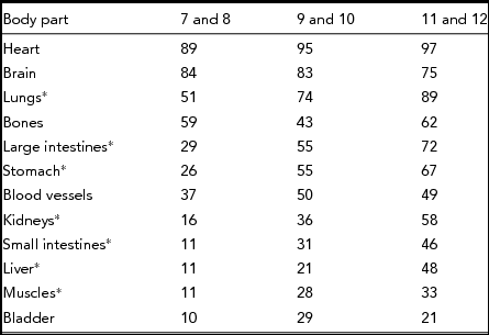
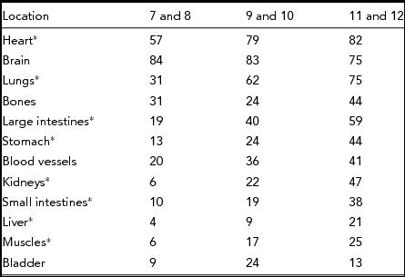
In order to confirm developmental progression with increasing age, the children were divided into three age groups: 7–8 years, 9–10 years, and 11–12 years. Tables 1 and 2 show the percentage frequencies for the identification and correct location of the individual body organs by each age group. The tables are ordered from the most identified body parts down to the least identified body parts. The most frequently identified and correctly located inside body parts were the heart and brain. The lungs, bones, and large intestines were the next most commonly identified and correctly located parts, however, identification and correct location of the small intestine, muscles, bladder, and spinal cord were slower to develop. Bones and blood vessels were identified by almost half of the children in all age groups but were less accurately located. Some body parts such as the appendix, tonsils, and pancreas, were not recognised or correctly located by many in any age group.
Percentage frequencies of body parts depicted by children across age groups.

*Significant differences on χ 2 test (p < .01).
Percentage frequencies of correct location of body parts by children across age groups.

*Significant differences on χ 2 test (p < .01).
Chi-square analyses were conducted separately for each body part to investigate the development in knowledge across age groups. Significant developmental trends are indicated by asterisks in the first column of each table. A significant developmental trend was found for the ability to correctly locate the heart, but not for its identification. A significant developmental progression was also found for the identification and location of the lungs, large intestine, stomach, kidneys, small intestine, liver, muscles, and spinal cord.
Two-way between groups analysis of variance (ANOVA) were conducted separately for number of internal body parts and the location of body parts as the dependent variable, and age categories and gender as the between subject variables. The results indicated that there were significant main effects for number of internal body parts identified, F2,189 = 51.69, p < .001, with a large effect size (partial eta squared = .31), and the correct location of body parts, F2,189 = 35.75, p = .001, also with a large effect size (partial eta squared = .28). ANOVAs revealed no gender or interaction effects for the number or location of body parts.
Bonferroni post-hoc tests indicated that all age groups differed significantly from each other for the number of body parts identified and location of internal body parts. The 11–12 year olds knew significantly more body parts and correct locations that the 9–10 years olds, who knew significantly more than the 7–8 year olds.
Function of Inside Body Parts
Data on the understanding of the function of major body parts was collected from a random subset of children (N = 54). A two-way between-groups ANOVA was conducted to explore the effects of age and gender on overall understanding of the function of the major body parts, which included the heart, brain, lungs, bones, stomach, liver, and bladder. Results indicated that there was a significant main effect for age, F2,54 = 9.52, p < .001, with a large effect size (partial eta squared = .28). No gender or interaction effects were found. Bonferroni post-hoc comparisons revealed that there was a significant difference between the overall understanding of organ function between the 7–8 year olds (M = 3.11, SD = 1.29), and the 11–12 years olds (M = 4.94, SD = 1.20), but not between either of these age groups and the 9–10 year olds (M = 4.39, SD = 1.39).
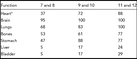
An inspection of children's knowledge of the function of individual organs indicated that there was an increase in knowledge across age groups (Table 3). Although Chi-squared analysis indicated that only knowledge of heart function differed significantly between the groups, Table 3 nevertheless shows clear developmental trends across age groups for nearly all major organs.
Percentage frequencies of children's knowledge of the function of major organs across age groups.

*Significant difference on χ 2 test (p < .01).
Discussion
This study provides further evidence that children's conceptualisation of the inside of the body follow a developmental progression similar to that already documented in human figure drawings research (e.g., Scott, Reference Scott1981). The present study corroborates an existing body of literature by also demonstrating that children's knowledge of the inside of the body improves with age (Amann-Gainotti & Antenore, Reference Amann-Gainotti and Antenore1990; Brumback, Reference Brumback1977; Garcia-Barros et al., Reference Garcia-Barros, Martinez-Losada and Garrido2011; Glaun & Rosenthal, Reference Glaun and Rosenthal1987; McEwing, Reference McEwing1996; Reiss & Tunnicliffe, Reference Reiss, Tunnicliffe and Behrendt2001). These studies have been consistent in demonstrating that, as age increases, children are able to identify more internal body parts and exhibit a greater awareness of the anatomical locations of different organs. Several of these studies have also suggested that children have a greater understanding of the interconnectedness of body organs with increasing age, and that they gradually develop a conceptualisation of the internal body as being made up of systems.
In the present study, children's knowledge increased with age for most body parts, with there being no gender differences in any age group. The present study found that the majority of children, even as young as 7 or 8 years, knew of major organs such as the heart, brain, and lungs, and had already started to develop an awareness of many other internal body parts such as the stomach, bones, large and small intestines, kidneys, and liver. By the time children were 11 or 12 years of age, at least half in the present study were able to identify such body parts. Figure 1 is an example of a drawing done by a child in the 9–10 year age range.
Human figure drawing done by a child.

The most readily identified organ in the present study was the heart, with nearly all children in the three age groups depicting this organ in their drawings. This is a relatively consistent finding across the literature (e.g., Brumback, Reference Brumback1977; Porter, Reference Porter1974; Reiss & Tunnicliffe, Reference Reiss, Tunnicliffe and Behrendt2001), with only a couple of studies finding the bones most frequently identified (Gellert, Reference Gellert1962; McEwing, Reference McEwing1996). The finding that the brain was identified by the majority of children across all age groups was not consistent with the finding of Glaun and Rosenthal (Reference Glaun and Rosenthal1987), however, who found that, in their sample, the brain was known by the majority only after 9 years of age. The lungs, stomach, and kidneys were also more readily depicted in the present study than in that of Glaun and Rosenthal, who reported knowledge of these organs was slow to develop in their sample of children. The present study, in comparison, found that over half of the 7 and 8 year olds, and nearly all the 11–12 year olds could name the lungs, and over half of the 11–12 year olds could also identify the kidneys.
Several studies (Gellert, Reference Gellert1962; Glaun & Rosenthal, Reference Glaun and Rosenthal1987; McEwing, Reference McEwing1996) have found that nearly all primary school children are familiar with the bones and blood, with these parts being regularly depicted. In the present study, however, bones and blood were only drawn by about half of the children. Nor were bones and blood identified more frequently by younger children as found by Brumback (Reference Brumback1977), but were identified by approximately half the children in the sample regardless of age group. Such discrepancies suggest that acquisition of body knowledge is to some extent context-based and not fully explained by development alone. Knowledge of specific parts may also reflect, for example, education, media coverage, and culture.
Children's awareness of the integration of internal body organs may be more developed than some earlier studies have shown. Amann-Gainotti and Antenore (Reference Amann-Gainotti and Antenore1990), for example, believed that such a concept posed major difficulties for the majority of school-aged children. Glaun and Rosenthal (Reference Glaun and Rosenthal1987) found that the inter-connectedness of the organs into body systems was recognised by only 38% of 10–11 year olds, whilst Cuthbert (Reference Cuthbert2000) observed that the majority of 7–11 year olds in his sample drew relatively small, unconnected, and freely suspended organs. These results are also consistent with those of Reiss and Tunnicliffe (Reference Reiss, Tunnicliffe and Behrendt2001) who reported that, whilst even young children knew about the heart and bones, most of their sample lacked an appreciation of organ systems. The present study, however, found that although young children of 7 and 8 years still placed most organs as free floating entities inside the body, a small percentage already demonstrated some knowledge that parts are connected. At 9 to 10 years of age, over half the children had some awareness of internal body systems, and by 11–12 years, the majority of children in the study were able to represent some functional systems.
The present study confirmed that children even as young as 7 years were able to realistically describe the function of several internal body parts. For example, most children 9 years and above were able to accurately describe the function of the lungs, in line with results reported by Schmidt (Reference Schmidt2001), as well as the brain, whilst at least half of all children could describe the general function of the bones and stomach. Children had the least knowledge of the function of the bladder and the liver, with some misconceptions being evident. One 9 year old, for example, indicated that the bladder was for ‘having babies’, and another reported that it ‘helps to digest our food’. Although the heart was the most commonly identified organ, interestingly the 7 and 8 year age group demonstrated most misconceptions about the function of this organ, frequently reporting it was for breathing. This misconception was also found by McEwing (Reference McEwing1996). By 9 years, however, the majority of children in the present study were able to provide a more accurate explanation of the function of the heart. As also found by McEwing, most children interviewed in the present study believed they could not live without any body part, and that all body parts were needed to survive, even though many children had only a vague idea of the function of the less frequently reported organs such as the bladder and liver.
Summary
The findings of the present study demonstrate that, as with children's drawings of the human figure, children's drawings of internal body organs follow similar developmental trends. In addition, and consistent with their ability to depict internal body parts, children's understanding of the function of body parts and knowledge of the existence of bodily systems, also increases developmentally. These findings are in broad agreement with other studies of children's inside-the-body drawings, with any differences between the present and earlier studies being possibly due to a more recent emphasis on health studies in the schools, for example, around alcohol, tobacco, and good nutrition, as well as greater resources generally for children about the human body. The availability of educational resources in influencing primary school-aged children's knowledge about their internal organs remains equivocal, however, and the authors had no knowledge of what educational materials the children in the present study may have had access to.
The results from the present study, together with those from previous studies, nevertheless have significant implications for paediatric medicine. As previously highlighted by McEwing (Reference McEwing1996), children's knowledge of specific body parts and their function should not be assumed, and health professionals need to take into account perceptions and attributions children may place on body parts. As a child's concepts of health and illness is influenced by an understanding of internal anatomy (e.g., Jones et al. Reference Jones, Badger and Moore1992), helpful communication between health professional and child rests on appropriate expectations of what a child of a particular age is capable of knowing about their own body.
Understanding that children's knowledge of their internal body parts progresses from a vague awareness of individual organs to a more accurate understanding of body systems over the years should therefore guide health professionals in better imparting medical information in a more developmentally appropriate manner. This may be particularly important in the case of surgical procedures where evidence from the present study suggests that children believe they need all body parts to survive. With respect to health care procedures, Schmidt (Reference Schmidt2001) similarly stressed the importance of health professionals understanding how much children may know about their body organs in order to correct misconceptions and increase compliance with treatment interventions.
The next step in children's internal body knowledge research is to look at the development of this knowledge amongst children who are culturally diverse, those who have chronic medical conditions, and those who fall outside the normal range of development. Understanding the similarities, as well as the differences, that might emerge amongst such groups of children will better inform health professionals, and assist them in communicating sensitively with their child patients. Finally, research to evaluate the effects of body education programmes may tell us more about whether children's knowledge of their internal organs can be improved with exposure to such information.