Implications
Improvements in hatchery efficiency and animal welfare are two major issues in the poultry industry. Avian embryos are greatly affected by light during embryogenesis. Previous studies have demonstrated that, light stimulation during incubation can result in improvements in hatchability, chick quality and post-hatch bird welfare. However, little is known about which wavelength of light will optimize these improvements. The present study demonstrated that in broiler chickens white and red light can improve hatchability, chick quality and post-hatch welfare, whereas the results from the use of green lights were similar to incubation without light.
Introduction
The poultry industry is always looking to find management techniques to improve efficiency and animal welfare. The main focus of improving hatchability is optimizing the setters and hatchers. Therefore, the majority of research in this area has focused on determining the best environmental conditions in these machines. Factors such as temperature, humidity, turning and even carbon dioxide concentrations during incubation have been the most researched areas. These factors are extremely important, however, there is evidence that exposing embryos to light during incubation can have an effect on hatchability (Cooper, Reference Cooper1972; Shafey and Al-Mohsen, Reference Shafey, Al-Batshan, Ghannam and Al-Ayed2002; Shafey, Reference Shafey2004; Archer and Mench, Reference Archer and Mench2014a and Reference Archer and Mench2014b; Archer, Reference Archer2015a) and decrease incubation time (Ghatpande et al., Reference Erwin, Boone and Barnett1995; Fairchild and Christensen, 2000; Shafey and Al-Mohsen, Reference Shafey, Al-Batshan, Ghannam and Al-Ayed2002; Tong et al., Reference Tong, McGonnell, Romanini, Bergoug, Roulston, Berckman, Exadaktylos, Guinebretière, Eterradossi, Garain and Demmers2015). Light exposure has been seen to improve animal welfare post-hatch as well (Archer et al., Reference Archer, Shivaprasad and Mench2009; Özkan et al., Reference Özkan, Yalçın, Babacanoğlu, Uysal, Karadaş and Kozanoğlu2012; Archer and Mench, Reference Archer and Mench2014a and Reference Archer and Mench2014b). Reductions in fear responses (Dimond, Reference Dimond1968; Archer and Mench, Reference Archer and Mench2014b and Reference Archer and Mench2016) and stress susceptibility (Archer et al., Reference Archer, Shivaprasad and Mench2009; Archer and Mench, Reference Archer and Mench2013 and Reference Archer and Mench2014b) have been observed as a result of light exposure during incubation. Özkan et al. (Reference Özkan, Yalçın, Babacanoğlu, Uysal, Karadaş and Kozanoğlu2012) concluded that light exposure during incubation resulted in chicks that were more prepared for novel situations based on stress hormone changes shortly after hatch.
Birds are very sensitive to different wavelengths of light and which can affect their physiology and behavior differently. Hluchý et al. (Reference Hluchý, Toman, Cabaj and Adamkovičová2012) observed that different wavelengths of light can impact embryogenesis differentially. Differences in hatch time have been attributed to the eggshell filtering of certain light spectrums (Ghatpande et al., Reference Erwin, Boone and Barnett1995). Furthermore, it has been observed that only some of the light reaches the embryo with an average of 99.8% of the light being absorbed by the shell (Shafey et al., Reference Shafey and Al-Mohsen2002 and Reference Shafey, Al-mohsen, Al-Sobayel, Al-Hassan and Ghnnam2005). Huth and Archer (Reference Huth and Archer2015) observed that broiler eggs saw an increase in hatchability when exposed to white light-emitting diode (LED) light, whereas white layer eggs showed no improvement in hatchability over eggs incubated in darkness and concluded that this may be related to how the broiler and layer eggs filter the light differently due to different shell pigmentation.
Archer (Reference Archer2016) observed that two different types of white light both increased hatch and were filtered similarly into more of the red frequency range. Hluchý et al. (Reference Hluchý, Toman, Cabaj and Adamkovičová2012) tested monochromatic lighting during incubation of broiler eggs and found red light produced a higher hatchability than blue, with white light having the highest overall hatchability. Archer (Reference Archer2016) observed that red light improved hatchability in white layer eggs, but not broiler eggs. Previous research has observed that exposing chicken (Rozenboim et al., Reference Rozenboim, Huisinga, Halevy and El Halawani2004; Zhang et al., Reference Zhang, Zhu, Wang, Li, Gao and Zhou2016) and turkey (Rozenboim et al., Reference Rozenboim, El Halawani, Kashash, Piestun and Halevy2003) embryos to green light during incubation resulted in chicks with greater BWs and breast muscle weights. Halevy et al. (Reference Halevy, Piestun, Rozenboim and Yablonka-Reuveni2006) concluded that this increased growth due to green light exposure was due to enhanced proliferation and differentiation of adult myoblasts and myofiber synchronization. Zhang et al. (Reference Zhang, Zhang, Wang, Wu, Qiao, Yue, Yao and Qi2014) also saw that green light exposure during incubation enhanced proliferation and differentiation of satellite cells. Green light exposure during incubation has also been observed to down regulate the green and red opsins, whereas red light enhances their expression (Rozenboim et al., Reference Rozenboim, Piestun, Mobarkey, Barak, Hoyzman and Halevy2013). Green light exposure during incubation has also been shown to improve feed conversion ratio (FCR) post-hatch in broilers (Zhang et al., Reference Zhang, Zhang, Qiao, Yue, Wu, Yao and Qi2011).
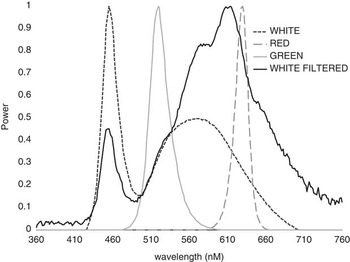
Little research has been done to determine how exposing avian embryos to different wavelengths of light can affect hatchability and also animal welfare post-hatch, therefore, we conducted an experiment to investigate this. Research has shown that eggs can filter light and that some light is filtered differently (Figure 1). The objective of this present study was to determine if utilizing monochromatic red and green LEDs could result in altered hatchability that has been previously seen in broiler eggs that are exposed to white LED light, and to determine how they affect post-hatch growth, fear and stress.
Light frequency spectrum for WHITE, RED and GREEN LED lights and the WHITE LED light spectrum filtered by broiler eggshells. RED and GREEN LED light spectrums are not changed by broiler eggshell.

Material and methods
General procedures
In total four replications were conducted to investigate the differential effects of providing white LED light, red LED light, green LED light or no illumination during incubation on hatchability, chick quality, and post-hatch growth, fear and stress. All methods were approved by the Texas A&M Institutional Animal Care and Use Committee (AUP # 2012-211 and # 2013-0256).
The study was conducted using Cobb500 broiler eggs (n=9216). Eight GQF 1500 incubators and eight GQF 1550 hatchers (GQF Manufacturing, Savannah, GA, USA) were used in each trial, and their front windows were blacked out with cardboard to prevent light intrusion into the machines. Two incubators were operated with the traditional dark method of incubation (0L : 24D, DARK), two incubators were outfitted with cool white (7500 K) LED strips (Superbrightleds WFLS-X3, Saint Louis, MO, USA; WHITE), two with red LED strips (Superbrightleds WFLS-RGBX2; RED, peak at 630 nm) and two with green LED strips (Superbrightleds WFLS-RGBX2; GREEN, peak at 520 nm). Treatment lighting was rotated between incubators to control for incubator differences. LED lights were on each level, with two strips running the length of the racks. The strips were attached to metal frames, which were in turn attached to the bottom of the rack above them. For the top rack, light strips were held up by a metal frame made to rest on the top rack. The lights were operated by a timer, with a 12L : 12D light schedule at 250 lux at egg level as measured using a light meter (Extech 401027; Extech Instruments, Nashua, NH, USA). Two egg trays were set on each rack with each tray holding 48 broiler eggs, for a total of six trays over three levels equaling 288 broiler eggs/incubator. The incubators were maintained at standard temperature and humidity levels of 99.5°F and 55% relative humidity. The eggs were incubated for 18 days, at which time they were moved into the hatchers which were maintained at standard temperature and humidity levels of 98.5°F and 65% relative humidity. The hatchers had no lights and the eggs were in complete darkness. Both incubators and hatchers were controlled by a digital theromostat and were checked several times a day. The temperature and humidity in each machine did not vary by >0.1°F or 2% relative humidity at any time. All of the chicks were weighed and counted at hatch. The quality of the live chicks was assessed, and they were categorized and counted as either no defect, having an unhealed navel, having leg abnormalities, weak, dirty, having traits a hatchery would cull or having any other abnormality. The remaining unhatched eggs were broken out, and counted as pipped, broken, infertile, early dead, mid dead and late dead.
After hatch analysis, 120 chicks from replication 1 from each treatment were set aside and reared for 45 days. The birds were managed according to the guidelines set forth in the Guide for the Care and Use of Agricultural Animals in Research and Teaching (Federation of Animal Science Societies, Reference Forkman, Boissy, Meunier-Salaün, Canali and Jones2010). They were housed in pens measuring 1×2 m with 20 birds/pen and placed in a random-block design within the house. They were fed ad libitum a standard starter, grower and finisher feed milled at the Texas A&M Poultry Research Center. Water was provided through nipple drinkers. The house was illuminated by incandescent bulbs and dimmed to an average of 20 lux at chick level using a light meter (Extech 401027; Extech Instruments) for the first 10 days of growth and then was lowered to 5 lux, and set to a 20L : 4D light schedule for the entire grow out.
Growth and feed conversion
The birds in each pen were weighed at days 0 and 45 and BW gain was calculated by subtracting day 0 weight from day 45 weights. Chicks were randomly allocated to pens. Feed was weighed before it was added to the feeder in each pen and residual feed was weighed back on bird weigh days so that feed intake could be calculated. Feed conversion ratio was calculated by dividing the total feed intake per pen by the total BW gain per pen and was corrected for mortality.
Fear tests
Isolation
To test the fear response of chicks an isolation test was performed. The isolation tests were performed at 10 days of age by randomly collecting 10 birds from a pen, bringing them to a separate area, and placing them in a 133 l uncovered plastic container. The birds were then individually placed in an unlidded 19 l bucket. A timer was set for 3 min, and the number of vocalizations produced by the bird during this time was counted. Later, the bird was placed in a separate holding container. After all 10 birds had been tested, they were returned to their pen, and 10 birds from the next pen were collected and tested. More vocalizations were considered to indicate more fearfulness (Forkman et al., Reference Ghatpande, Ghatpande and Khan2007; Archer and Mench, Reference Archer and Mench2014b).
Tonic immobility
Tonic immobility (TI) was conducted at 5 weeks of age by again collecting the 10 random birds from a pen, bringing them to a separate area, and placing them in a holding container. Methods were modified from previous research by Jones (Reference Jones1986) and Archer and Mench (Reference Archer and Mench2014a). A 21 cm wide by 22 cm high by 30 cm long wooden cradle with the sides sloping out at a 108° angle from the base was obtained, covered in a black cloth and placed on a table. Each bird was individually taken and placed on its back in the cradle. The head of the bird was covered with one hand, whereas the breast was held with the other for ~15 s to induce TI, after which time contact was removed and a timer was started. If the bird righted itself within 15 s, the timer was reset and the above procedure was performed again. If again the bird righted itself in under 15 s, it was recorded as a time of 0. Otherwise, the latency of each bird to right itself (or attempting to right itself) was recorded, with a maximum of 10 min. After all 10 birds had been tested they were returned to their pen, and the 10 birds from the next pen were collected and tested. Longer latency for birds to right themselves were considered to indicate more fearfulness (Jones, Reference Jones1986). The same birds were used in both fear tests and there is no evidence that these two tests would affect each other as they were carried out several weeks apart.
Stress measures
Physical asymmetry
Physical asymmetry of each bird was measured as per Archer et al. (Reference Archer, Shivaprasad and Mench2009) at 45 days immediately after they were euthanized. In brief, using a calibrated Craftsman IP54 Digital Caliper (Sears Holdings, Hoffman Estates, IL, USA), the middle toe length, metatarsal length and metatarsal width were measured for both the right and left legs. The composite asymmetry score was calculated by taking the sum of the absolute value of left minus right of each trait, then dividing by the total number of traits. Thus the formula for this trial would be (|L−R|middle toe length+|L−R|metatarsallength+|L−R|metatarsalwidth)/3=composite asymmetry score.
Corticosterone and serotonin
At 42 days, blood samples were collected from 12 birds/treatment (these birds were different from those used for the humoral immunity or fear tests). The area around the wing vein was sanitized with 70% alcohol, and in preparation, the inside of a 3 ml syringe was lined with a small amount of heparin. Between 1 and 2 ml of blood were collected from each bird. The blood was injected into a plasma separation gel and lithium heparin vaccutainer (BD 368056, BD, Franklin Lakes, NJ, USA), which was temporarily stored in an ice bath. Once all samples had been taken, the vaccutainers were spun down in a Beckman GS-6R centrifuge (Beckman Coulter, Brea, CA, USA) for 15 min at 4000 r.p.m. to separate the cells from the plasma. The plasma was poured off into 2 ml microcentrifuge tubes and stored at −19°C until further analysis. Plasma corticosterone and serotonin concentrations were measured using a commercially available ELISA kit (Corticosterone: ADI-901-097; Enzo Life Sciences, Farmingdale, NY, USA; Serotonin: ADI-EKS-715; Enzo Life Sciences). The inter- and intra-assay %CV were both under 5% for both types of assays. Increases in corticosterone (Cockrem, Reference Cockrem2007) and decreases in serotonin (Nejad et al., Reference Nejad, Kim, Lee and Sung2016) indicated increases in stress.
Humoral immunity titers
To examine the effects the secondary immune response to an antigen, at 2 weeks of age and then again 1 week later, 12 birds/treatment (these birds were different from those used for the corticosterone/serotonin or fear tests) were injected i.m. in the leg with 0.1 ml of 1 ng keyhole limpet hemocyanin (KLH)/0.1 ml of saline. These birds were marked with livestock marker and were not used in any other testing. Three days following the second injection a 1 ml blood sample was collected from the wing vein of all KLH-injected birds. The blood samples were centrifuged and plasma removed and stored at 4°C until analysis. The plasma was analyzed for anti-KLH immunoglobulin (IgG) titer using a commercially available ELISA kit (Alpha Diagnostic Intl. Inc., San Antonio, TX, USA). The inter- and intra-assay %CV were both under 5%. Stress has been shown to decrease humoral immunity (Archer and Mench, Reference Archer and Mench2014b).
Statistical analysis
General linear models were used to investigate treatment, trial, treatment×trial effects on hatchability, embryo mortality, chick quality. Analysis of variance was used to investigate treatment effects on composite asymmetry, isolation, TI, corticosterone, serotonin, weight gain and feed conversion. The LSD test was used to test all planned comparisons. All of the assumptions of GLM were tested (Shapiro–Wilk’s test for normality, Levene’s test for homogeneity of variance). No transformations were needed to meet assumptions. All analyses were performed using SAS 9.3 for Windows (SAS Institute Inc., Cary, NC, USA). Significant differences were determined at P<0.05. No effects were observed on trial or treatment×trial interaction so only treatment effects will be discussed.
Results
Hatchability and chick quality
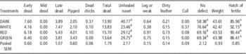
The embryo mortality, chick quality, hatchability data are presented in Table 1. No differences (P>0.05) were observed between treatments in early, mid and late embryo mortality. No differences (P>0.05) in the number of pipped eggs, dead chicks in the hatch tray or overall dead embryos was observed between treatments. Differences were observed (P<0.05) in chick quality with the DARK treatment having more chicks with unhealed navels resulting in less chicks classified as ‘no defect’ chicks overall than all the other treatments; however, no differences (P>0.05) between treatments were observed in the percentage of chicks that had leg issues, dirty feathers or had to be culled at hatch. At hatch there was no difference observed between treatments in chicks weights (P>0.05). The WHITE and RED treatments had a higher (P>0.05) hatch of fertile percentage than both the DARK and GREEN treatments.
The embryo mortality (%), chick quality (%), weight (g) and hatchability (%) of broilers incubated under no light, white, red or green light

A,BDifferent superscript letters within column are significantly different (P<0.05).
Growth and feed conversion
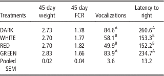
Growth and feed conversion data can be found in Table 2. No difference in initial weight, 45 days weight or feed conversion were observed between treatments (P>0.05).
The weight (kg) and feed conversion ratio (FCR) at 45 days of age, number of vocalizations during 3 min isolation (#/3 min) and latency to right during tonic immobility (s) of broilers incubated under no light, white, red or green light

A,BDifferent superscript letters within column are significantly different (P<0.05).
Fear tests
Results of the isolation and TI fear tests are presented in Table 2. The DARK and GREEN treatments vocalized more during isolation (P<0.05) and had longer latency to right themselves during TI (P<0.05) than WHITE and RED treatments.
Stress measures
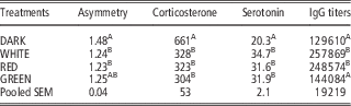
The results of the stress measures are presented in Table 3. The DARK treatment had greater (P<0.05) composite asymmetry than the WHITE and RED treatments. The GREEN treatment was not different (P>0.05) in composite asymmetry than any other treatment. The DARK treatment had higher plasma corticosterone (P<0.05) and lower plasma serotonin (P<0.05) than all other treatments. The DARK and GREEN treatments had lower (P<0.05) anti-KLH IgG titers than the WHITE and RED treatments.
The composite asymmetry scores (mm), plasma corticosterone concentrations (pg/ml), plasma serotonin (ng/ml) and anti-keyhole limpet hemocyanin immunoglobulin (IgG) titers (unit/ml) of broilers incubated under no light, white, red or green light

A,BDifferent superscript letters within column are significantly different (P<0.05).
Discussion
The wavelength of the light that the broiler eggs in this present study were exposed to affected the hatchability of the fertile eggs. Specifically, white light and monochromatic red light increased the hatchability when compared with the dark incubated eggs. The improved hatchability with white light in this present study agrees with previous findings (Archer and Mench, Reference Archer and Mench2014a and Reference Archer and Mench2014b; Archer, Reference Archer2015b and Reference Archer2016). Hluchý et al. (Reference Hluchý, Toman, Cabaj and Adamkovičová2012) had similar findings to this present study with red light increasing hatchability, however, Archer (Reference Archer2016) did not see an increase in hatchability in broiler eggs exposed to red light during incubation. Archer (Reference Archer2016), however, did see an increase in hatchability in white layer eggs that were exposed to the same red light in this present study. It is possible with the increased replication and egg numbers in this present study compared with Archer (Reference Archer2016) that this present study was able to observe a difference missed in that previous study. Green light did not improve hatchability in this present study and appears not to be the wavelength of light responsible for increased hatchability. It is possible that hatchability is related to the stimulation of the opsins in the retina and extra retinal photoreceptors as green light exposure during incubation down regulates the green and red opsins, whereas red light enhances their expression (Rozenboim et al., Reference Rozenboim, Piestun, Mobarkey, Barak, Hoyzman and Halevy2013). This requires further research as the two have not been linked at this point.
Chick quality was also improved by exposing eggs to white and red light during incubation in this present study which agrees with previous research (Archer and Mench, Reference Archer and Mench2014a and Reference Archer and Mench2014b; Archer, Reference Archer2015b and Reference Archer2016). Most of this improved chick quality was due to increased navel maturation resulting is less unhealed navels and navel tags. Navel development has been shown to be influenced by light exposure during incubation, with the accelerated growth caused by light exposure resulting in improved maturation of the navel over dark trials (Erwin et al., Reference Fairchild and Christensen1971). Green light again did not have the same effect as red or white light and does not appear to be the wavelength responsible for the increased hatch rate. This again could be related to opsin expression as a result of light exposure during incubation.
The weight of the chicks at hatch was not affected by the lighting treatments during incubation. This does agree with previous research which observed increased weight as a result of incubating eggs with green light (Rozenboim et al., Reference Rozenboim, El Halawani, Kashash, Piestun and Halevy2003 and Reference Rozenboim, Huisinga, Halevy and El Halawani2004; Zhang et al., Reference Zhang, Zhu, Wang, Li, Gao and Zhou2016). Archer (Reference Archer2016) did not see a difference between white, red or dark incubated broiler chicks in weight at day of hatch.
Post-hatch the effect of exposing eggs to any wavelength of light did not cause increased 45 days weights or feed conversion. This does not agree with previous research which observed increased BWs (Rozenboim et al., Reference Rozenboim, El Halawani, Kashash, Piestun and Halevy2003 and Reference Rozenboim, Huisinga, Halevy and El Halawani2004; Zhang et al., Reference Zhang, Zhu, Wang, Li, Gao and Zhou2016) and feed conversion (Rozenboim et al., Reference Rozenboim, Piestun, Mobarkey, Barak, Hoyzman and Halevy2013) when eggs were exposed to green light during incubation. Possible explanations for the lack of effect in this present study are that, this present study used a different wavelength of green light (520 nm v. 560 nm) and a different illumination schedule (12-h light/12-h dark v. 15-min light/15-min dark) than the previous studies. This further indicate that specific wavelengths may be important in avian development and furthermore that illumination schedules and other factors may also play a role. It has been documented that timing and onset of light exposure during incubation is important for post-hatch effects (Archer et al., Reference Archer, Shivaprasad and Mench2009; Chiandetti et al., Reference Chiandetti, Galliussi, Andrew and Vallortigara2013; Archer and Mench, Reference Archer and Mench2014a and Reference Archer and Mench2014b).
Providing light during incubation has previously been observed to improve bird welfare post-hatch by reducing fear responses and decreasing stress susceptibility (Archer et al., Reference Archer, Shivaprasad and Mench2009; Özkan et al., Reference Özkan, Yalçın, Babacanoğlu, Uysal, Karadaş and Kozanoğlu2012; Archer and Mench, Reference Archer and Mench2013, Reference Archer and Mench2014a and Reference Archer and Mench2014b; Huth and Archer, Reference Huth and Archer2015). This present study confirmed the previous research findings that white light exposure during incubation can improve welfare by reducing fear responses and decreasing stress susceptibility. According to Ratner (Reference Ratner1967), fear of predation is a major component in fear behavior of prey animals, and is linked to predator avoidance behavior. Both of the fear tests used in this present study (isolation and TI) demonstrated that white and red light exposure during incubation results in a decreased fear response. In this present study four measures of stress were used (composite asymmetry, corticosterone, serotonin and humoral immunity) and all four measures demonstrated that white and red light exposure during incubation decreases stress susceptibility. The reduction in fear in stress is very important as itillustrates a clear improvement in animal welfare. The serotonin and fear responses show that the animals were in a better ‘emotional’ state, which resulted in lowered stress hormones, which led to improved immune function and symmetry. This present study is the first time that red light exposure during incubation has been observed to improve bird welfare post-hatch. Green light exposure during incubation did not affect fear response, composite asymmetry or humoral immunity. Green light did affect corticosterone and serotonin concentrations in a similar manner to white and red light. Although related, the stress measures used in this present study are measuring stress in different manners so it is possible that green light may have some positive post-hatch benefits in stress susceptibility reduction, however, it is apparent that white and red light have the greatest benefit on animal welfare post-hatch.
The eggshell itself is an important factor in how light effects the embryo as it absorbs most of the light it is exposed to (Shafey et al., Reference Shafey and Al-Mohsen2002 and Reference Shafey, Al-mohsen, Al-Sobayel, Al-Hassan and Ghnnam2005). Huth and Archer (Reference Huth and Archer2015) noted that white light is filtered in a manner that the wavelengths of light are shifted to the red hue. Figure 1 illustrates this point again and further demonstrates that monochromatic red and green light are not filtered or shifted like white light. As demonstrated by Rozenboim et al. (Reference Rozenboim, Piestun, Mobarkey, Barak, Hoyzman and Halevy2013) green and red light affect the opsins in the photoreceptors differently. It is possible that effect off different wavelengths on the opsins could be the mechanisms or part of the mechanism that is altering physiology during embryogenesis, that has resulted in the observed increases hatchability and decreases fear response and stress susceptibility. We know that light stimulation during incubation alters brain development (Rogers and Krebs, Reference Rogers and Krebs1996) and hormone levels (Zeman et al., Reference Zeman, Gwinner, Herichová, Lamošová and Koštál1999) during development. Both of these have been noted to change behavior (Rogers and Workman, Reference Rogers and Workman1989) and physiology (Nelson and Demas, Reference Nelson and Demas1997) as well. Therefore, these as well as the effect on opsins merit future research to determine the mechanism(s) that are responsible for all the effects observed in this present study.
Whatever the underlying mechanism for the positive effects observed in this present study, the use of white or red light during the incubation of broiler eggs appears to be a worthwhile management technique. Utilizing these wavelengths of light during incubation can increase the number of chicks and more of those chicks are healthy with no defect. Furthermore, this present study illustrates that those chicks will have improved welfare by having reduced fear and a better ability to deal with stressors during production. The addition of light to the practice of incubating commercial poultry needs to be considered as it can improve efficiency and animal welfare simultaneously.
Acknowledgement
The author would like to thank Tyson Foods for donating the eggs used in this study and the undergraduates who helped with the study as well.