Introduction
The occurrence of multiple origins of similar phenotypic features (homoplasy) has long been intriguing to evolutionary biologists and paleontologists, because it provides an explicit opportunity to explore both intrinsic (developmental) and extrinsic (ecological) factors, as well as the issue of predictability, in evolution (Blake Reference Blake1980; Harvey and Pagel Reference Harvey and Pagel1991; Wake Reference Wake1991; Moore and Wilmer Reference Moore and Willmer1997; Gould Reference Gould2002; Hall Reference Hall2003; Vermeij Reference Vermeij2006; Wiens et al. Reference Wiens, Brandley and Reeder2006; Powell Reference Powell2007; Dick et al. Reference Dick, Lidgard, Gordon and Mawatari2009; Eroukhmanoff et al. Reference Eroukhmanoff, Hargeby, Arnberg, Hellgren, Bensch and Svensson2009; Losos Reference Losos2011, Reference Losos2017; Agrawal Reference Agrawal2017; Blount et al. Reference Blount, Lenski and Losos2018; Allmon Reference Allmon2020). Repeated evolution of a feature is often considered indicative of functional utility, even if the function of the feature is presently unknown. Alternatively, it may be the result of developmental constraints of various sorts, which may limit or channel variation. Properly interpreting the repeated evolution of an organismal feature in fossil taxa requires understanding of (1) phylogenetic context, (2) ecological/functional context, (3) external environmental conditions, and (4) morphogenetic patterns and processes leading to the formation of the feature (Fraser et al. Reference Fraser, Gorelick and Rybczynski2015; Lamsdell et al. Reference Lamsdell, Congreve, Hopkins, Krug and Patzkowsky2017; Cole et al. Reference Cole, Wright and Ausich2019). Phylogenetic context is needed to make several critical inferences, including whether the structure is homologous or analogous (i.e., whether it was present due to common ancestry or appeared independently). Knowledge of ecological, functional, and environmental context is required to assess potential hypotheses of functional utility and selection. Morphogenetic information (i.e., how the feature is constructed through ontogeny) is required to assess likelihood of parallelism (shared genetic predisposition resulting in related lineages evolving independently in similar ways).
The appearance of a feature in distantly related clades is typically taken as indicative of an adaptive response (notwithstanding exceptions; e.g., Gould and Lewontin Reference Gould and Lewontin1979; Anderson and Allmon Reference Anderson and Allmon2018 and references therein), though this observation does not necessarily provide evidence of the possible function(s) (Geary et al. Reference Geary, Staley, Müller and Magyar2002; Tseng Reference Tseng2013). Directly establishing utility through hypothesis formation and experimentation (the “paradigm method” [Rudwick Reference Rudwick2018]; e.g., Arnold Reference Arnold1983; Wainwright and Reilly Reference Wainwright and Reilly1994) may help determine whether the feature under investigation is indeed adaptive, exaptive, or nonaptive, and if it is adaptive, how selection may have acted to establish the feature (Wake Reference Wake1991; Wake et al. Reference Wake, Wake and Specht2011). The ecological context can also help determine the function of a feature by identifying what environments selected for that feature (e.g., Harvey and Pagel Reference Harvey and Pagel1991; Vermeij Reference Vermeij2002; Gemmell et al. Reference Gemmell, Trewick, Crampton, Vaux, Hills, Daly, Marshall, Beu and Morgan-Richards2018).
A feature appearing in multiple species or clades is homoplastic when the common ancestor is inferred to have not possessed the feature and its development may be the result of natural selection, developmental or morphological constraint, or atavism (Wake Reference Wake1991; Wagner Reference Wagner1995; Vermeij Reference Vermeij2002). Convergence occurs where a structure is homoplastic due to independent origin under selection. Parallelism is a special case of convergence in which a feature arises multiple times in lineages with a common genetic or structural predisposition toward development of the feature (Blount et al. Reference Blount, Lenski and Losos2018). Like convergence, parallelism implies the absence of the feature in the common ancestor of the taxa in question, but invoking parallelism suggests that common descent is a result of positive constraint (Gould Reference Gould2002). Homoplasy, convergence, and parallelism may be most easily understood as a nested set, with both convergence and parallelism representing cases of homoplasy that are likely aptations. Parallelism represents “homology of underlying generators” (Gould Reference Gould2002: p. 1080; Saether Reference Saether1983; Wake Reference Wake1991), and is therefore homoplastic at the level of the organism (possessing the feature), but represents homology at the level of the shared genetic/developmental factors (whether these are confirmed through genetic sequencing or inferred to be present). Parallelism has been invoked when taxa possessing a feature are close relatives (Scott Reference Scott1891; Gould Reference Gould2002). Genetic or structurally constrained parallelism may be difficult to distinguish from phylogenetic niche conservatism (Wiens and Graham Reference Wiens and Graham2005; Wiens et al. Reference Wiens, Ackerly, Allen, Anacker, Buckley, Cornell, Damschen, Davies, Grytnes and Harrison2010; Peterson Reference Peterson2011), in which features are related to a particular environmental context. Organismal features that are present as the result of shared common ancestry may sometimes facilitate incontrovertible independent invasions of similar environments (e.g., Rosenberg Reference Rosenberg1996), identifying a clear case of parallelism. If descendant species share a common environment, they may also be subject to similar selective pressures and independently evolve a feature through parallelism or convergence.
Parallelism may be identified through a variety of investigative strategies. Direct identification of the common genetic changes that serve as precursors to the evolution of a feature in related lineages would, of course, be the strongest line of evidence (Blount et al. Reference Blount, Lenski and Losos2018). Unfortunately, the genetics underpinning gastropod shell morphology are not yet well understood (Liu et al. Reference Liu, Davey, Banerjee, Han, Yang, Aboobaker, Blaxter and Davison2013; Jackson and Degnan Reference Jackson and Degnan2016; Kocot et al. Reference Kocot, Aguilera, McDougall, Jackson and Degnan2016; Maderspacher Reference Maderspacher2016), and fossils are not amenable to such analyses. An associative approach is frequently taken, in which frequent development of a feature within closely related lineages is considered suggestive of parallelism (Gould Reference Gould2002; Vermeij Reference Vermeij and Briggs2005). Parallelism would also be suggested by repeated evolution in closely related but not sister species (e.g., confamilials) within a similar environmental and ecological context (e.g., Jones et al. Reference Jones, Culver and Kane1992). Evaluating the ontogeny or construction of a feature is an additional means of assessing whether there are common genetic mechanisms predisposing clade members to evolve in a particular manner.
It is also possible that a feature may be evolved initially in parallel, and then further improved upon in one lineage and not another. In that case, parallelism may have contributed to the history of the evolution of a feature in the two lineages and may remain the proper term for describing how the feature originated, but the present developmental pathways would represent an autapomorphy of one sister lineage and retention of the plesiomorphic condition for the other. Therefore, the same feature may be described as the product of parallelism, although the construction of that feature could suggest convergence. This distinction can only be resolved if the fossil record of the lineage preserves this information and is placed in proper phylogenetic context. Absent this information or other evidence (such as experimental evidence of ecophenotypy), we suggest that if one wishes to distinguish parallelism as a special form of convergence, it is preferable to describe a feature that appears to show differences in development or construction as convergent, and not specifically a case of parallelism, even if the species are closely related.
Gastropods show homoplasy in many shell structures (e.g., Hubendick Reference Hubendick1952; Gould Reference Gould1971; Gosliner and Ghiselin Reference Gosliner and Ghiselin1984; Allmon and Geary Reference Allmon and Geary1986; Gosliner Reference Gosliner1991; Van Damme and Pickford Reference Van Damme and Pickford1995; West and Cohen Reference West and Cohen1996; Wagner Reference Wagner2001; Vermeij Reference Vermeij2001, Reference Vermeij and Briggs2005, Reference Vermeij2007; Geary et al. Reference Geary, Staley, Müller and Magyar2002; Strong and Glaubrecht Reference Strong and Glaubrecht2002; Collin and Cipriani Reference Collin and Cipriani2003; Price Reference Price2003; Albrecht et al. Reference Albrecht, Wilke, Kuhn and Streit2004; Wagner and Erwin Reference Wagner and Erwin2006; Ponder et al. Reference Ponder, Colgan, Healy, Nutzel, Simone, Strong, Ponder and Lindberg2008; Chirat et al. Reference Chirat, Moulton and Goriely2013). For this reason, shell characters are often excluded from phylogenetic analyses of extant taxa (e.g., Ponder and Lindberg Reference Ponder and Lindberg1997; Kantor et al. Reference Kantor, Fedosov, Puillandre, Bonillo and Bouchet2017). The pattern (number and orientation) of shell layers in gastropods can nevertheless be phylogenetically informative (West and Cohen Reference West and Cohen1996), indicating conservation of genetic elements. Changes in morphological structure of biominerals can also be interpreted to reflect different genetic programs (Jackson and Degnan Reference Jackson and Degnan2016), although closely related mollusks may also exhibit variation (Carter and Clark Reference Carter, Clark and Broadhead1985). In this paper, we investigate the evolutionary origins of a particular shell feature that occurs in many lineages of caenogastropods: extreme parietal callusing. This feature is convergent, in that the last common ancestor of these lineages lacked it. More detailed analysis, which shows independent evolution of the extreme parietal callus employing the same new (to the lineage) constructional pattern or microstructure in its fabrication, is strongly suggestive of parallelism (Seilacher Reference Seilacher1970; Reif et al. Reference Reif, Thomas and Fischer1985; Briggs Reference Briggs and Briggs2005).
Extreme Parietal Callus
Knowledge of the processes by which gastropods form their shells is poor compared with bivalves, but growing (e.g., Bøggild Reference Bøggild1930; Kohn et al. Reference Kohn, Myers and Meenakshi1979; Bandel Reference Bandel and Carter1990; Fleury et al. Reference Fleury, Marin, Marie, Luquet, Thomas, Josse, Serpentini and Lebel2008; Cusack et al. Reference Cusack, Guo, Chung and Kamenos2013; Kocot et al. Reference Kocot, Aguilera, McDougall, Jackson and Degnan2016). The gastropod shell usually consists of three or four layers: an outer periostracum and two or three calcareous layers—an outer prismatic layer, a middle cross-lamellar layer, and, where present, an inner nacreous layer (Bøggild Reference Bøggild1930; Hyman Reference Hyman1967; Hickman Reference Hickman2013; Salinas and Kisailus Reference Salinas and Kisailus2013). Understanding of the parietal callus (Cox Reference Cox and Moore1960: I107) is especially sparse. The overall morphology of some taxa with extensive callus has been described (e.g., Savazzi Reference Savazzi1992; Vermeij Reference Vermeij and Briggs2005), but the process of its formation remains understudied (but see Irie and Morimoto Reference Irie and Morimoto2016). The callus is clearly laid down by a piece of the mantle associated with the foot when it is extended from the shell aperture. A number of gastropod groups, including Cypraeidae and Olividae, lay down a thin glaze of shell material over the outer layer of the shell (Vermeij Reference Vermeij and Briggs2005), and the callus is presumably formed by a similar mechanism.
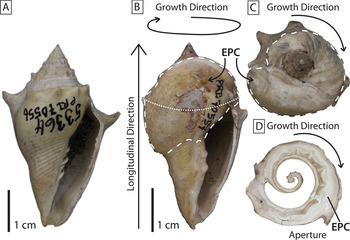
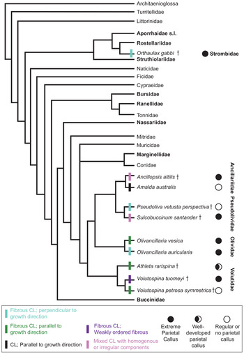
The callus is deposited by a fold of the mantle on the inner (parietal) lip of the aperture, sometimes extending across the body whorl or even onto the spire. “Extreme parietal callus” (EPC) is defined herein as callus that covers more than 50% of the ventral shell surface and that represents substantial thickening (as opposed to a thin glaze lacking complex structure) (Fig. 1). The EPC is observed in a number of distantly related taxa (Table 1, Fig. 2), and varies in thickness, extent, and appearance. In some taxa it is a terminal growth feature, while in others it is present throughout ontogeny. It is known to have evolved in at least 13 caenogastropod families and at least 30 genera (Supplementary Table 1).
An example of the gross morphology of extreme parietal callus (EPC) development. A, A specimen of Volutospina petrosa symmetrica (Eocene of the U.S. Gulf Coastal Plain) that does not construct an EPC. B, A specimen of Volutospina tuomeyi (Eocene, U.S. Gulf Coastal Plain) with an EPC (outlined by the dashed white line) that covers more than 50% of the ventral shell surface as well as the spire and even spines from previous body whorls. The dotted white line represents the general horizon from which cross sections were examined in this study C, View down the longitudinal axis of V. tuomeyi to show the extent of the spire callus and thickness of secondary callus outlined by the dashed white line. D, A cross section taken from V. tuomeyi shows the development of the EPC over the spines.

Phylogeny of Caenogastropoda showing occurrence of extreme parietal callusing. Families where the extreme parietal callus (EPC) is known to occur in at least one species are in bold font. The microstructures employed in the construction of the callus are indicated by colored cross bars (For reference to colors indicated in the key, the reader is directed to the online version.) Filled circles indicate the EPC is present in the species, open circles indicate no EPC, and a partially filled circle was used to indicate enlarged (but not extreme) callus. †Indicates extinct species. The EPC is uncommon in all families examined (see Supplementary Table 1); however, only calloused and control (examined non-EPC) species are shown. Therefore, this figure should not be interpreted as indicating the EPC is primitive for any clade. Phylogeny modified from the strict consensus morphological tree of Ponder et al. (Reference Ponder, Colgan, Healy, Nutzel, Simone, Strong, Ponder and Lindberg2008), using the phylogenies of Simone (Reference Simone2005), Kantor et al. (Reference Kantor, Fedosov, Puillandre, Bonillo and Bouchet2017), Strong et al. (Reference Strong, Puillandre, Beu, Castelin and Bouchet2019), and Friend (Reference Friend2020). A single representative in Kantor et al. (Reference Kantor, Fedosov, Puillandre, Bonillo and Bouchet2017) places Volutidae outside Nassariidae. Either position does not impact our interpretation. CL, crossed-lamellar.

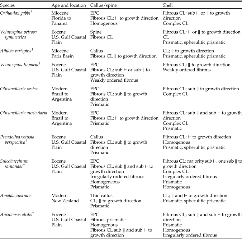
Synopsis of the microstructures employed in the construction of callus, extreme parietal callus (EPC), and shell of each species examined herein. For each species, the microstructural types for callus and shell are each listed from the most commonly used (thickest layers) to least commonly used (thinnest layers). CL = crossed lamellar; “⊢” = perpendicular; “∥” = parallel. †Indicates an extinct species.

Our approach to analyzing convergent evolution of the EPC is twofold. First, we describe the microstructure and ultrastructure of gastropod shells and callus as a proxy for the underlying genetic programming of gastropod shell development. The primitive and dominant microstructure employed in caenogastropod shell material is aragonitic crossed-lamellar (Ponder et al. Reference Ponder, Colgan, Healy, Nutzel, Simone, Strong, Ponder and Lindberg2008). We document the development of shell wall (body chamber of gastropod); thin, moderate, and extreme parietal callus; and spine morphology in 10 specimens representing five gastropod families known to develop an EPC: Strombidae, Volutidae, Pseudolividae, Ancillariidae, and Olividae (taxonomy of olivoids follows Kantor et al. Reference Kantor, Fedosov, Puillandre, Bonillo and Bouchet2017). We then investigate the ecological, environmental, and temporal context for the repeated homoplastic evolution of the EPC, and we perform two experiments to test the functional adaptation of the EPC as ballast for gastropods living in high-energy environments or subject to overturning by predator species.
Methods
Microstructural Imaging
We examined 10 fossil and modern gastropod species from four phylogenetically closely related families, Volutidae, Pseudolividae, Olividae, and Ancillariidae, with a fifth family, Strombidae, serving as an outgroup (Table 1, Fig. 2). A note on the use of the species name Ancillopsis altilis: In order to re-rank the genus Ancillopsis and place it within the Ancillariidae, Palmer and Brann (Reference Palmer and Brann1966) recombined the species Ancillaria altile as Bullia altilis, thereby repositioning B. altilis within the family Nassariidae. More recent opinions (Allmon Reference Allmon1990; Vermeij Reference Vermeij1998; W.D.A. unpublished data; but see Pacaud and Cazes Reference Pacaud and Cazes2014) support the placement of B. altilis within the Ancillariidae (instead of Nassariidae) based on the presence of spiral bands around the fasciole and body whorl, a lack of spiral sculpture, and a lack of a columellar fold, which is typical in species of Nassariidae (Allmon Reference Allmon1990; Vermeij Reference Vermeij1998). For this study, we use the prior species name Ancillopsis altilis (Conrad, Reference Conrad1832) to refer to B. altilis (sensu Palmer and Brann Reference Palmer and Brann1966), and we consider A. altilis part of the Ancillariidae.
One specimen was selected for each species from the collections of the Paleontological Research Institution (PRI; Ithaca, NY). Adult specimens were selected, based on shell size, and only specimens composed of original shell material were included. Six of the species examined develop an EPC, one produces a moderate callus, and the remaining three produce a thin callus (Table 1). To examine gastropod microstructure, we cut sections parallel to the gastropod growth direction and perpendicular to the coiling axis (Fig. 1) with a rock saw. These shell slices were sometimes etched briefly with glacial acetic acid or mechanically abraded with metal dental tools or abrasive paper to reduce saw marks and to better expose the microstructure. The shell sections were then mounted and sputter coated in a ~5 μm coat of gold to prepare them for scanning electron microscopy (SEM) analysis.
Initial observations of shell microstructure were made on a Jeol JCM-5000 Neoscope scanning electron microscope housed at PRI. The majority of the analyses were conducted on a Hitachi Science Systems S-3400N scanning electron microscope in the lab of Ivano Aiello at the California State University Moss Landing Marine Laboratory. The SEM was run under variable pressure, usually allowing 15 to 20 Pa of air pressure to reduce specimen charging that can be a common issue with large specimens such as these gastropod shell sections. Voltage of 15 kV and a current of 50 amperes were applied to each specimen and secondary electrons were detected. SEM observation also confirmed original preservation for the specimens examined. We did not see indications of diagenesis; microstructural crystals were distinct, with regular or only slightly irregular boundaries, and no intergrowths, overgrowths, or blebs were observed between crystals (Knoll et al. Reference Knoll, Landman, Cochran, Macleod and Sessa2016).
Superficial morphological resemblance with underlying structural differences may indicate convergent origins of a feature for a similar function (Averof and Patel Reference Averof and Patel1997; Hickman Reference Hickman2013). In contrast, independent origins of a feature within closely related taxa employing similar underlying structures or constructional arrangements would suggest convergence through parallelism. Here we interpret EPC development as convergent, except where underlying microstructures employed in the construction of the EPC in closely related taxa employ similar microstructural organization, particularly where this common EPC microstructural organization is not the standard crossed-lamellar microstructure most commonly used in caenogastropod shell construction.
Evaluation of Functional Hypotheses
Environment
A literature survey of the environment and ecology of fossil and modern species with an EPC was conducted for the five families included in this study. Species that possess an EPC but not sampled for microstructural analysis were also included (Supplementary Table 2). Geologic depositional environments of fossils were based on primary literature interpretations of sediment types and structures. If an interpretation of the environment was not provided or was too broad for our purposes (e.g., “marine”), then additional research into the reported formation, member, and locality was conducted using MacroStrat and primary literature. For the modern species, collections that reported the marine environment were used. Those that reported only water depth greater than 10 m but less than 200 m were considered shallow subtidal, those that reported 0 m to some depth greater than 10 m were coded peritidal to shallow subtidal. Reported depositional environments were summarized into eight marine environments that can be readily distinguished using the sedimentary record. Each species was considered as a single occurrence within each unique reported depositional environment (i.e., species A might be counted once for shallow subtidal and once for lagoonal, etc.). Diet, tiering relative to the sediment–water interface, and motility were coded for each species using the Paleobiology Database (PBDB) and primary literature.
Wave Tank Experiment
To test the hypothesis that the EPC functions as ballast to maintain aperture-down orientation, we developed a series of tests using the wave tank at State University of New York (SUNY) Geneseo. This tank measures approximately 2 m by 2 m. One specimen of Volutospina tuomeyi (Conrad, Reference Conrad1853) (EPC) was compared with one specimen of Volutospina petrosa symmetrica (Conrad, Reference Conrad1853) (no EPC, control). Each specimen was filled with a wax plug to represent the density of the visceral mass (simulated to be withdrawn into the shell) and to reduce turbulence from the shell aperture. A modern turritelline gastropod visceral mass was dissected from its shell and used to calculate the density of a typical gastropod body. Air was removed from the experimental shells by submerging them with the aperture oriented toward the water surface before placement on the sediment. Experimental conditions varied aperture orientation and shell orientation to wave direction (Supplementary Table 3). Specimens were placed in the center of the tank below the wave break to reflect their fossil and modern environmental distributions in the shallow subtidal. We chose a wave frequency of ~100 per minute and reset the sediment incline to the same approximate angle between each experimental run. Video analysis was conducted to record any change in shell orientation and the degree of translational movement. Each trial lasted 3 to 5 minutes. Fisher's exact test was applied to evaluate statistically significant differences in extent of motion during the trial and final specimen orientation (aperture up or down).
Free-Fall Experiments
To test the hypothesis that the EPC functions as a dive weight to assist in more rapid sinking or to orient the shell aperture-down when resettling on the seafloor, we conducted a drop experiment. Barnett (Reference Barnett1981) showed that EPC-bearing V. tuomeyi were more likely to land aperture-down than non-EPC-bearing congenerics. For our study, the same two Volutospina specimens from our wave study were used, with wax again representing the visceral mass. Initially a fish tank 32 cm in height and then a 0.94-m-tall plastic trash container were used to conduct this experiment, but the horizontal motion of the shell was such that it would make contact with the edge of the container and tumble to the bottom. We therefore made use of a swimming pool to conduct the experiment without these edge effects, and we report the results of this methodology. The two specimens were held slightly below the surface of the pool to allow air to escape before being gently released. Five trials were conducted at each aperture starting orientation (orientated toward the bottom of the pool or toward the water surface), and the experiment was replicated at 0.93 m depth and 2.3 m depth to allow for more time in free fall. The time from release until contact with the bottom of the pool was recorded as well as initial and final shell orientation with regard to aperture position (Supplementary Table 4). Fisher's exact test was used to evaluate the significance of results for the orientation dataset. Student's t-test was used to compare the mean and confidence intervals of settling time for each experimental-depth trial.
Results
Microstructure of the EPC
We observed a wide range of microstructures, including homogenous, simple prismatic, spherulitic prismatic, crossed-lamellar (CL), complex crossed-lamellar (CCL), weakly ordered fibrous, and irregularly ordered fibrous (Table 1). Homogenous microstructure is composed of aragonite granules with highly ordered crystallographic orientation but little physical organization (Bøggild Reference Bøggild1930; Bandel Reference Bandel1979, Reference Bandel and Carter1990; Carter and Clark Reference Carter, Clark and Broadhead1985; Chateigner et al. Reference Chateigner, Hedegaard and Wenk2000; Avery and Etter Reference Avery and Etter2006). The lack of physical organization of homogenous granules and limited organic matrix is less metabolically expensive than construction of other highly ordered microstructural types (Taylor and Layman Reference Taylor and Layman1972; Palmer Reference Palmer1981, Reference Palmer1983, Reference Palmer1992; Belyea and Carter Reference Belyea, Carter and Carter1990; Avery and Etter Reference Avery and Etter2006). Prismatic microstructure is composed of parallel first-order prisms (Bandel Reference Bandel1979, Reference Bandel and Carter1990; Carter and Clark Reference Carter, Clark and Broadhead1985). Spherulitic prismatic is constructed from first-order prisms with second-order prisms that radiate outward (Bandel Reference Bandel1979, Reference Bandel and Carter1990; Hedegaard Reference Hedegaard1997; Ponder and Lindberg Reference Ponder and Lindberg1997; Chateigner et al. Reference Chateigner, Hedegaard and Wenk2000). CL microstructure is constructed from third-order prisms, often composed of twinned aragonite crystals, that are arranged (as sheets or bundles) into second-order lathes (Bøggild Reference Bøggild1930; Bandel Reference Bandel1979, Reference Bandel and Carter1990; Hedegaard Reference Hedegaard1997; Chateigner et al. Reference Chateigner, Hedegaard and Wenk2000; Rodriguez-Navarro et al. Reference Rodriguez-Navarro, Checa, Willinger, Bolmaro and Bonarski2012). Adjacent first-order lamellae are composed of second-order lathes with an opposing (subperpendicular; often 70° to 90°) orientation (Bandel Reference Bandel1979, Reference Bandel and Carter1990; Chateigner et al. Reference Chateigner, Hedegaard and Wenk2000, Rodriguez-Navarro et al. Reference Rodriguez-Navarro, Checa, Willinger, Bolmaro and Bonarski2012). Complex crossed-lamellar (CCL) is composed of first-order lamellae with more than two dip directions and often appears as interweaving second-order lathes (Carter and Clark Reference Carter, Clark and Broadhead1985; Hedegaard Reference Hedegaard1997). To form the highly organized CL microstructure, gastropods must generate an energetically expensive organic framework to control crystal deposition (Wainwright et al. Reference Wainwright, Biggs, Currey and Gosline1976; Palmer Reference Palmer1983; Currey Reference Currey, Trueman and Clarke1988). The density of prisms in microstructures like CL and prismatic can also be more energetically expensive and could limit growth and shell layer thickness (Taylor and Layman Reference Taylor and Layman1972; Palmer Reference Palmer1983). Weakly ordered fibrous is composed of elongate, needle-like prisms organized into loosely packed fibrous bundles with little additional first- or second-order orientation. The spherulitic irregularly ordered prismatic microstructure observed in microconchids is constructed of elongate crystals radially arranged and irregularly oriented (Vinn Reference Vinn2013). We observed for the first time in gastropods an irregularly ordered fibrous microstructure, which has a crystallite arrangement similar to irregular prismatic, but with elongated, needle-like prisms.
Strombidae
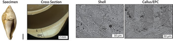
The earliest stromboidean gastropods appear in the Early Jurassic (Ponder et al. Reference Ponder, Colgan, Healy, Nutzel, Simone, Strong, Ponder and Lindberg2008) and for this study serve as an outgroup to represent the development of shell and EPC in a clade that did not evolve the EPC through parallelism. Orthaulax gabbi (Dall, Reference Dall1890) is a Miocene strombid species ranging from Florida to Panama (PBDB). The sampled specimen constructs the shell from fibrous CL microstructure (Fig. 3), and a thin (8 μm) prismatic layer separates the shell and callus. Each shell layer is composed of first-order lamellae oriented either subparallel or subperpendicular to the growth axis; this orientation alternates for each successive layer (Fig. 3). The transition between shell layers takes place over a distance on the order of 100 μm (0.1 mm). In this space, the first-order lamellae from the two layers thin toward the intersection and then interfinger with one another to form a CCL microstructure (Fig. 3). The second-order lamellae shift in orientation over this distance, moving, for example, from an orientation that is subparallel to the growth direction to subperpendicular to the growth direction. CCL appears as braid or plait between each successive shell layer.
Representative specimen, cross section, and shell and callus microstructure from Strombidae. Orthaulax gabbi. The top of the shell image features crossed-lamellar (CL) microstructure showing the subperpendicular orientation of second-order lamellae within each first-order lamellae. The bottom and middle shows the complex CL microstructure formed at the transition between shell layers. The callus image features complex CL formed at the transition between callus layers. Specimen scale bar, 1 cm. EPC, extreme parietal callus; S, shell.

The innermost callus layer of O. gabbi is constructed of homogenous material characterized by no second-order organization. Third-order lamellae are rectangular and 2–4 μm in length. The remainder of the callus is composed of CL microstructure with first-order lamellae that are parallel to the longitudinal shell direction and perpendicular to the growth direction and therefore oriented perpendicular to the shell surface (Fig. 3). First-order lamellae measure 30 μm in thickness and are generally continuous but do thicken, thin, and sometimes pinch out and disappear. Second-order lamellae are poorly developed bundles (2 μm in thickness) loosely organized into lathes made of a single layer of fibrous bundles. The orientation of the lathes abruptly shifts between each successive first-order lamellae to a subperpendicular orientation (Fig. 3). Third-order lamellae are needle shaped, ~10 μm in length. In the callus, there are thin (sub–5 μm), continuous gaps in the microstructure that are parallel to the shell layers but do not disrupt the orientation of first-, second-, or third-order lamellae. Third-order lamellae either terminate and then originate, or decrease in density, to create a more spacious region of shell microstructure but maintain the same orientation on either side of these gaps.
Volutidae
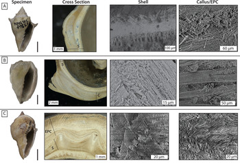
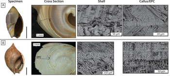
Volutid gastropods diverged from the olivoid clade before the Aptian (Early Cretaceous) (Ponder et al. Reference Ponder, Colgan, Healy, Nutzel, Simone, Strong, Ponder and Lindberg2008; PBDB 2019). Volutospina petrosa symmetrica is a fossil species of volutid gastropod from the Eocene of the U.S. Gulf Coastal Plain (Fig. 4A). This species does not construct any substantial callus and is sister to V. tuomeyi, which develops an EPC (Friend Reference Friend2020). The innermost shell layer of V. petrosa symmetrica is constructed of a thin (0.15 mm) prismatic to homogenous layer. There is an abrupt gap before the next ultrastructural shell layer (0.4 mm), which is composed of fibrous third-order lamellae organized into thin second-order lathes that make up first-order CL lamellae organized parallel to the growth direction. This layer transitions to another shell layer (0.6 mm) in which, again, needle-like third-order lamellae are organized into second-order bundles or thin (25 μm) lathes. Each of the first-order lamellae is constructed of either a thin lathe oriented perpendicular to the growth direction or fibrous bundles oriented parallel to the longitudinal direction (Fig. 4A). The alternating bundles and lathes form space in the shell, which is noticeable as a darker region in the cross-sectional image (Fig. 4A). This more open shell texture is bounded by another shell layer (0.55 mm) composed of tightly packed third-order needles aligned in thin second-order lathes that form first-order lamellae that are subparallel to the growth direction. In this layer of the shell, there are thin (sub–5 μm), continuous gaps in the microstructure where third-order lamellae either terminate and then originate, or decrease in density, to create a more spacious region of shell microstructure. These horizons are parallel to the shell layers, but the gaps do not disrupt the orientation of first-, second-, or third-order lamellae and do not result in the development of a new ultrastructural layer. The third-order needles maintain the same orientation on either side of these gaps. The external-most shell layer (0.25 mm) is composed of first-order lamellae oriented perpendicular to the growth direction and perpendicular to the shell edge. Second-order lamellae appear as thin lathes composed of rectangular third-order lamellae. At the outer shell edge (60 μm thickness), the first-order lamellae bend and bifurcate at an angle subperpendicular relative to the shell edge, forming a spherulitic prismatic microstructural layer. The lamellae then realign to meet the shell edge as a 25 μm layer of prismatic crystals. A microscale crack was observed in the external prismatic layer and CL layer with first-order lamellae perpendicular to the shell edge (Fig. 5A). The crack dissipated ~0.5 mm into the shell as it was deflected by the change in direction of first-order CL structure. Volutospina petrosa symmetrica develops a thin callus, but its spines are composed of material with fibrous CL microstructure (Fig. 4A).
Representative specimens, cross sections, and shell and callus microstructure from Volutidae. A, Volutospina petrosa symmetrica. Shell image features crossed-lamellar microstructure composed of alternating bundles and lathes (darker region) in contact with another shell layer composed of tightly packed third-order needles/second-order lathes. Rightmost microstructure image depicts a poorly organized layer within the CL microstructure of the specimens spine. B, Athleta rarispina. Shell image depicts thin, second-order lathes of needle-like third-order lamellae. The callus image features secondary lamellar bundles. C, Volutospina tuomeyi. The cross section shows the growth of the callus layers over the spines of the shell wall. The shell microstructural image displays fibrous crossed-lamellar structure where fibrous third-order lamellae are organized into thin second-order lathes. The extreme parietal callus (EPC) image shows third-order fibrous needles that are poorly organized into thin second-order lathes along with regions of chaotic fibrous needles. Specimen scale bar, 1 cm. S, shell; C, callus.

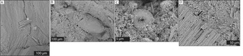
Phenomena observed within the microstructure of various specimens. A, The external 0.5 mm shell layers of Volutospina petrosa symmetrica contain a microcrack that dissipates when it reaches a transition in the orientation of first-order lamellae. B, A siliciclastic grain incorporated in the shell microstructure of Sulcobuccinum santander. C, A coccolith incorporated between the prismatic external shell and irregularly ordered fibrous microstructure of the callus of S. santander. D, Broader view of the contact shown in C, with the coccolith indicated by a black arrow. The prismatic external shell (bottom left) abruptly contacts the irregularly ordered fibrous microstructure of the callus (top right).

Athleta rarispina (Lamarck, Reference Lamarck1811) is a Miocene species from the Paris Basin of France and an outgroup to the two Volutospina species from the Eocene of the U.S Gulf Coastal Plain (Friend Reference Friend2020) (Figs. 2, 4B). The shell of A. rarispina is constructed of an interior layer of blocky prismatic, in some places almost homogenous, microstructure. The majority of the shell thickness is composed of first-order lamellae of CL orientation, organized parallel to the growth direction. These lamellae are constructed from alternating layers of thin, second-order lathes of needle-like third-order lamellae (Fig. 4B). The CL lamellae transform into an 80 μm blocky prismatic layer that forms the exterior of the shell. The callus of A. rarispina is in direct contact with the outermost prismatic layer of the shell.
While A. rarispina does produce a robust callus, it does not meet our definition of the EPC, as it fails to substantially thicken the shell. The callus of A. rarispina is composed of thick (up to 1 mm) microstructural layers composed of CL first-order lamellae made of second-order fibrous bundles composed of needle-like third-order lamellae (Fig. 4B). This is similar to the construction of the shell microstructure, except the bundled nature of the second-order lamellae compared with the thin lathes in the shell creates more space within the callus structure. The fibrous-bundle layers appear as thicker (30–40 μm) darker regions in the shell cross-sectional photograph and alternate with lighter, thin (15 μm) regions of more densely packed third- and second-order lamellae that make up platy CL (Fig. 4B). When examined in cross section, the spine of A. rarispina is formed from 200-μm-long, tightly packed, rectangular third-order lamellae organized in thin second-order lathes. The first-order lamellae are oriented parallel to the growth direction and perpendicular to the external edge of the spine.
Volutospina tuomeyi is an EPC-forming fossil species from the Eocene of the U.S. Gulf Coastal Plain (Fig. 4C). Volutospina tuomeyi develops an EPC that clearly overlaps the shell and spines; filling in the voids between the spines but without dissolution of any prior shell in the process. The shell is composed of many thin (0.625–0.75 mm) layers of weakly ordered fibrous microstructure as well as fibrous CL microstructure. Fibrous third-order lamellae are organized into thin second-order lathes (Fig. 4C). First-order lamellae are commonly oriented parallel to the growth direction and perpendicular to the shell edge. The spines are formed from continuous shell layers in which the lamellae lengthen toward the shell edge to create thicker layers (Fig. 4C). In the spines, the lamellar orientation becomes subperpendicular to the shell edge due to the addition of wedge-shaped lamellae.
Similar to the shell wall, the EPC of V. tuomeyi is constructed of multiple thin layers (20–40 μm) of fibrous CL microstructure. In contrast to the shell, the third-order fibrous needles are poorly organized into thin second-order lathes (Fig. 4C). These lathes are discontinuous and oriented subparallel to the growth direction and subparallel to the shell edge to form first-order lamellae. Among the lathes are regions of chaotic fibrous needles without first-, second-, or third-order organization. The more external callus layers are constructed of these fibrous packages, with first-order lamellae often oriented perpendicular or subperpendicular to the growth direction and perpendicular to the shell edge.
Olividae
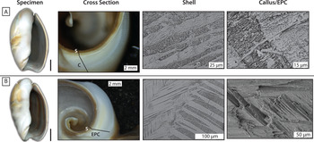
Olivancillaria vesica (Gmelin, Reference Gmelin1791) is a modern gastropod ranging from southeastern Brazil to Argentina in the family Olividae (Fig. 6A) (Jurberg Reference Jurberg1970; Teso and Pastorino Reference Teso and Pastorino2011). Olivancillaria vesica constructs its shell from fibrous third-order lamellae organized into thin second-order lathes that form first-order lamellae that are subparallel to the growth direction. The first-order lamellae are ~20 μm thick, but they sometimes split or branch into 5 μm units (Fig. 6A). Unlike in other species, the internal shell surface does not have a prismatic layer; rather, the CL microstructure appears to dissolve, forming the surface of the body chamber. This might represent secondary dissolution of the inner shell wall. At the outer edge of the shell, the CL microstructure abruptly terminates and a thin (6 μm) prismatic horizon is deposited.
Representative specimens, cross sections, and shell and callus microstructure from Olividae. A, Olivancillaria vesica. The shell microstructural image shows pinching and expansion of first-order lamellae in crossed-lamellar (CL) microstructure. The callus microstructural image features a thin (1.5 μm) prismatic horizon separating wedges of CL callus. The CL orientation does not change across these prismatic horizons. B, Olivancillaria auricularia. The shell microstructural image shows a transition between shell layers where the orientation of first-order lamellae changes. During this transition, the second-order lathes bend and braid into the new orientation over an ~50 μm interval of complex CL. The extreme parietal callus (EPC) image shows fibrous third-order lamellae organized into tightly packed second-order lathes. Specimen scale bar, 1 cm. S, shell.

The callus of O. vesica is composed of CL microstructure, constructed of the same first-, second-, and third-order lamellae as the shell. The first-order lamellae are organized subparallel to the growth direction. These lamellae are not continuous in thickness, but pinch and swell throughout the callus and thicken from the internal surface in contact with the shell wall toward the external callus edge. Layers within the callus are indicated by thin (1.5 μm) prismatic horizons that emerge tangential to the shell and extend through the callus to demarcate wedges of callus material. There are more of these prismatic horizons within the interior portion of the callus (Fig. 6A). The CL lamellae do not change in orientation across these prismatic horizons (unlike the interaction of CL and prismatic microstructure in the callus of Amalda; see “Ancillariidae”).
Olivancillaria auricularia (Lamarck, Reference Lamarck1811) is a modern gastropod with an EPC; its range is from southeastern Brazil to Argentina in the family Olividae (Jurberg Reference Jurberg1970; Teso and Pastorino Reference Teso and Pastorino2011). The shell is constructed of CL microstructure composed of fibrous third-order lamellae arranged into thin second-order lathes that form discrete first-order lamellae (Fig. 6B). Each shell layer is composed of pairs of alternating first-order lamellae oriented at a subperpendicular angle. At the transition between shell layers, the overall orientation of first-order lamellae switches from perpendicular to the growth direction to parallel to the growth direction. Approaching this transition, the first-order lamellae begin to branch apart or thin substantially to ~2 μm limbs (Fig. 6B). During the transition between shell layers, the second-order lathes bend and braid into the new orientation over an ~50 μm interval of CCL, a pattern observed in strombids and other olivoids. The external shell is composed of a 10-μm-thick prismatic horizon that rapidly emerges from the CL lamellae. The callus is deposited on top of this prismatic layer, with a thin gap separating the shell and callus and no evidence for dissolution.
The EPC of O. auricularia is composed of fibrous third-order lamellae organized into tightly packed second-order lathes that form first-order lamellae (Fig. 6B). Like the shell, these first-order lamellae form distinct bands oriented perpendicular to the growth direction and perpendicular to the shell margin. Thin prismatic horizons separate callus layers, just as in congener O. vesica.
Pseudolividae
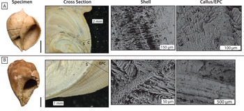
Pseudoliva vetusta perspectiva (Conrad, Reference Conrad1860) is a fossil species from the early Eocene of the U.S. Gulf Coastal Plain and represents a control (non-EPC) species for comparison with its congener, Sulcobuccinum santander (Gardner, Reference Gardner1945) (Fig. 7A). The innermost shell layer (8 μm) of P. vetusta perspectiva is constructed from prismatic microstructure oriented perpendicular to the inner shell edge, which then grades into a thin (~20 μm) homogenous layer. Homogenous material is constructed of 0.5–1.5 μm third-order rectangular lamellae with loose second-order organization parallel and perpendicular to the internal shell surface. The majority of the shell is constructed of a multitude of CL layers (15–180 μm) with transitions indicted by changes in third-order lamellae packing density. Fibrous, needle-like third-order lamellae (4–16 μm) in length are organized into second-order bundles (12 μm) and extremely thin (2–3 μm) lathes. Alternating first-order lamellae are constructed of either bundles or lathes; thin lathes are oriented perpendicular to the growth direction and subparallel to the longitudinal direction and alternate with first-order lamellae composed of thicker bundles oriented perpendicular to the growth direction but at an acute angle to the lathes. These fibrous bundles produce a great volume of space within the shell microstructure (Fig. 7A). The external-most shell layer is formed from warped third-order lamellae from a CL layer that reorient through a spherulitic prismatic transition to form a thin (10 μm) prismatic layer perpendicular to the shell edge.
Representative specimens, cross sections, and shell and callus microstructure from Pseudolividae. A, Pseudoliva vetusta perspectiva. The shell image depicts alternating first-order lamellae; thin layers made of tightly packed lathes alternate with lamellae composed of fibrous bundles that appear as more spacious regions within the shell microstructure. The callus is primarily composed of crossed-lamellar (CL) microstructure constructed of fibrous needles of third-order lamellae organized into thin lathes. B, Sulcobuccinum santander. The shell microstructural image shows thin fibrous prismatic horizons (30 μm thick) extending from bottom left to top right, separating CL layers and marking a change in the CL lamellar orientation between subsequent layers. The extreme parietal callus (EPC) microstructural image shows multiple CL layers of variable thickness separated by thin prismatic or fibrous homogenous layers. Specimen scale bar, 1 cm. S, shell; C, callus.

The callus of P. vetusta perspectiva is deposited directly on the external-most shell layer, without interweaving or dissolution. The innermost callus layer is a series of thin (15 μm) fibrous prismatic layers. The remainder of the callus is constructed of alternating layers of fibrous CL microstructure (~50–120 μm) and prismatic layers (~50 μm). Fibrous needles of third-order lamellae (15 μm length) are organized into thin lathes (up to 25 μm) oriented subparallel to the growth direction and shell edge (Fig. 7A). When these layers are interrupted by prismatic horizons, the third- and second-order orientation of fibrous CL is not altered and continues in the same orientation on the other side. Additionally the callus contains thin continuous gaps formed by terminations or density decreases of third-order laminae, similar to those described for O. gabbi and V. petrosa symmetrica.
Sulcobuccinum santander (for discussion of generic assignment, see Vermeij Reference Vermeij1998) is an EPC-forming Eocene gastropod from the U.S. Gulf Coastal Plain (Fig. 7B). The inner edge of shell is a 10 μm prismatic layer, closely packed to form the smooth inner surface. The next most interior layer is homogenous, composed of small polygons up to 4 μm in length with no second- or third-order orientation. Diagenesis is ruled out, considering the well-preserved nature of the rest of the shell layers. The remainder of the shell is constructed of multiple layers (100–400 μm thick) composed of fibrous CL and irregularly ordered fibrous material. In the CL layers, needle-shaped third-order lamellae (~30 μm length) are organized into second-order thin lathes that are loosely organized into first-order lamellae. First-order structure alternates throughout the shell from an orientation subperpendicular to the growth direction to one subparallel to the growth direction, always oriented subperpendicular to the shell margin. Irregularly ordered fibrous layers are composed of 0.5–5 μm rectangular blades with no second- or third-order orientation. Thin fibrous prismatic horizons (30 μm thick) appear between CL layers and mark a change in the orientation between subsequent layers (Fig. 7B). The external layer is prismatic and fully terminates before the addition of the callus layer. Within the chaotic fibrous layer of the shell, we observed an amorphous clast (0.27 mm by 0.14 mm) lacking carbonate microstructure (Fig. 5B). A single coccolithophore plate was observed resting between the external-most S. santander shell and the subsequent callus, suggesting: (1) the shell was exposed before the callus was secondarily added, and (2) callus was added rapidly and without dissolution, as evidenced by the perfect preservation of this coccolith plate (Fig. 5C,D).
The EPC of S. santader is composed of multiple layers of irregularly ordered fibrous microstructure, fibrous CL microstructure, and prismatic horizons, which range in thickness from tens to hundreds of micrometers (Fig. 7B). Individual filaments have a rectangular profile and are 20–30 μm in length. These fibers are organized into thin second-order lathes. These lathes construct first-order lamellae at varying densities, sometimes well packed and in other layers, more loosely ordered. Lamellae are oriented subparallel and subperpendicular to the growth direction. There is no emergent pattern of layer thickness, orientation, or density of lamellae packing throughout the callus. At the transition from one callus layer to the next, platy prismatic or fibrous homogenous texture separates the CL layers and allows for a change in orientation without distortion of the lamellae. At the external edge of the callus, the third-order lamellae shorten and reorient to form a prismatic layer ~20 μm thick.
Ancillariidae
Amalda australis (Sowerby, Reference Sowerby1830) is a modern gastropod from New Zealand that develops a callus similar to other olivoid gastropods, but does not form an EPC (Fig. 8A). The innermost layer (12 μm thick) is constructed of blocky prismatic microstructure. The remainder of the shell is composed of three layers of CL microstructure constructed from rectangular third-order lamellae arranged in thin second-order lathes that stack to form 10- to 15-μm-wide first-order lamellae. The orientation of first-order lamellae are subperpendicular to one another and perpendicular or subperpendicular to the shell edge. Shell layers alternate from perpendicular to the growth direction to parallel to the growth direction (Fig. 8A). The transition between shell layers is achieved through thinning of first-order lamellae and change in orientation of the second-order lathes over a 40-μm-thick shell region, the same CCL interwoven pattern observed in O. gabbi. CL lamellae bend to form a spherulitic prismatic microstructure and then join to form a 20-μm-thick prismatic horizon as the external-most shell surface.
Representative specimens, cross sections, and shell and callus microstructure from Ancillariidae. A, Amalda australis. The shell microstructural image depicts crossed-lamellar (CL) microstructure composed of thin second-order lathes. The callus microstructure features one of the thin prismatic horizons which mark a transition in CL orientation. B, Ancillopsis altilis. The shell microstructural image depicts from bottom to top: an irregular fibrous layer, a thin blocky prismatic layer, and a layer of loosely organized fibrous needles. The extreme parietal callus (EPC) microstructure shows the interior EPC layers composed of alternating thin layers (5–40 μm) of fibrous prismatic and homogenous shell material. Specimen scale bar, 1 cm. S, shell; C, callus.

The callus of Amalda is overlain on the prismatic shell surface. The CL microstructure of the callus is initially composed of fibrous third-order lamellae that are organized into thin lathes that form first-order lamellae. The initial CL horizon exhibits a slight bend in the long axis of the third- and second-order lamellae. The remainder of the callus is well organized, composed of CL microstructure subperpendicular to the shell edge and parallel to the growth direction. Within the callus, CL microstructure abruptly meets thin (8 μm) prismatic horizons that mark a transition in CL orientation on either side (Fig. 8A).
Ancillopsis altilis is a fossil species from the Eocene of the U.S. Gulf Coastal Plain previously assigned to Nassariidae but more properly placed in Ancillariidae (Vermeij Reference Vermeij1998; W.D.A. unpublished data). The interior portion of the shell is constructed of fibrous prismatic and homogenous layers (Fig. 8B). The majority of the shell is composed of CL microstructure constructed from needle-like third-order lamellae arranged into either second-order fibrous bundles or thin lathes organized into first-order lamellae. These lamellae are oriented subparallel and subperpendicular to the growth direction. Additional shell layers completely lack structure and are composed of chaotic fibrous needles. The external-most shell layer is composed of a dense blocky prismatic texture that is composed of blunt lamellae that are relatively well packed. The transition to the callus is marked by additional thinner prismatic layers composed of loosely packed, fibrous lamellae.
The callus of A. altilis is formed of layers of homogenous, prismatic, and fibrous CL microstructures that vary in thickness on a scale of tens to hundreds of micrometers (Fig. 8B). The initial callus is composed of alternating thin layers (5–40 μm) of fibrous prismatic and homogenous shell material with no clear alternating pattern. As the thickness of the callus layers expands, the layers are composed of fibrous CL microstructure made of third-order lamellae of fibrous needles organized into thin second-order lathes. Thin lathes are stacked into first-order lamellae with the orientation of fibers subparallel and subperpendicular to the growth direction and to the shell margin. At the outermost callus, the CL fibers merge to form a prismatic layer (~10 μm thick) perpendicular to the shell edge.
Functional Ecology
Environment
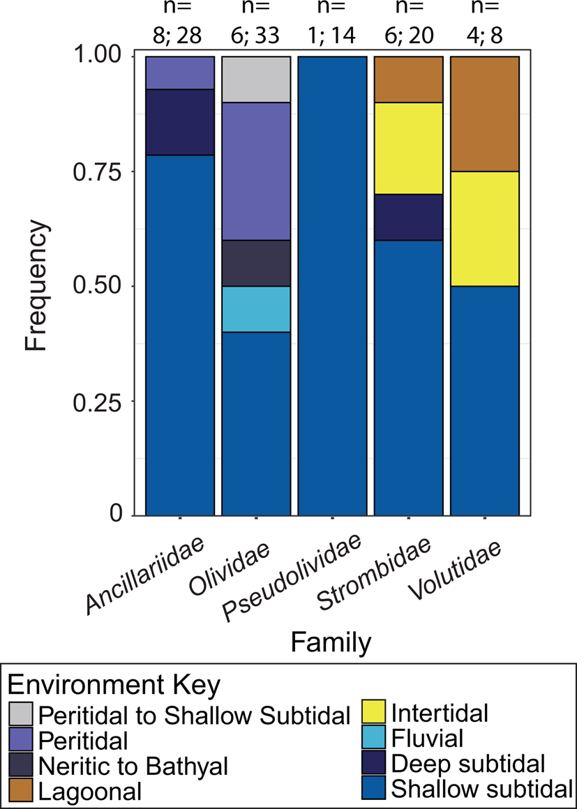
A literature survey of the environment of deposition and ecology of species with an EPC from the five families included in this study indicates these species were primarily from shallow subtidal, intertidal, and lagoonal environments (Fig. 9). Modern species of Olividae are even known to swim or surf in the swash zone (Wilson Reference Wilson1969; Farmer Reference Farmer1970; Brown Reference Brown1971, Reference Brown2001; Cyrus et al. Reference Cyrus, Rupert, Silva, Graf, Rappaport, Paladino, Peters, Marine, Station, Grande, Cruz and Rica2012). The one instance of an olivid found in a bathyal environment is thought to be an instance of postmortem transport, as it deviates from the group's documented environmental preference for shallow subtidal habitats
The environmental distribution of extreme parietal callus–bearing species from the five families finds the majority of occurrences in the shallow subtidal, intertidal, and peritidal environments. n = number of species evaluated; number of unique localities. (For reference to colors indicated in the key, the reader is directed to the online version.) Data available in Supplementary Table 2.

Wave Tank
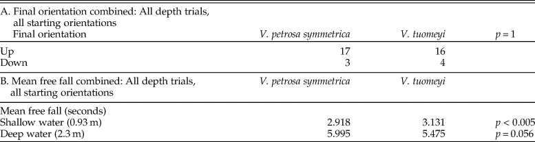
We find significant evidence (Fisher's exact test p = 0.045) that specimens with an EPC experience little to no wave transport in a high-energy environment (Table 2). In four trials, V. tuomeyi rolled over to an aperture-down position and then remained stationary; in the remaining trials, the specimen did not move. In contrast, in six trials, V. petrosa symmetrica rolled actively in the swash zone until the trial was ended or until the specimen was transported below the wave base. From this limited dataset, we suggest the EPC might assist in stabilizing the shell in a high-energy environment. We found no evidence that the presence of an EPC results in a change in resting shell orientation in a high-energy environment. There was no significant difference in final shell orientation; the majority of trials ended with both species oriented in the starting orientation, with their apertures facing away from the sediment, toward the water surface (Table 2).
Results of wave tank experiment on Volutospina petrosa symmetrica and Volutospina tuomeyi suggest a significant role for extreme parietal callus (EPC) as a shell stabilizer in a high-energy environment. We find no significant evidence in final orientation for any experimental treatment. Supplemental Table 3 shows each experimental trial individually. Analyses were conducted on combined data.

Drop Tank
We find no significant difference in final orientation for specimens with or without an EPC following a free fall through the water column (Fisher's exact test p = 1) (Table 3). In the majority of trials, most specimens reached the bottom of the pool in the “aperture-up” position, irrespective of the presence of an EPC or starting orientation (Table 3A, Supplementary Table 4). A significant difference in free fall (Fisher's exact test p < 0.005) was observed in the deep-water trial, with V. tuomeyi reaching the bottom on average in 5.5 seconds (95% confidence interval [CI] = 0.311) compared with V. petrosa symmetrica's average of 6 seconds (95% CI = 0.730) (Table 3B). This suggests a role for the EPC as a “dive weight,” assisting the gastropod in reaching the sediment faster than species without an EPC. However, in the shallow-water trial, V. petrosa symmetrica settled to the bottom of the pool faster than V. tuomeyi (Fisher's exact test p = 0.056). These two trials produced conflicting results and must be interpreted with caution, considering only one specimen from each species was used and therefore might not be representative of the species as a whole.
Results of pool drop. A, There is no significant difference in final orientation for any experimental treatment (Fisher's exact test p =1). B. Volutospina tuomeyi was found to free fall significantly faster than Volutospina petrosa symmetrica in deep-water trials (Fisher's exact test p < 0.005). Supplemental Table 4 shows each experimental trial individually. Analyses were conducted on combined data.

Discussion
Microstructure and Evolution
The vast majority of species in the five caenogastropod families considered in this study do not display an EPC, suggesting each lineage has generally converged on the development of an EPC. Microstructural analyses of the shell wall (as opposed to spine or callus), callus, and EPC within and between the families allowed us to document specific cases of parallel evolution of the EPC within Olivancillaria and parallel evolution of the EPC between Pseudolividae and Ancillariidae. We suggest that the genus Olivancillaria converged on EPC development when compared with other olivoid gastropods and that EPC development in Volutidae is also convergent when compared with other families considered in this study.
Stromboidean gastropods diverged from the Volutidae and olivoid clades in the Early Jurassic (Ponder et al. Reference Ponder, Colgan, Healy, Nutzel, Simone, Strong, Ponder and Lindberg2008). The shell wall and EPC of O. gabbi are constructed of fibrous CL microstructure. The organic content required for the highly controlled deposition of first-, second-, or third-order lamellae in CL microstructure is considered to be energetically expensive (Taylor and Layman Reference Taylor and Layman1972; Wainwright et al. Reference Wainwright, Biggs, Currey and Gosline1976; Palmer Reference Palmer1983; Currey Reference Currey, Trueman and Clarke1988).For the purposes of this study, the microstructure of the strombid gastropod O. gabbi is considered representative of the ancestral condition, as CL microstructure (with small layers of prismatic) is the most common microstructure observed in caenogastropods (Bøggild Reference Bøggild1930; Bandel Reference Bandel1979, Reference Bandel and Carter1990; Hedegaard Reference Hedegaard1997; Chateigner et al. Reference Chateigner, Hedegaard and Wenk2000; Ponder et al. Reference Ponder, Colgan, Healy, Nutzel, Simone, Strong, Ponder and Lindberg2008; Rodriguez-Navarro et al. Reference Rodriguez-Navarro, Checa, Willinger, Bolmaro and Bonarski2012). Formation of callus with the same microstructural organization as the shell wall is likely the default microstructure for most gastropods producing shell material through normal mantle-controlled deposition. This is because producing callus material in a different microstructural organization requires the presence of genetic programming identifying the callus region and also signaling that a different biomineralization pathway is to be employed. The alternative, that the callus is remotely biomineralized (Checa Reference Checa2000) and therefore less organized, is not tenable, as mantle tissue is extensive in olivoids (the majority of species examined herein).
Parallel and Convergent Evolution in Olivancillaria
Olivancillaria is a member of the family Olividae, which is the outgroup to the Ancillariidae + Pseudolividae clade within Olivoidea (Kantor et al. Reference Kantor, Fedosov, Puillandre, Bonillo and Bouchet2017) (Fig. 2). Olivancillaria was recently taxonomically reassessed, but the position of the genus within Olivoidea remains phylogenetically unresolved (Teso and Pastorino Reference Teso and Pastorino2011; Kantor et al. Reference Kantor, Fedosov, Puillandre, Bonillo and Bouchet2017). Of the eight living species of Olivancillaria, all are callus-bearing and all but three produce an EPC. In Olivancillaria, the conserved use of highly ordered fibrous CL microstructure in the shell and callus/EPC of both species examined here suggests parallelism in the development of an EPC. Each species likely evolved an EPC independently, and our observation of similarity in biomineralization suggests shared genetic pathways. An interpretation of homologous EPC development within Olivancillaria might also be demonstrated by future phylogenetic work.
Both Olivancillaria species in this study form CCL microstructure during shifts in first-order lamellar orientation at the transitions between discrete shell layers. This braiding pattern is possibly homologous to the shell construction observed in strombids, which also employ this strategy to alternate CL orientation between shell layers. The well-ordered microstructure in the shell wall and callus/EPC of Olivancillaria suggests high levels of genetic control over biomineralization in this clade, in contrast with other olivoids (Ancillopsis, Sulcobuccinum), which make use of poorly ordered microstructure in EPC construction. These differences in microstructure lead us to conclude that the development of the EPC in Olivancillaria as a clade is a result of convergent evolution. Olivancillaria developed an EPC independently from its olivoid relatives and uses a unique, highly controlled shell programming pathway to generate the EPC.
Parallel Evolution of the EPC between Pseudolividae and Ancillaridae
Both Pseudolividae species use multiple (10+) layers of weakly ordered fibrous CL in their shell-wall and callus construction, but S. santander uses more poorly ordered homogenous and irregularly ordered fibrous material to construct an EPC. These poorly organized microstructures require less metabolic energy for deposition, possibly allowing for more rapid shell construction (Taylor and Layman Reference Taylor and Layman1972; Palmer Reference Palmer1981, Reference Palmer1983, Reference Palmer1992; Belyea and Carter Reference Belyea, Carter and Carter1990; Avery and Etter Reference Avery and Etter2006). The presence of a siliciclastic grain within the shell wall (Fig. 5B) suggests the retraction of the mantle and periodic exposure of the propagating shell margin to the environment whereby sediment becomes entrained in the shell matrix. In both species, a prismatic horizon and a micron-scale gap separates the shell from the initiation of callus/EPC deposition (Fig. 5C,D). The observation of a coccolith plate between the shell and callus layers (Fig. 5C,D) is also further evidence for exposure of the shell-wall surface before subsequent deposition of the callus without any resorption of the underlying shell.
In the pseudolivids’ sister family, the Ancillariidae, the species A. altilis (Eocene, U.S. Gulf Coastal Plain) makes great use of homogenous and fibrous prismatic microstructure in multiple (10+) layers to form the EPC, in contrast with the highly organized callus formation in its confamilial A. australis (modern, New Zealand).The use of a mixture of homogenous, fibrous prismatic, and fibrous CL in multiple orientations and their phylogenetic position in sister families suggests parallel evolution of the EPC in A. altilis and S. santander. Each constructs an EPC from a multitude of layers and poorly organized microstructure that differentiates it from well-ordered shell-wall and callus construction of confamilials. The similarity in A. altilis and S. santander microstructural development of the EPC suggests shell modification through a shared developmental pathway that results in shell wall and EPC constructed of multiple layers (10+) of poorly ordered microstructure. The diversity of microstructure (seven types; Table 1) employed by olivoids represents a more general expression of shared genetic pathways for employing diverse or less-controlled microstructures during biomineralization.
The interpretation of parallel evolution of the EPC in Ancillopsis and Sulcobuccinum hinges on the phylogenetic placement of A. altilis within the Ancillariidae (instead of Nassariidae) (see Allmon Reference Allmon1990; Vermeij Reference Vermeij1998). If Ancillopsis was within Nassariidae, which is phylogenetically more distant, with an Early Cretaceous divergence point, then its shared microstructure with Sulcobuccinum would more likely represent general convergence, given a more distant shared ancestry. This example illustrates how important phylogeny is for questions regarding evolutionary ecology.
Convergence by Volutidae
Volutidae converge on the production of the EPC. Volutidae diverged from the olivoids before the Aptian (Early Cretaceous) (Ponder et al. Reference Ponder, Colgan, Healy, Nutzel, Simone, Strong, Ponder and Lindberg2008). In Kantor et al. (Reference Kantor, Fedosov, Puillandre, Bonillo and Bouchet2017), genetic analyses of a specimen of Melo melo place Volutidae outside Nassariidae, which is in contrast with the Ponder et al. (Reference Ponder, Colgan, Healy, Nutzel, Simone, Strong, Ponder and Lindberg2008) morphology-based phylogenetic placement shown in our Figure 2. Either phylogenetic placement does not change our interpretation of convergence of Volutidae and olivoids on EPC construction. The first known occurrence of an EPC in both olivoids and volutids is from the Paleogene, suggesting no shared ancestral development of this morphological structure. Volutospina tuomeyi constructs both shell wall and callus from weakly ordered fibrous and fibrous CL microstructure, a unique deviation from the more ordered microstructures observed in the shell wall and callus of its confamilials (Fig. 4). The weakly ordered EPC microstructure of V. tuomeyi might be derived from the fibrous CL microstructure used to form shell and spines observed in both V. petrosa symmetrica and A. rarispina. The microstructure of the EPC in V. tuomeyi is different from the homogenous, irregularly ordered fibrous, and fibrous prismatic observed in olivoids. Volutid gastropods independently acquired an EPC following a different morphogenic process from their sister clade, the olivoids, which we interpret as reflecting independent evolution of the genetic programming and convergence of EPC production strategies (Jackson and Degnan Reference Jackson and Degnan2016).
All of the EPC-bearing species examined in this study express anomalous shell-wall growth (Table 1). In these species the first-order lamellae of CL microstructure are oriented both parallel to the growth direction in some shell-wall layers and perpendicular to the growth direction in other shell-wall layers. Specimens with normal secondary callus development only show shell-wall CL first-order lamellae in one orientation; either the parallel or perpendicular growth orientation plane. We interpret the use of diverse (often poorly ordered) microstructure in EPC development and variable orientation of shell-wall lamellae only in EPC-bearing specimens as indicative of some ancestral, shared genetic programming in which there is also a deviation of first-order lamellae organization in shell-wall biomineralization when the EPC is developed.
Functional Ecology
The majority of the gastropod species with an EPC from the five families considered in this study live in shallow subtidal, intertidal, and peritidal habitats and are actively mobile and epifaunal or semi-infaunal (Fig. 9, Supplementary Table 2). Members of Ancillariidae, Olividae, Pseudolividae, and Volutidae with an EPC are carnivores, while the Strombidae with an EPC are grazers or omnivores. The shallow subtidal environment is above storm-wave base and influenced by both fair-weather and storm waves. Semi-infaunal and epifaunal gastropods living in this environment require adaptations for wave energy and exposure to shell-crushing and shell-drilling predators.
One adaptive hypothesis that would be applicable across the diversity of shell shapes exhibited by families with EPC-bearing species is use as a sink for excess carbonate ions or a buffer against variations in carbonate saturation state (Alyakrinskaya Reference Alyakrinskaya2005). If the EPC is involved in such chemical regulation, it would be expected to be highly plastic within EPC-bearing taxa and to show signs of resorption or redissolution (e.g., Kohn et al. Reference Kohn, Myers and Meenakshi1979). We did not observe abrupt prism terminations or dissolution surfaces within the callus microstructure and therefore do not support a chemical regulation function for the EPC.
The results of the pool drop experiment do not support the presence of the EPC for function in orienting the shell if dropped by a potential predator or swept off the level-bottom habitat by storm waves. Unlike studies of Athleta (Volutospina herein) by Barnett (Reference Barnett1981), our work revealed conflicting differences in settling rate and no increase in aperture-down orientation by EPC-bearing species of V. tuomeyi compared with callus-free V. petrosa symmetrica. We interpret our opposing observation (contra Barnett Reference Barnett1981) as the result of our methodological approach, which provided more vertical distance to accommodate natural settling behavior. This experiment was limited by application to only one EPC family (and one specimen representing each species) and might yield different results in other families with different shell morphology. In addition, this study was conducted in a quiet pool without wave turbulence. A study that could include the influence of wave energy at depth would improve the validity of the results. Finally, while the density of the visceral mass was reconstructed using wax, our experimental approach did not consider how the weight of the callus might interact with behavioral deployment of the foot or mantle of the gastropod to assist in settling velocity and final orientation or for surfing behaviors (Wilson Reference Wilson1969; Farmer Reference Farmer1970; Brown Reference Brown1971, Reference Brown2001; Cyrus et al. Reference Cyrus, Rupert, Silva, Graf, Rappaport, Paladino, Peters, Marine, Station, Grande, Cruz and Rica2012).
Wave tank experiments did suggest a role for the EPC as ballast. The test specimen of V. tuomeyi with an EPC was found to remain stationary or to reorient aperture-down and cease rolling, while the control specimen of V. petrosa symmetrica did not reorient and settle when exposed to gentle wave activity, but rather continued to move until the end of the trial. As ballast, the EPC would function either to maintain proper orientation or to right the animal when overturned in surf or when dropped by predators (Palmer Reference Palmer1977; Barnett Reference Barnett1981). Improper orientation can affect mobility and can also increase the animal's susceptibility to foot-nipping predators as it rights itself (Scott Reference Scott1902; Palmer Reference Palmer1977). Olivoids and volutids in particular have shell morphologies adapted toward moving through soft substrata, but which do not rest stably on the surface (Graus Reference Graus1974). Therefore, parietal ballast may be adaptive for moving at or above the sediment–water interface, including the swimming or surfing behaviors exhibited by members of Olividea (Wilson Reference Wilson1969; Farmer Reference Farmer1970; Brown Reference Brown1971, Reference Brown2001; Cyrus et al. Reference Cyrus, Rupert, Silva, Graf, Rappaport, Paladino, Peters, Marine, Station, Grande, Cruz and Rica2012). Broad parietal expansions of the shell have been hypothesized to provide stability in the genus Cassis to prevent overturning in currents or waves (Graus Reference Graus1974). In Olivancillaria urseus, callus development was shown to vary environmentally; wider specimens with robust callus development were observed in higher wave-energy environments (Carranza and Norbis Reference Carranza and Norbis2005). These examples alongside our experimental results and environmental analysis suggest a possible adaptation for orientation in higher-energy environments (Fig. 9).
As an EPC is defined relatively simply, it is very possible that it does not have a common function across all species. Embellishment of the molluscan shell is frequently ascribed to predation resistance (e.g., Vermeij Reference Vermeij1974, Reference Vermeij1987, Reference Vermeij and Briggs2005, Reference Vermeij2015; Palmer Reference Palmer1979). Gastropods are subject to predation by complete ingestion, apertural entry, shell crushing, spire breakage, aperture breakage (peeling), drilling, and partial consumption (Vermeij Reference Vermeij1987, Reference Vermeij2015; Alexander and Dietl Reference Alexander, Dietl, Kelley, Kowalewski and Hansen2003). As a means of resisting predation, the EPC may increase gape necessary to consume the shell, but spines or varices may do so more efficiently (Bertness and Cunningham Reference Bertness and Cunningham1981; Miller and LaBarbera Reference Miller and LaBarbera1995) and are present but covered by volutids that bear an EPC. In some cases, the EPC partially obstructs the aperture (e.g., V. tuomeyi) or umbilicus (e.g., S. santander; also some naticids [Cernohorsky Reference Cernohorsky1971]), but the occlusion is very limited. In some species, the EPC may partially cover the spire and therefore may reinforce the spire directly and also increase the gape necessary for a spire-breaking predator to grip the shell (e.g., Edgell and Neufeld Reference Edgell and Neufeld2008). Many EPC-bearing taxa are, however, already low spired. The EPC may also serve to interfere with predator grip, but non-EPC-bearing olivoids are already known to have slippery shells (Vermeij Reference Vermeij1993, Reference Vermeij and Briggs2005, Reference Vermeij2015; Kohn Reference Kohn and Savazzi1999).
While we did not conduct functional experiments to analyze the development of the EPC as a postcapture, anti-predatory defense, we did make observations of shell microstructure that can contribute to this discussion. First, we acknowledge that localized shell thickening provides a mechanism for increasing size to limit durophagous predation but represents an inefficient mechanism compared with the more material- and energy-efficient approach of constructing spines or varices (Bertness and Cunningham Reference Bertness and Cunningham1981; Miller and LaBarbera Reference Miller and LaBarbera1995). Shell thickening is also cited as an anti-predatory defense against shell peeling and drilling; however, the placement of the EPC on the ventral surface does not support a role in the direct prevention of peeling behaviors. The lack of consistent microstructure used for the construction of the EPC within or between families does not provide conclusive support for a single type of anti-predatory defense. CL microstructure has been shown to redirect crack propagation from radial to tangential layers, away from the interior shell wall, potentially a defense against durophagy (Taylor and Layman Reference Taylor and Layman1972; Currey and Taylor Reference Currey and Taylor1974; Currey Reference Currey1976; Currey and Kohn Reference Currey and Kohn1976; Jackson et al. Reference Jackson, Vincent and Turner1988, Reference Jackson, Vincent and Turner1990; Kuhn-Spearing et al. Reference Kuhn-Spearing, Kessler, Chateau, Ballarini, Heuer and Spearing1996; Kamat et al. Reference Kamat, Su, Ballarini and Heuer2000). The “plywood”-like nature of CL, with its frequent changes in the direction of first-order lamellae and second-order lathes, redirects propagating cracks, resulting in a loss of energy when bridging between layers (Cook and Gordon Reference Cook and Gordon1964; Currey and Kohn Reference Currey and Kohn1976; Currey Reference Currey1977; Kamat et al. Reference Kamat, Su, Ballarini and Heuer2000; Avery and Etter Reference Avery and Etter2006; Salinas and Kisailus Reference Salinas and Kisailus2013). Homogenous microstructure has been shown to be resistant to mechanical abrasion, and is therefore cited as a possible defense against drilling predation, but is inferior at preventing crack propagation (Gabriel Reference Gabriel1981; Belyea and Carter Reference Belyea, Carter and Carter1990; Avery and Etter Reference Avery and Etter2006). The location of the EPC on the ventral surface of the animal suggests the EPC is not a drilling predation deterrent.
For surface-dwelling gastropods such as these families, defense against crushing predators may be the most important factor controlling shell form (Palmer Reference Palmer1980; Bertness and Cunningham Reference Bertness and Cunningham1981). A parietal callus may further distribute crushing forces if the animal is being crushed along the minimal (dorsoventral) cross section (i.e., if a predator consuming the animal whole has its mouth around the smallest portion of the shell). Thickening the shell in any way would provide added strength (Currey and Kohn Reference Currey and Kohn1976; West and Cohen Reference West and Cohen1996; Avery and Etter Reference Avery and Etter2006), and possessing more shell layers, even of poorly organized microstructure would provide additional crushing resistance (Currey Reference Currey, Trueman and Clarke1988; West and Cohen Reference West and Cohen1996; Avery and Etter Reference Avery and Etter2006). The limited organic matrix, and therefore less metabolically expensive microstructure, could allow for rapid deposition of shell layers (Taylor and Layman Reference Taylor and Layman1972; Palmer Reference Palmer1981, Reference Palmer1983, Reference Palmer1992; Currey Reference Currey, Trueman and Clarke1988; Belyea and Carter Reference Belyea, Carter and Carter1990; Avery and Etter Reference Avery and Etter2006). Our observation of anomalous shell-wall growth associated with EPC development suggests there is a connection in the underlying genetic program that allows for the rapid evolution of the EPC and also results in reduced organic matrix in shell development. The combination of these traits may provide an energy-efficient mechanism for shell thickening in high wave energy and/or high predation intensity environments.
Conclusions
The EPC arose convergently at least three times and through parallel processes two times in our examination of caenogastropod evolution. Strombidae, Volutidae, and Olivancillaria each converge on EPC development as evidenced by a different microstructural approach; different environmental, temporal, and ecological contexts for each family; and distant familial relationships. We find evidence for two cases of parallel development of the EPC. Species within the genus Olivancillaria likely form an EPC through parallel genetic pathways evidenced by the constrained CL microstructure shown in callus and EPC development. The more closely related Ancillariidae and Pseudolividae likely form an EPC using parallel developmental pathways that generally decrease the organic regulation of shell construction. Understanding the evolutionary origin of a structure is a useful tool in evaluating hypothesized homoplasies, because it may reveal differences that allow for the discrimination between parallelism and convergence. Through examination of the morphology, differences can be found in what appear to be the “same” structure. This is especially crucial for gastropod phylogenetics and evolutionary studies, because of the common occurrence of homoplasy in shell structure.
The exact genetic and developmental mechanisms of EPC morphogenesis remain unknown until studies of living snails are undertaken and the genetics underlying shell formation become better understood. We are, however, able to describe EPC formation as consisting of the iterative addition of layers of shell material without resorption, sometimes with microstructure similar to the non-callus shell-wall material of the organism, and sometimes with the inclusion of layers with less microstructural organization than the shell-wall microstructure. Although several studies have related mantle region to shell type (e.g., Sud et al. Reference Sud, Poncet, Saihi, Lebel, Doumenc and Boucaud-Camou2002), it is unknown whether these studies can be extrapolated across taxa. It is difficult to judge homologies if the developmental pathway is not understood, and it is therefore possible that many shell structures previously believed to be homologous (e.g., the strombid “wing” or muricid spines) may actually be convergent if the role of the mantle in their formation is not understood. The relationships between environmental conditions, growth pattern, and final callus morphologies are not well understood, with the limited data on terminal growth calluses in gastropods indicating great variability even within a single species (Irie and Morimoto Reference Irie and Morimoto2016).
The persistence of the EPC throughout the fossil record (Paleocene to today) and convergent appearance in at least three lineages demonstrates that it is not deleterious and is likely advantageous. We suggest a function for both ballast for shell orientation and defense against shell crushing. A combined use of phylogenetics, environmental and temporal distribution, and experimentation results in our complex interpretation of the EPC and provides a framework for future studies of homoplasy and functional ecology in a phylogenetic context (Mahler et al. Reference Mahler, Weber, Wagner and Ingram2017).
Acknowledgments
Thanks to J. Lamsdell and C. Congreve for organizing this Phylogenetic Paleoecology Special Issue. L. Tackett and an anonymous reviewer provided helpful comments and suggestions. L. Skibinski, D. Friend, and C. Visaggi provided access to samples for destructive analyses. D. Friend also provided guidance on volutid and olivoid phylogenetic relationships. Initial SEM imaging was performed in the PRI BioLab which was supported by donations from D. Meyer, J. McPheeters, P. Bartels, and Cornell University's NanoBiotechnology Laboratory. I. Aiello and G. Bongey provided access to and assistance on the SEM at Moss Landing Marine Laboratories. J. Over provided access to the wave tank at SUNY Geneseo. M. Agelvis assisted in the wave tank experiment. A. Carsrud assisted with SEM imaging. The Pietsch family is acknowledged for access to a pool and logistical assistance in the free-fall experiment. Y. Peerthum collected an initial dataset of environmental distribution of EPC families. We are also grateful to G. P. Dietl, L. C. Ivany, M. E. Kosloski, A. R. McCune, P. M. Mikkelsen, E. Petsios, U. E. Smith, B. D. Solomon, and J. Voltzow for valuable discussion and assistance throughout this project, and to H. Ford and U. E. Smith for sectioning specimens. This work was supported in part by National Science Foundation grant EAR-0719642 to W.D.A., by an Einhorn Discovery grant from Cornell University to L.M.M., and by a San José State University College of Science Research Scholarship and Creative Activity award to C.P.