Introduction
Epistaxis is an important medical problem for doctors as well as patients, because of its high incidence. Some authors report that 60 per cent of the population will require treatment for epistaxis at some point in their lives. Haemorrhage arises most often from the anterior part of the nasal septum, especially Little's area.Reference Pallin, Chung, McKay, Emond, Pelletier and Camargo1
In most cases of epistaxis, conservative treatment is adequate.Reference Shaheen2 If the haemorrhage persists or relapses despite conservative therapy, surgical treatment is indispensable, for example, exfoliation or incision of the nasal mucosa, and nasal septoplasty or resection of the nasal mucosa.Reference Barsan3–Reference Juselius6
However, suturing is one of the simplest and most effective surgical treatment methods, although little used.Reference Adornato7–Reference ZhengHua, Gang, BingWei and JiaWen10 This method can be applied only in those particular, but often seen, cases in which the haemorrhage is located in the anterior part of the nasal septum and the pulsating artery is obvious. The use of a trans-septal suture with two punctures has been much reported. However, many authors describe it as insufficient in cases of haemorrhage from Little's area. This region is located in the anterior part of the nasal septum, where vascularisation arises from three arteries – the sphenopalatine, anterior ethmoidal and facial – and from three directions – posterior, postero-superior and antero-inferior.Reference Anggard11–Reference Ritter14
Materials and methods
Over the years 2007–2009, four male patients were admitted as ENT emergencies because of isolated arterial haemorrhage from Little's area. These patients' ages ranged from 42 to 69 years, with a medium age of 54.32 years. The haemorrhage was located on the left in three patients and on the right in one. In all patients, anterior packing was attempted but was ineffective as the epistaxis was too profuse.
Because of their significant, persistent blood loss, the patients were informed of the new treatment method of trans-septal mattress suturing, and their conscious consent was obtained.
The nasal cavities were disinfected using Hibitane®-soaked cotton balls. Regional mucosal anaesthesia was administered to both sides using 2 per cent lidocaine, followed by infiltration of lidocaine for local anaesthesia.
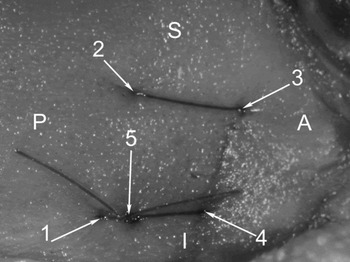
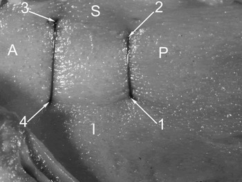
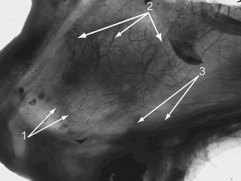
Trans-septal mattress suturing was then performed using four punctures placed in the following order: infero-posterior, supero-posterior, antero-superior and antero-inferior (Figures 1 and 2). Polyfilament Vicryl 4-0 sutures were used. The origins of Little's area vascularisation were identified on an anatomical specimen, and suturing was deemed adequate by comparison (Figure 3). The suture drawing strength required to blanch the haemorrhage site varied between patients.
Photograph of human head specimen, showing the nasal septum from the right side (on the side of bleeding) with applied suture. S = superior; P = posterior; A = anterior; I = inferior; 1 = first puncture; 2 = second puncture; 3 = third puncture; 4 = fourth puncture; 5 = knot

Photograph of human head specimen, showing the nasal septum from the left side (on the opposite side of bleeding) with applied suture. S = superior; P = posterior; A = anterior; I = inferior; 1 = first puncture; 2 = second puncture; 3 = third puncture; 4 = fourth puncture

The human nasal septum specimen (consisted of the septal cartilage and bi-side preserved mucosa) prepared by dehydration in 25, 50 and 100 per cent acetone solutions and treatment with methyl salicylate was held against the source of strong light to show the vascular pattern. 1 = vessels originating from superior labial artery; 2 = vessels originating from ethmoidal artery; 3 = vessels originating from sphenopalatine artery

Results
In all four patients, intra-operative haemostasis was instantaneous.
Post-operatively, the patients were observed for up to six months (range six to 24 months, arithmetic mean 9.12 months). No complications or relapses were encountered.
Discussion
The described trans-septal mattress suture technique addresses the anatomical vascularisation pattern of the anterior nasal septum. No previously reported suture technique has resulted in cessation of blood flow to Little's area; such techniques completely block only the antero-inferior sources of vascularisation (i.e. the superior labial artery branches), leaving posterior and postero-superior vascularisation incompletely blocked. Because of this problem, Adornato described applying not only a single suture below the bleeding vessel, by the fundus of the nasal cavity, but also using chemical or electro-cautery on both sides to target the distal branches supplying the haemorrhage site.Reference Adornato7 In our cases, chemical or electro-cautery was not necessary.
In our patients treated for persistent epistaxis using trans-septal mattress suturing, we did not encounter relapse of haemorrhage or any other complication (e.g. haematoma or nasal septum perforation); others have also confirmed these results.Reference Adornato7–Reference ZhengHua, Gang, BingWei and JiaWen10 Only one nasal septum haematoma has been described following treatment of epistaxis with suturing, but this occurred in a study population of several hundred patients.Reference Cantor9 Adornato reported that epistaxis relapsed much more rarely and less intensively in patients treated with suturing plus chemical and electro-cautery, compared with patients treated by more conventional methods.Reference Adornato8 ZhengHua et al. described complete effectiveness of Little's area suturing as treatment for epistaxis, with no complications reported.Reference ZhengHua, Gang, BingWei and JiaWen10 Interestingly, these authors used a different technique based on placing the suture through the mucosa on one side, rather than using a ‘through and through’ approach. However, we believe that the trans-septal mattress suture technique is much easier than ZhengHua and colleagues' method, because the mucous membrane in Little's area is thin and sensitive; furthermore, the latter method has been reported to cause iatrogenic mucosal damage.
Mattress suturing can be performed in adults using only local anaesthesia. However, in children general anaesthesia is indispensable, as the patient's cooperation is not usually forthcoming.Reference Adornato7, Reference Adornato8