


Hyperprolinemia type II (HP-II) is a rare autosomal recessive disorder caused by biallelic pathogenic variants in ALDH4A1 gene encoding for the enzyme L-Δ-1-pyrroline-5-carboxylic acid dehydrogenase. Reference Van De Ven, Gardeitchik, Kouwenberg, Kluijtmans, Wevers and Morava1 HP-II is characterized by a variable phenotype including intellectual disability (ID), epilepsy, and behavioral and psychiatric problems. Reference Van De Ven, Gardeitchik, Kouwenberg, Kluijtmans, Wevers and Morava1–Reference AlQurashi, Alawam, Alhaddad and Alrebh6 Developmental regression and encephalopathy triggered by infection have also been described. Reference Van De Ven, Gardeitchik, Kouwenberg, Kluijtmans, Wevers and Morava1,Reference Walker, Mills, Peters and Merton2,Reference Kaur, Paria, Saini, Suthar, Bhatia and Attri5–Reference Selkoe8 Movement disorders have not been commonly described in individuals with HP-II. Here, we present a young child with HP-II manifesting with ataxia, status epilepticus, and dystonia in the setting of a febrile illness, expanding the phenotypic spectrum of this disorder.
A 22-month-old boy was admitted with repetitive head movements in the context of parainfluenza upper respiratory tract infection. The medical history was notable for being a first born to non-consanguineous parents of Indian ancestry; his mother had febrile seizures, and his maternal aunt had unexplained ID who succumbed at 18 years of age. Prenatal and perinatal risk factors included pregnancy-induced hypertension, oligohydramnios, intrauterine growth restriction, and prematurity; he was born at 32 weeks of gestation with a birth weight of 1,120 g and required approximately 5 weeks of hospitalization for the management of low birth weight. Early developmental milestones were age-appropriate. At 12 months of age, following a mild upper respiratory tract infection that did not require hospitalization, the patient developed regression of expressive language, with loss of five previously acquired words. These words were not regained. At the time of presentation, his physical exam revealed undernutrition (weight 8.8 kg, below 2nd centile; height 78 cm, below 2nd centile), normal head circumference (46 cm, 10th centile), frontal bossing, and hypertelorism. He was alert and followed simple commands. Cranial nerve examination was normal. Appendicular hypotonia with preserved deep tendon reflexes were identified. Head titubation, truncal, and gait ataxia were observed. He began experiencing generalized tonic-clonic seizures that progressed to refractory status epilepticus managed with multiple antiseizure medications and mechanical ventilation. Subsequently, he developed episodes of dystonic posturing with no ictal correlates on continuous electroencephalogram (EEG).
Brain MRI showed diffusion restriction in bilateral globus pallidus (GP), anterior commissure, hypothalamus, mammillothalamic tracts, and T2/Fluid-attenuated inversion recovery (FLAIR) hyperintensity in the anterior fornix (Figure 1). EEG revealed intermittent generalized bursts of epileptiform discharges, and a disorganized background comprised of high amplitude delta–theta waves. CSF analysis was non-contributory. Homocysteine and vitamin B12 levels were normal. Plasma amino acid analysis by ultra-performance liquid chromatography showed high proline level >3000 µmol/L (reference range 118–372 µmol/L). Urine organic acids analysis revealed large urinary excretion of N-(pyrrole-2-carboxyl) glycine and glutarate (97 mmol/mol creatinine) and mild elevation of 2-hydroxyglutarate, with no methylmalonic acid. Repeat urine organic acid analysis 6 months later confirmed persistent elevation of N-(pyrrole-2-carboxyl) glycine, with normalization of glutarate and 2-hydroxyglutarate. Exome sequencing revealed compound heterozygous variants, c.1033G>A and c.679-6G>A, in the ALDH4A1 gene, inherited biparentally and located in trans. Both variants were classified as likely pathogenic, and this genetic finding, together with the biochemical profile, confirmed the diagnosis of HP-II. He was managed with pyridoxine 20 mg/kg/day and levetiracetam; no dietary intervention was implemented. The ataxia resolved within a week, and dystonia did not recur after 24 h of its onset. At the last follow-up, he continued on pyridoxine and levetiracetam and remained seizure-free. He had gradual developmental gains, but expressive language delay persisted.
Brain MRI. Diffusion-weighted images showing subtle diffusion abnormalities in the internal GP, anterior commissure, hypothalamus, and mamillothalamic tracts (A–C). Mild T2/FLAIR signal changes (D). Subtle T2/FLAIR hyperintensity along the anterior fornix bilaterally and the cerebral peduncles (E,F). GP = globus pallidus.

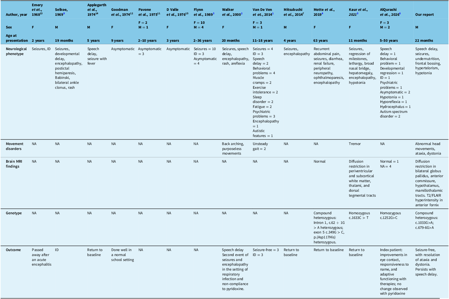
HP-II is a rare inherited metabolic disorder with limited reports in the literature, and the full spectrum of clinical manifestations has not been described. HP-II is caused by the deficiency of L-Δ-1-pyrroline-5-carboxylic acid dehydrogenase which catalyzes the conversion of pyrroline-5-carboxylate (P-5-C), derived from proline or ornithine, to glutamate 9,10 (Supplementary Figure 1). This results in the accumulation of proline and P-5-C and increased urinary excretion of proline, hydroxyproline, P-5-C, and glycine. Reference Walker, Mills, Peters and Merton2,Reference Mitsubuchi, Nakamura, Matsumoto and Endo7,9 The phenotypes and genotypes seen in individuals with HP-II are summarized in Table 1, with more detailed information in Supplementary Table 1. The clinical manifestations reported in HP-II encompass seizures (60%), ID (22%), encephalopathy (17%), speech delay (14%), behavioral problems (14%), and psychiatric problems (11%). Thirty percent of the reported individuals were asymptomatic. Reference Van De Ven, Gardeitchik, Kouwenberg, Kluijtmans, Wevers and Morava1–Reference Selkoe8,11–15 Movement disorders have been poorly characterized in HP-II. Although unsteady gait, back arching, purposeless movements, and tremor have occasionally been reported, Reference Van De Ven, Gardeitchik, Kouwenberg, Kluijtmans, Wevers and Morava1,Reference Walker, Mills, Peters and Merton2,Reference Kaur, Paria, Saini, Suthar, Bhatia and Attri5 well-characterized ataxia and dystonia have not been established as prominent manifestations.
Clinical characteristics and genotype of the cases with HP-II reported in the literature

Table 1. Long description
A table comparing clinical characteristics and genotypes of HP-II cases reported in the literature. The table has 15 columns and 8 rows, including headers. Columns are labeled: Author, year, Sex, Age at presentation, Neurological phenotype, Movement disorders, Brain MRI findings, Genotype, Outcome. Each row provides data for different cases.
The publications are organized in columns, and the clinical characteristics, neuroimaging findings, genetic results, and outcome are organized in rows.
The paper by Emery et. at, 1968 reports 1 female, age at presentation 2 years. Neurological phenotype includes seizures and intellectual disability. Movement disorders not available. Brain MRI not available. Genotype not available. Outcome reports the patient passed away after an acute encephalitis.
The paper by Selkoe, 1969 reports 1 male, age at presentation 19 months. Neurological phenotype includes seizures, developmental delay, encephalopathy, postictal hemiparesis, Babinski, bilateral ankle clonus, and rash. Movement disorders not available. Brain MRI not available. Genotype not available. Outcome reports intellectual disability.
The paper by Applegarth et. al, 1974 reports 1 male, age at presentation 5 years. Neurological phenotype includes speech delay, and seizure with fever. Movement disorders not available. Brain MRI not available. Genotype not available. Outcome reports return to baseline.
The paper by Goodman et. al, 1974 reports 1 female, age at presentation 9 years. Neurological phenotype describes asymptomatic. Movement disorders not available. Brain MRI not available. Genotype not available. Outcome reports the patient has done well in a normal school setting.
The paper by Pavone et. al, 1975 reports 2 females and 1 male, age at presentation 2–10 years. Neurological phenotype includes asymptomatic 3 patients. Movement disorders not available. Brain MRI not available. Genotype not available. Outcome not available.
The paper by D Valle et. al, 1976 reports 1 female, age at presentation 3 years. Neurological phenotype describes asymptomatic. Movement disorders not available. Brain MRI not available. Genotype not available. Outcome not available.
The paper by Flynn et. al, 1989 reports 10 females and 4 males, age at presentation 2 to 36 years. Neurological phenotype includes seizures in 10 patients, intellectual disability in 3 patients, asymptomatic 4 patients. Movement disorders not available. Brain MRI not available. Genotype not available. Outcome not available.
The paper by Walker et. al. 2000 reports the case of 1 female, age at presentation 20 months, the neurological phenotype includes seizures, speech delay, encephalopathy, rash, areflexia, and movement disorder described as back arching and purposeless movements. Brain MRI not available. Genotype not available. Outcome reports speech delay, second event of seizures and encephalopathy in the setting of respiratory infection and non-compliance to pyridoxine.
The paper by Van De Ven et. at, 2014 reports 3 females and 1 male patient, age at presentation 11 to 18 years. The neurological phenotype included seizures in 4 patients, intellectual disability in 3 patients, speech delay in 2 patients, behavioral problems in 4 patients, muscle cramps in 2 patients, exercise intolerance in 2 patients, sleep disorder in 2 patients, fatigue in 2 patients, psychiatric problems in 3 patients, encephalopathy in 1 patient and autistic features in 1 patient. Movement disorder disorder described as unsteady gait in 2 patients. Brain MRI not available. Genotype not available. Outcome reports seizure freedom in 3 patients, and intellectual disability in 3 patients.
The paper by Mitsubuchi et. al, 2014 reports 1 male, age at presentation 4 years. Neurological phenotype included seizures and encephalopathy. Movement disorder not available. Brain MRI not available. Genotype not available. Outcome reports return to baseline.
The paper by Motte et. al, 2019 reports 1 female, age at presentation 63 years. Neurological phenotype includes recurrent abdominal pain, seizures, diarrhea, renal failure, peripheral neuropathy, ophthalmoparesis, encephalopathy. Movement disorder not available. Brain MRI normal. Genotype reports a compound heterozygous variant in Intron 1, c.62 + 1G > A heterozygous and exon 5 c.349G > C, p.(Asp117His) heterozygous. Outcome reports return to baseline.
The paper by Kaur et. al, 2021 reports a female, age at presentation 11 months. Neurological phenotype includes seizures, regression of milestones, lethargy, broad nasal bridge, hepatomegaly, encephalopathy, hypotonia. Movement disorder described as tremor. Brain MRI reports diffusion restriction in periventricular and subcortical white matter, thalami, and dorsal tegmental tracts. Genotype reports homozygous variant c.1633C > T. Outcome reports return to baseline.
The paper by AlQurashi et. at, 2026 reports 3 females and 2 males, age at presentation 5–50 years. Neurological phenotype includes speech delay in 1 patient, behavioral problem in 1 patient, developmental regression in 1 patient, intellectual disability in 1 patient, psychiatric problems in 1 patient, asymptomatic 2 patients, hypotonia 1 patient, hyporeflexia 1 patient, hydrocephalus 1 patient, autism spectrum disorder 2 patients. Movement disorder not available. Brain MRI reports normal results in 1 patient, not available in 4 patients. Genotype reports homozygous variant c.1252G>C. Outcome reports the index patient had improvements in eye contact, responsiveness to name, and adaptive functioning with therapies but no change observed with pyridoxine.
Our report describes 1 male, age at presentation 22 months. Neurological phenotype includes speech delay, seizures, undernutrition, frontal bossing, hypertelorism, hypotonia. Movement disorder described as abnormal head movements, ataxia, dystonia. Brain MRI reports diffusion restriction in bilateral globus pallidus, anterior commissure, hypothalamus, mamillothalamic tracts. T2/FLAIR hyperintensity in anterior fornix. Genotype reports compound heterozygous variants c.1033G>A and c.679-6G>A. Outcome reports seizure freedom, with resolution of ataxia and dystonia, and persistence of speech delay.
HP-II = hyperprolinemia type II; F = female; M = male; ID = intellectual disability; NA = not available.
The seizures in HP-II are thought to be caused by pyridoxal phosphate (PLP) inactivation by the accumulation of P-5-C.16 PLP is essential for numerous enzymatic reactions in amino acid and neurotransmitter metabolism, and its deficiency can disrupt the metabolism of neurotransmitters such as γ-aminobutyric acid (GABA), dopamine, and serotonin.17 It is possible that such a neurotransmitter imbalance may contribute to the dystonia in our patient. Additionally, proline accumulation was shown to reduce glutamate uptake, Na+-K+-ATPase activity, and adenosine triphosphate (ATP) levels, inducing excitotoxicity and energy deficit in a rat model.18 We hypothesize that energy failure may explain the cytotoxic edema observed in the GP on MRI, which in turn may account for the patient’s dystonia. However, we acknowledge that the dystonia may instead represent an acute symptomatic manifestation of metabolic decompensation and/or bilateral basal ganglia injury, rather than a primary or chronic feature of HP-II. Alternative contributors were also considered: the patient experienced refractory status epilepticus, which may have led to hypoxic-ischemic injury and contributed to the bilateral basal ganglia changes seen on MRI. Follow-up MRI was not done, as the child remained clinically stable.
Gait instability has been described in two patients with HP-II; however, the nature of the gait disturbances was not elaborated further. Reference Van De Ven, Gardeitchik, Kouwenberg, Kluijtmans, Wevers and Morava1 One of these patients was a 13-year-old girl with speech delay, seizures, exercise intolerance, and ID. The second patient was a 12-year-old girl with seizures, exercise intolerance, and behavioral problems. Their genotype was unavailable. Reference Van De Ven, Gardeitchik, Kouwenberg, Kluijtmans, Wevers and Morava1 PLP is an essential cofactor for the conversion of glutamate to GABA through glutamate decarboxylase;19 we hypothesize that PLP inactivation in HP-II may lead to GABA deficiency. Cerebellar Purkinje cells are GABAergic neurons, and the loss of their GABAergic inhibition can lead to abnormal firing pattern,20 theoretically explaining the ataxia in our patient.
The literature on neuroimaging features of HP-II is scarce, with two cases reporting normal findings, Reference Motte, Fisse and Grüter4,Reference AlQurashi, Alawam, Alhaddad and Alrebh6 and one case describing T2/FLAIR signal abnormalities and diffusion restriction affecting the periventricular and subcortical white matter, thalamus, and dorsal tegmental tracts. Reference Kaur, Paria, Saini, Suthar, Bhatia and Attri5 This last patient was an 11-month-old girl presenting with seizures, encephalopathy, hypotonia, and tremor but had a different genotype than our case. Our patient also had a different MRI pattern with diffusion restriction involving GP, anterior commissure, hypothalamus, mammillothalamic tracts, and fornix, which has not been previously described.
HP-II should be considered in patients presenting with fever-induced seizures and other clinical features like unexplained encephalopathy, ID or developmental delay, ataxia, and dystonia. Our case expands the clinical and neuroimaging spectrum in children with HP-II.
Supplementary material
The supplementary material for this article can be found at https://doi.org/10.1017/cjn.2026.10685.
Author contributions
A.U.C. contributed to the conceptualization, preparation of figures and tables, writing of the first draft, and reviewing of the final manuscript. H.S.F. contributed to the conceptualization, writing of the first draft, preparation of figures, and reviewing of the final manuscript. S.Y. contributed to the conceptualization, writing of the first draft, and reviewing of the final manuscript. P.K. contributed to the preparation of figures and reviewing of the final manuscript. J.C. contributed to the review and critique of the final manuscript. L.Y. contributed to the review and critique of the final manuscript. L.A. contributed to the review and critique of the final manuscript. C.G. contributed to the conceptualization and reviewing of the final manuscript.
Funding statement
None.
Competing interests
C.G received grants from the Dystonia Medical Research Foundation and the Center for Brain and Mental Health at SickKids. She received consulting fees from Medtronic Inc., payments or honoraria for lectures, presentations, speakers bureaus, manuscript writing, or educational events from Medtronic Inc. and Acadia, participated in Data Safety Monitoring Board or Advisory Board from Medtronic Inc., and had a leadership or fiduciary role in the Education committee from the Movement Disorders Society.
All other authors have nothing to disclose.
*References [9–20] are present in the supplementary material.



 Open access

