Introduction:
We will focus on the pathophysiology and diagnosis of neuropsychiatric diseases based on the assessment of histochemistry and immunofunctions.
– First we give a brief overview on the immunology of the affective and schizophrenic disorders.
– Measuring CSF parameters by conventional, in neurology well established methods, will be demonstrated to be a valuable approach to psychiatric diseases.
– In addition, we will report a novel approach applying flow cytometry technology used in haematology to characterize immunocompetent cells in the major compartments, the peripheral blood and the CSF.
Overview on the immunologic aberrations reported in neuropsychiatric diseases:
Epidemiological, clinical and post mortem and cytokine studies indicate that inflammatory and immune reactions are involved in the pathomechanisms of affective and schizophrenic spectrum disorders Reference Davis, Stewert, Friedman, Buchsbaum, Harvey and Hof(1-Reference Licinio and Wong5). Epidemiological studies indicate infections to increase the risk of psychiatric disorders during pregnancy, around delivery or later during childhood and are caused by different pathogenetic mechanisms Reference Meyer, Nyffeler, Schwendener, Knuesel, Yee and Feldon(6-Reference Bechter and Bogerts9). In general this requires three factors Reference Rioux and Abbas(10)): genes, environment and immune system.
Improvement of psychiatric disorders after treatment with viro-static or antibiotic drugs support the view of infectious agents involved in the pathophysioloyi. Therapy resistant cognitive impairment in psychotic cytomegaly virus (CMV) seropositive patients improved during antiviral therapy. Since the antiviral treatment was effective only in patients seropositive for CMV, it has been suggested a latent CMV infection to be involved in psychiatric disorders Reference Dickerson, Boronow, Stallings, Origoni and Yolken(11,Reference Torrey, Leweke, Schwarz, Mueller, Bachmann, Schroeder, Dickerson and Yolken12) . Comparable, improvement of depression has been reported after successful treatment of maxil-lofacial Streptococcus infection Reference Bechter(13)). Toxoplasma gondii has been described in relation to psychiatric diseases Reference Torrey and Yolken(14)). The increased frequency of autoimmune disorders in psychosis pedigrees Reference Eaton, Byrne, Ewald, Mors, Chen, Agerbo and Mortensen(15)) supports the hypothesis of an autoimmune pathophysioogy. New treatment regimes were applied in psychoses, e.g., add-on therapy with Cox-2 blockers Reference Müller, Riedel, Scheppach, Brandstätter, Sokullu and Krampe(16-Reference Müller, Strassnig, Schwarz, Ulmschneider and Riedel18), immune modulatory treatment Reference Bechter, Herzog, Schreiner, Brinkmeier, Aulkemeyer and Weber(19,Reference Bechter, Schreiner, Herzog, Breitinger, Wollinsky, Brinkmeier, Aulkemeyer, Weber and Schüttler20) or neuroprotective treatment Reference Ehrenreich, Hinze-Selch, Stawicki, Aust, Knolle-Veentjer and Wilms(21)). Immune suppressive therapy was proposed Reference Knight, Menkes, Highton and Adams(22)).
At present major discussions are based on different models: neurodevelopmental alteration induced by immune inflammatory mediators or infectious agents Reference Meyer, Nyffeler, Schwendener, Knuesel, Yee and Feldon(6)), a specific role of macrophages in depression Reference Smith(23)), cytokine abnormalities and inflammation interacting with neurodegeneration Reference Myint and Kim(24,Reference Leonard25), imbalance of Th1 and Th2 type immunity and related glutamate abnormalities Reference Müller and Schwarz(26)) or mild encephalitis (ME) underlying a subgroup of psychiatric spectrum disorders Reference Bechter(27)).
A specific and sensitive approach to detect CNS inflammation is the analysis of cerebrospinal fluid (CSF) Reference Wildemann, Oschmann and Reiber(28)). Investigation of paired CSF and blood samples are considered as the gold standard Reference Sindic, M-P and Goffette(29,Reference Reiber and Peter30). The cknowledge based interpretation of disease-related CSF data patterns allows an important contribution of CSF analysis to differential diagnosis in neurological diseases. Investigation, however, of psychiatric disorders by CSF analysis is recommended up to now to exclude meningo-encephalitis and plays a minor role in psychiatric diagnosis and research.
Conventional CSF analysis in psychiatric disorders:
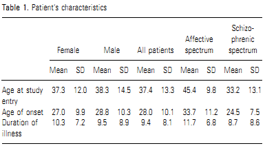
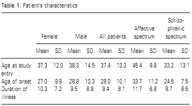
To show the advantage to perform CSF analysis in the diagnostic procedure of psychiatric patients, we investigated psychiatric inpatients of the schizophrenic and affective spectrum (Table 1) for blood-CSF barrier (BCB) function, the humoral and cellular intrathecal immune response and interpret the humoral immune response in Reibergrams using a recently developed statistic approach for evaluation of CSF protein data in patient groups. Determining intrathecal antibody synthesis directed against defined antigens and intrathecal autoantibody production we made a first approach to proof the assumed low level immune responses and to explain moderate blood-CSF barrier dysfunctions. We investigated also the neopterin-tryptophan-kynurenine pathways involved in various infectious and autoimmune disorders Reference Dotevall, Fuchs, Reibnegger, Wachter and Hagberg(31-Reference Hagberg, Dotevall, Norkrans, Larsson, Wachter and Fuchs33). Neopterin production and tryptophan degradation are closely linked to Th1-type immune activation and measurements of these metabolic changes should therefore allow some insight into the potential role of immune activation in patients with schizophrenic and affective spectrum disorders.
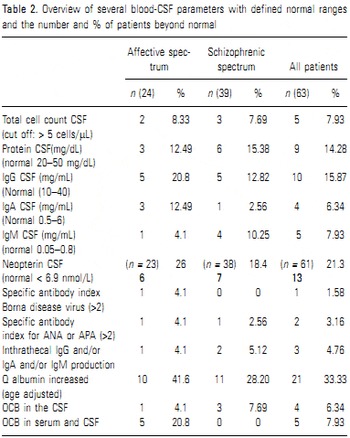
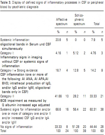
The parameters indicating inflammatory processes are outlined in detail (Table 2 and Table 3):
Intrathecal humoral immune reaction
We detected in 9/63 (14%) patients an intrathecal humoral immune reaction. Possible interpretations are: 1. acute inflammatory process or 2. Chronic inflammatory process or 3. scar from a former inflammatory process. Acute inflammatory processes have been excluded from this study and none of the cases showed data patterns arguing for a scar from an earlier acute inflammatory process. Chronic inflammatory processes include multiple sclerosis and neuroborellioses, but none of the patients fitted the requirements for such diagnosis. So these cases could be diagnosed as chronic low level CNS inflammation (LLCI). A LLCI response fit in the Mild Encephalitis (ME) hypothesis and LLCI is hypothesized to represent the pathogenic core in a subgroup of affective and schizoprenic spectrum disorders.
Systemic signs of inflammation:
In further 5/63 cases (8%), all of the affective spectrum (of those 21%), signs of systemic inflammation were observed. 3 cases did not display CNS involvement (identical oligoclonal bands (OCBs) in CSF and blood) one case had AA-AI and QAlb increase and another case CNS involvement seemed to be likely. This observation of identical OCBs in CSF and blood is found in many inflammatory neurological diseases such as herpes simplex virus encephalitis or GBS polyradiculitis Reference Wurster, Zettl, Lehmitz and Mix(34)).
Infectious or autoimmune etiologies:
Based on the finding of CSF abnormalities we identified in 4/63 (6%) patients specific infectious (streptococcus, BDV) or autoimmune associated (unclassified autoimmune disorder close to classification as SLE, Sjogren syndrome) etiologies, in one case with considerable treatment consequences Reference Bechter, Bindl, Horn and Schreiner(35)). Since the antibody screening panel was limited an improved differential diagnostic power from an extended test-panel of autoautoantibodies and infectious agents could be expected. However, even in acute meningoencephalitis the causal agent is identified in only about 50% of cases Reference Bloch and Glaser(36)). Furthermore, autoantibodies are not highly predictive for autoimmune disease, but often detectable years before symptoms onset (37.38).
Albumin Quotients and BCB dysfunction:
In 21/63 (33%) patients wedetectedanincreased QAlb withvalues 20% abovetheage related limit which indicate a BCB dysfunction. In 15/63 (24%) patients this was an isolated finding without any other CSF abnormalities.
Proposal of a model of pathophysiological processes in psychiatric diseases:
The patients we have included in the study suffered from chronic illness (mean duration 12 years in affective and 9 years in schizophrenic disorders) and often inefficient therapy, but lumbar puncture to investigate CSF was in most of the cases performed for the first time. The aim of the study was to deal with sensitivity borders of current CSF analytic methods to unravel a hypothesized low level chronic inflammatory (LLCI) process or mild encephalitis (ME) subgroup in major psychiatric disorders and to sharpen the definition of LLCI and ME. In a comparable well characterized disease, the HIV encephalitis (HIVE), the definition problems can be demonstrated: histopathologic criteria of HIV encephalitis correlate well with distinct clinical criteria Reference Cherner, Cysique, Heaton, Marcotte, Ellis and Masliah(39)). Many HIV cases present some inflammatory findings in post mortem brains but do not fulfil HIVE criteria, and for this group histopathology remains unclassified. Nevertheless, HIV cases with minor degree histopathological alterations have an increased psychiatric morbidity with low degree CNS inflammation. There is ample evidence from epidemiological studies that pathogens increase the risk of psychoses. Histopathologic alterations in post mortem brains in psychosis were interpreted as low level CNS inflammation Reference Uranova, Orlovskaya, Vikhreva, Zimina, Kolomeets, Vostrikov and Rachmanova(40-Reference Steiner, Bielau, Brisch, Danos, Ullrich and Mawrin43) and genetic findings point also to neuroinflammation Reference Hanson and Gottesman(44,Reference Wong, Dong, Maestre-Mesa and Licinio45). In recent studies, as outlined in the second part of the presentation on T-cells, we found in a subgroup of patients with spectrum psychoses by CSF cell subtyping a pattern similar to that in patients with meningoencephalitis Reference Maxeiner, Rojewski, Schmitt, Tumani, Bechter and Schmitt(46)). Furthermore, T-cell clonal expansion indicates a specific intrathecal immune reaction Reference Oleszak, Zhang, Lin, Lu, Chang and Tsygankov(47)). Summary: We tested the relevance of CSF analysis Reference Reiber and Peter(48)) with respect to low level chronic inflammation and the mild encephalitis hypothesis of schizophrenic and affective spectrum disorders. In 63 patients, with treated for years with various approaches but poor therapy response we found in 9 (14%) patients an intrathecal humoral immune response as a sign of a low level CNS inflammatory process. An isolated blood CSF barrier dysfunction was found in 15 (24%) patients, caused likely by a reduced CSF flow. In 5 (8%) patients, (all affective spectrum disorders) representing 21% of this subgroup, we found signs of systemic inflammation (identical OCBs in CSF and blood) and CNS involvement was observed in 2 of the patients according to CSF or MRI findings. Overall, 41% of patients displayed CSF abnormalities indicating CNS involvement.
A novel approach applying cytoflow technology used in haematology to characterize immunocompetent cells in the major compartments, the peripheral blood and the CSF: Overview on immunological research in neuropsychiatric diseases:
After having proven by standardized methods the involvement of neuroinflammation in a subgroup of patients suffering from psychiatric disorders, we now wanted to develop more specific approaches to the assessment of immunofunctions. Recent research has investigated the frequency and function of immuncompetent cells in peripheral blood of psychiatric patients and to a minor extend in the CSF Reference Nikkilä, Müller, Ahokas, Miettinen, Rimón and Andersson(49-Reference Oleszak, Lin, Chang, Zhang, Herzog, Bhattacharjee, Rudner, Platsoucas and Bechter51). Up to date, results are inconclusive even in high frequent subsets such as CD4+ und CD8+ cells and vary with psychiatric diagnosis Reference Atanackovic, Kroger, Serke and Deter(52-Reference Cazzullo, Saresella, Roda, Calvo, Bertrando, Doria, Clerici, Salvaggio and Ferrante54). However, it is important for the understanding of the immunopathology of diseases to assess the frequency and the activation status of T cells since it allows to conclude for example the sensitivity of cells to cytokines. IL-10 acts immunostimulatory as well as immunosuppressive depending on the activation of the target cell Reference Ye, Huang, Hao, Xu, Yu, Van Den Hurk and Xiang(55)). A low frequency cell type, characterized by CD25+, have regulatory function and synthesize interferon gamma, IL-4, IL-5, IL-10 and IL-12 Reference McDyer, Li, John, Yu, Wu and Ragheb(56,Reference Walker57). The CD25+ cells have been investigated rarely Reference Maes, Bosmans, Suy, Vandervorst, De Jonckheere and Raus(58)), but in autoimmune diseases, the cells have been described as key players Reference Morris and Waldmann(59,Reference Baecher-Allan and Hafler60) . One marker for activation, CD69 Reference Lindsey, Lowdell, Marti, Abbasi, Zenger, King and Lamb(61,Reference Sancho, Gomez and Sanchez-Madrid62), has been investigated in psychiatric diseases in the peripheral blood Reference Mihaylova, Deruyter, Rummens, Bosmans and Maes(63)), and was successfully used in the diagnosis of multiple sclerosis in the CSF and peripheral blood Reference Kosmaczewska, Bilinska, Ciszak, Noga, Pawlak, Szteblich, Podemski and Frydecka(64,Reference Perrella, Carrieri, De Mercato and Buscaino65). In addition CD4 + CD25+ was found with different frequencies in the peripheral blood and CSF Reference Feger, Luther, Poeschel, Melms, Tolosa and Wiendl(66)). CD45 RO Reference Tchilian and Beverley(67,Reference Holms68) has been used to monitor multiple sclerosis patients Reference Okuda, Okuda, Apatoff and Posnett(69)). In psychiatric patients it was investigated rarely Reference Craddock, Lockstone, Rider, Wayland, Harris, McKenna and Bahn(70)).
Outline of an approach:
In a study we investigated patients with neurological diseases and psychiatric disorders. We applied cytoflow techniques which allows with high sensitivity to detect six cell surface markers per cell. The aim was to detect aberration in immunosurveillance and to use the characterization as a differential diagnostic tool in psychiatric and neurological diseases. The comparison to neurological patients intended to find similarities to conclude by analogy to comparable pathophysiologies.
Simultaneously CSF and PBMC were collected from 45 patients from the departments of psychiatry and neurology. 17 patients were diagnosed with major psychoses (MPD), 16 with non-inflammatory neurological diseases (NIND), 7 with meningitis (MEN) and 5 with chronic inflammatory diseases (CIND). Samples were stained with monoclonal antibodies directed against CD4, CD8, CD25, CD45, CD69, CD127. The surface markers characterized the main T cell populations (CD4 and CD8), the T regulatory T cells (CD25 CD127), the activation status (CD69) and the maturation status (CD45). The results are briefly summarized and subsequently discussed focusing on the relevance for neuroinflammation.
Summary of results:
Significant differences (P < 0.05) were observed in the peripheral blood (PB) for CD4+ cells for patients with meningitis (MEN): The frequency was 45.8% but was lower in NIND: 33.06% or CIND: 31.03%. Significant differences were found in PB for the CD4 + CD45RO+ cell population. The frequency for the cell type in meningitis patients was24.08% but 11.12% in CIND patients. For the CD4 + CD25 + population in the peripheral blood we found in MEN a frequency of 0.89% in CIND: 0.28% and in MPD: 0.39%. In CSF the CD4 + cell population was in MEN patients 5.15% vs. in MPD 10.18%. A significantly higher frequency of CD4 + CD25 + CD127dim, representing Tregs were observed in PB of MEN (0.25%) vs. CIND (0.09%).
Patient's chara cteristics
Discussion of selected results: CD4+/CD8+ subset
For the T lymphocyte subsets CD4+ and CD8+ cells, in PB different frequencies were reported for psychiatric disorders, varying in part with the diagnosis. In patients with anxiety disorder reduced CD8+ T cell counts as compared to healthy controls was observed Reference Atanackovic, Kröger, Serke and Deter(71)), elevated CD4+ and CD8+ counts in patients with posttraumatic stress disorder Reference Boscarino and Chang(72)). Psychiatric patients on long term multiple drug therapy displayed a decreased CD4+ frequency Reference Cosentino, Fietta, Caldiroli, Marino, Rispoli, Comelli, Lecchini and Frigo(73)). In the PB of schizophrenic patients a lower frequency of CD4+ T cells but a higher CD8+ frequency as compared to healthy controls was reported Reference Cazzullo, Saresella, Roda, Calvo, Bertrando, Doria, Clerici, Salvaggio and Ferrante(74)). When comparing in our study the frequency of CD4+ in the PB of patients with neurological diseases and psychiatric patients, we observed a slightly higher frequency of this subset in psychiatric patients than in NIND or CIND patients but lower than in MEN patients (differences nonsignificant). This may indicate a moderate systemic reaction. In the CSF of psychiatric patients, the frequency of CD4+ cells was higher (non-significant) than in all other groups. Since CD4+ cells are capable to release proinflammatory cytokines, which have been reported to be involved in the pathogenesis of psychiatric diseases, the slight increase of CD4+ cells in the studies psychiatric patients may indicate a low-inflammatory status Reference Pollmacher, Haack, Schuld, Reichenberg and Yirmiya(75-Reference Schiepers, Wichers and Maes77).
CD25+ subset:
The alpha chain of the IL-2 receptor, termed CD25, is involved in the pathway leading to the production of inflammatory cytokines such as INF-gamma, IL-4, IL-5, IL-10 and IL-12 Reference McDyer, Li, John, Yu, Wu and Ragheb(78)) In patients with major depression, a higher number and percentage of CD25+ cells has been reported Reference Maes, Bosmans, Suy, Vandervorst, De Jonckheere and Raus(79)). In autoimmune diseases over expression of IL-2Ralpha was detected Reference Baecher-Allan and Hafler(60,Reference Morris and Waldmann80). Although CD25negative Treg cells, defined as Foxp3 positive cells, have been described, the major fraction of Treg cells are characterized as CD25+ Reference Yi, Zhen, Jiang, Zheng and Zhao(81)).
We observed significant differences in CD25+ cell populations in the PB between the patients groups. In contrast, we did not see a difference in the CSF. This indicates a systemic reaction in meningitis, leading to significant changes in this cell population in the periphery but not to the same extend in the CSF or to a more transient elevation in the CSF than in the periphery. This can also be monitored by the ratio of PBMC/CSF of cell frequencies and stresses the difference in function between the blood CSF barrier as described for small molecules by Q albumin Reference Wildemann, Oschmann and Reiber(28,Reference Bloch and Glaser36) and the immunological blood CSF barrier.
Patient's chara cteristics

Display of defined signs of inflammatory processes in CSF or peripheral blood by psychiatric diagnosis

CD45+subset:
The leukocyte common antigen CD45 is a tyrosine phosphatase expressed in various isoformes. The isoforms are linked to autoimmune diseases, pointing to an immunomodulatory function of the enzyme. CD45RO, in opposite to CD45RA, is expressed on the memory T cell type Reference Holms(68,Reference Tchilian and Beverley82) Together with other T cell markers, determination of CD45RO on CSF-cells has been used to monitor MS patients Reference Okuda, Okuda, Apatoff and Posnett(69,Reference Svenningsson, Andersen, Hansson and Stemme83). In PB of psychiatric patients, CD45RO+ on CD4+ or CD8+ cells were reported to be non-significant lower in schizophrenic patients than in matched normal controls Reference Craddock, Lockstone, Rider, Wayland, Harris, McKenna and Bahn(84)). Patients in this study with CIND displayed the lowest frequency of CD4+CD45RO+ cells in PB and CSF as compared to all other groups. In psychiatric patients we observed the highest frequency, indicating a post-acute downregulation in a chronic state (CIND) and a persistent high (non-significant) CD4 activation status in psychiatric patients in the CSF but not in the PB.
CD69+ subset:
CD69 is considered a surrogate marker for T cell activation by mitogen or antigen stimulation Reference Marzio, Mauel and Betz-Corradin(85,Reference Sancho, Gomez and Sanchez-Madrid86). Changes in frequency were reported in neuropsychiatric diseases Reference Perrella, Carrieri, De Mercato and Buscaino(65,Reference Mihaylova, Deruyter, Rummens, Bosmans and Maes87,Reference Bueno, Vaz, Oliveira, Machado, Livramento, Mielli and Ueda88,Reference Kosmaczewska, Bilinska, Ciszak, Noga, Pawlak, Szteblich, Podemski and Frydecka89). In our patient the non-significant lower frequency of CD4 + CD69 + lymphocytes in the CSF in chronic inflammatory neurological diseases as compared to other patients groups may indicate that the activation markers CD69 is transiently elevated in the initial phase on CSF cells only, since defined as a very early marker, and later in the course of the disease the downregulation of this marker is higher in CIND patients then in the other patient groups.
Consequences for diagnostic procedures:
We now tried to use the information on T cell frequencies in the compartments CSF and peripheral blood to approach a classification of neuropsychiatric disorders by immunological guidance.
As an example the distribution of T regulatory cells as characterized by CD4+ CD127dim surface markers is shown in Figure 1 . To apply other surface markers to peripheral blood and CSF derived cells will likely reveal clusters when applying to a larger and less heterogeneous patient study group.
Another accumulation analysis of cell distribution only in the peripheral blood revealed 6 out of 17 MPD displaying a ratio of CD4 +/CD8+ cells in PB > 3.2, but only 2 NIND patients..
Overall summary:
The consequences of CSF analysis of these psychiatric patients are as follows:
1) In combination with an interdisciplinary clinical approach CSF analysis validated in neurology provides an underestimated tool for differential diagnosis in psychiatric diseases.
2) We were able to demonstrate in a small number of patients suffering from chronic psychiatric disorders a substantial and lasting improvement by enhancing the diagnostic sensitivity by CSF analysis.
3) Similarities in CSF analysis and lack of significant differences may be due to moderate specificity of psychiatric symptoms for affective and schizophrenic spectrum disorders, although the observation of systemic inflammation in affective spectrum disorders only may be supported by a study detecting autoantibodies in bipolar but not in schizophrenic patients Reference Padmos, Bekris, Knijff, Tiemeier, Kupka and Cohen(90)).
4) The subgroup of psychiatric patients with abnormal CSF and serum data or with intrathecal pathologies are candidates for further investigation for immunofunctions with regards to LLCI or ME pathology.
5) To validate immuno-and inflammatory markers Reference Dantzer, Capuron, Irwin, Miller, Ollat and Perry(91-Reference Padmos, Hillegers, Knijff, Vonk, Bouvy and Staal94) for the diagnostic procedure in psychiatric disorders Reference Mössner, Mikova, Koutsilieri, Saoud, Ehlis and Müller(95)), it is necessary to combine the investigation with well established CSF and serum markers of known pathophysiological function. The class of histochemical markers is proposed to be supplemented by T-and B-cell typing as outlined in the presentation.
Proposal for further investigation:
6) Identical OCBs in blood and CSF were found. The observation remains in psychiatric disorders without an explanatory model.
7) The impact of substance class and dose of medication on the CSF flow rate needs to be investigated.
8) Astroglia may play a role in patients with an isolated BCB-D without signs of a humoral or cellular immune response but with increased CSF neopterin.
9) Detailed work has to be done on the term neuroinflammation and neurodegeneration Reference Gao and Hong(96)) and on the difference from acute inflammation Reference Medzhitov(97)). LLCI respectively ME in psychiatric spectrum disorders share similarities with unrecognized encephalitis. The proposed new term para-inflammation may only partially cover the pathophysiological and nosological entities.
Acknowledgement:
We thank Dagmar Vogel, CSF Laboratory and Dr. R. Kilian, Clinic for Psychiatry and Psychotherapy II, Ulm University for help, the Margarete Ammon Stiftung for major and the Stiftung Propter Homines, Vaduz -Fürstentum Liechtenstein for financial support. There are no conflicts of interest between the authors. There are no financial interests, personal relationships or affiliations which could have influenced the work.