Introduction
Fascioliasis is a parasitic disease caused by the flukes, Fasciola hepatica and F. gigantica (Trematoda: Fasciolidae). A wide range of herbivorous mammals are reservoir hosts, such as sheep, cows and goats (Chen & Mott, Reference Chen and Mott1990). Fascioliasis is a major public health problem in animal populations worldwide and is responsible for important economic losses in animal husbandry. The condition costs the country of Perú an estimated loss of $US11 million annually (Leguía et al., 1989), and also causes the loss of an important protein source for the local population. The prevalence of the infection in cattle approximates 100% in many endemic areas (Leguía et al., 1989; Marcos et al., Reference Marcos, Maco, Terashima, Samalvides, Espinoza and Gotuzzo2004).
Fasciola hepatica infection has two defined clinical phases – acute and chronic, seen in both humans and animals (Dalton, Reference Dalton1999). The acute phase is characterized by the migration of larvae from the duodenum through the intestinal wall, peritoneal cavity and across Glisson's capsule, entering the liver parenchyma and finally reaching the biliary ducts. Histologically, fascioliasis produces subcapsular haemorrhages; hepatic degeneration; infiltration into the parenchyma of eosinophils, lymphocytes and macrophages; fibrosis; venous thrombosis; appearance of necrotic cords with giant cells and granulation tissue; and occasionally the occurrence of granuloma-containing parasite eggs (Dalton, Reference Dalton1999; Hamir & Smith, Reference Hamir and Smith2002). This phase lasts between 2 and 4 months and presents the most serious complications of the disease as reported in humans. In this phase subcapsular haemorrhages (Loja et al., Reference Loja, Alvizuri, Vilca, Aviles and Sanchez2003), multiple hepatic abscesses, hepatic necrosis and the destruction of liver parenchyma can be found (Kim et al., Reference Kim, Lim, Kim, Lee and Lim1999), occasionally complicated by the presence of severe anaemia (Vilchez, Reference Vilchez1983). The chronic state, which occurs months to years after infection, develops when adult parasites deposit eggs in the biliary ducts (MacLean, Reference MacLean1999). Clinically, patients with chronic-phase illness typically present with non-specific or vague abdominal symptoms (some of which result in surgical exploration) when adult parasites obstruct the biliary ducts or cause hepatic dysfunction (Hadden & Pascarelli, Reference Hadden and Pascarelli1967; Jiménez et al., Reference Jiménez, Loja, Ruiz, Maco, Marcos and Aviles2001).
Better understanding of the pathology caused by the parasite during chronic infection is needed where most cases are asymptomatic and remain undiagnosed (Marcos et al., Reference Marcos, Maco, Terashima, Samalvides and Gotuzzo2002). Latent infection shows an extended time period. Some reports carried out in endemic areas demonstrated the occurrence of F. hepatica infection in children and adults with liver cirrhosis of unknown origin (Almendras-Jaramillo et al., Reference Almendras-Jaramillo, Rivera-Medina, Seijas-Mogrovejo and Almendras-Jaramillo1997; Heredia et al., Reference Heredia, Bordas, Mondelo and Rodes1984; Sanchez-Sosa et al., Reference Sanchez-Sosa, Rojas-Ortega, Reed-San Roman and Torres-Santana2000; Marcos et al., Reference Marcos, Maco, Castillo, Terashima, Zerpa and Gotuzzo2005). An increased risk of biliary stones in chronic advanced states of fascioliasis in rats has also been described (Valero et al., Reference Valero, Santana, Hernandez and Mas-Coma2003).
The present study was designed to determine the presence or absence of an association between the intensity of infection (number of parasites per infected liver) and pathological findings in hepatic F. hepatica infection in cattle from endemic areas of Perú.
Materials and methods
Animals
Over a 1-week time period, liver tissues were obtained from consecutive cattle with fascioliasis from a popular slaughterhouse in Lima, Perú. Diagnosis was made by gross examination and detection of adult parasites in the biliary ducts, immediately after the sacrifice of the animal. Livers obtained from two cattle negative for infection were included as control tissue. Under the supervision of an experienced veterinarian, livers with hydatidosis, bovine lymphomatosis, tuberculosis or other common diseases were excluded. Tissues were refrigerated at 4°C until transported (in less than 6 h) to the Pathology Laboratory of the Faculty of Veterinary Sciences and Zoology of Universidad Peruana Cayetano Heredia (UPCH).
Tissue examination
Following necropsy, tissues were grossly examined and the findings documented based on an established protocol (De Aluja et al., Reference De Aluja, Constantino and Aldrete2002). Between 9 and 12 biopsies were randomly taken from each liver – 3–4 per hepatic lobe. After excluding other parasitic infections, the biliary ducts were identified and carefully dissected. Multiple biopsies were taken since the hepatic disease in fascioliasis does not always affect an organ in a homogeneous pattern (Regev et al., Reference Regev, Berho and Jeffers2002). Each hepatic biopsy was fixed in 10% buffered formalin for 48 h at room temperature and processed in paraffin. Samples were stained with haematoxylin and eosin (H & E) and observed using an optical microscope. To prevent intra- and inter-observer errors (Soloway et al., Reference Soloway, Baggenstoss and Schoenfield1971), tissues were examined by two independent observers (P.Y.; J.S.). The first two observers had no knowledge of the gross examination findings or of the number of parasites per liver. When observers finished reading the biopsies, gross and microscopic results were compared.
Gross examination
In order to describe the gross findings, a record was established that included the determination of number of parasites and histopathological findings of the hepatic capsule (haemorrhagic, thickness, milk stains), hepatic surface (marbled, shiny, granulated, resistant), tissue colour (white, red, pink), tissue consistency (soft, hard), liver edge (sharp, round), thickness of the biliary ducts (degrees from 1 to 3), calcifications within the biliary ducts, abscesses, and the presence of tumours. Reports of findings of the capsule, surface, consistency, calcifications in the biliary ducts and abscesses were shown as a proportion of the affected area of liver. Percentages that corresponded to less than 30% were arbitrarily termed slight; between 30 and 70%, moderate; and more than 70%, severe.
Histological description
Histological categorization of human fascioliasis had previously been carried out through the study of 60 hepatic biopsies from infected patients (Picoaga et al., Reference Picoaga, Lopera and Montes1980). A previous study allowed the creation of 30 potential variables divided into five groups: (1) inflammatory findings (portal lympho-histiocytosis, intralobullar eosinophilia, Kupffer cell hyperplasia, portal eosinophilia, diffuse and portal neutrophilia, microabscess with neutrophils, microabscess with eosinophils, diffuse lympho-histiocytosis, granuloma formation and vasculitis); (2) necrosis (small area, diffuse, haemorrhagic, cellular oedema and vacuole degeneration); (3) cholestasis (intracellular, biliary, periductal fibrosis, bile duct proliferation, epithelial hyperplasia of bile ducts); (4) regeneration (cellular, reparative fibrosis, nodular, granuloma formation, degree of fibrosis); and (5) other additional findings (hepatic congestions, localization of eggs and presence of tumours).
Evaluation of fibrosis
Fibrosis was evaluated by a semi-quantitative morphological method (Oliveira et al., Reference Oliveira, María de Souza and Andrade2004). The extracellular matrix was evaluated under an optical microscope after H & E staining, while connective tissue was examined after employing Masson's trichrome stain (Wu & Danielsson, Reference Wu and Danielsson1995). Degree of fibrosis was graded as follows: Stage I, fibrosis of the portal area with few focal areas, sometimes with proximity to parasitic lesions, leaving most of the parenchyma uninvolved; Stage II, fibrosis observed along the hepatic parenchyma, with very thin septae largely separated from another, with some being incomplete; Stage III, prominent and longer septae widely distributed within the liver, with nodules of regeneration.
Statistical analysis
Pathological findings were presented in percentages. The statistical significance between histological variables (in percentages) and hepatic cirrhosis (Stage III) was evaluated by means of χ2 or Fischer's exact test. The correlation analysis was performed using Kendall's tau-b coefficient (direction of causation being unknown) where ordinal data are applied to square and non-square tables with the objective of measuring the association between quantitative (number of Fasciola) and categorical (degree of fibrosis) variables. Positive values indicate a direct association. In order to maximize the estimation of the number of Fasciola we established the histological diagnosis of cirrhosis and milder forms of fibrosis, and a Receiver Operating Characteristic (ROC) was constructed. The area beneath the curve was calculated between 1 (perfect test) and 0.5 (useless test). The ROC curve provided the probability of a positive test. A value of P < 0.05 was considered statistically significant. Analysis of variance (ANOVA) was performed to compare whether or not the number of Fasciola parasites showed an association with the gross and microscopic results. Degree of agreement between the two pathology observers was calculated by percentage of accordance (>80% valid) and the Cohen Kappan coefficient was calculated, with 1 as perfect concordance (Bakeman & Gottman, Reference Bakeman and Gottman1986).
Results
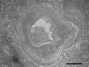
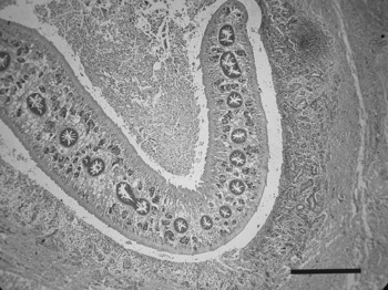
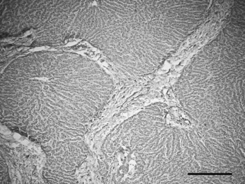
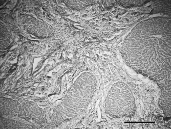
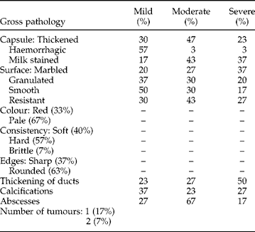
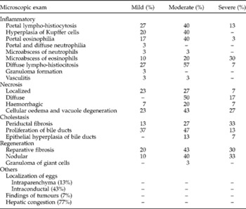
Liver tissues were obtained from 24 F. hepatica infected cattle and two uninfected cattle serving as controls. Of the 24 livers, 16 were diagnosed with cirrhosis according to anatomical–pathological findings (fig. 1–4). In accordance with the degrees of fibrosis, 17% were classified as Stage I, 17% Stage II and 66% Stage III. All infected livers had at least one degree of hepatic fibrosis. The cirrhosis variable was significantly associated with the number of infecting Fasciola, when there were more than 40 (P = 0.02). The correlation analysis indicated that the number of Fasciola was related to degree of fibrosis (+0.296; P = 0.042). Cirrhotic cases (n = 16) showed a parasite density of 116 ± 30, greater than the group without cirrhosis (stages I and II = 56 ± 28 flukes; P = 0.2). In ANOVA analysis, microscopic findings associated with the increase of Fasciola were: hyperplasia in the biliary ducts (η2: 0.529, P = 0.012) and microabscess of neutrophils (η2: 0.49, P = 0.024); and from gross examination: resistant surface (η2: 0.517, P = 0.015) and thickening of biliary duct (η2: 0.694, P = 0.001). Pathological characteristics of the hepatic biopsies by gross examinations and microscopy are shown in tables 1 and 2, respectively. Based on the ROC curve analysis, the cut-off point considered to be the most sensitive for the diagnosis of cirrhosis was 38 Fasciola, with a sensitivity of 94%, specificity of 75%, and an area below the curve of 0.77.
Focal necrosis in hepatic parenchyma (magnification × 25). (H & E stain). Scale bar represents 500 μm.

Adult F. hepatica parasite found in an intrahepatic biliary duct. Epithelial hyperplasia of the duct can be seen (magnification × 25). (H & E stain). Scale bar represents 500 μm.

Increased hepatic stroma with early atrophy and regeneration (magnification × 25). (H & E stain). Scale bar represents 500 μm.

Liver cirrhosis, severe fibrosis with replacement of hepatic parenchyma. Nodularity of parenchyma showing atrophy (magnification × 25). (H & E stain). Scale bar represents 500 μm.

Gross hepatic findings and severity in 24 F. hepatica infected cattle.
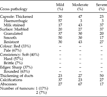
%, Percentage expressed as positives/total number of livers.
Microscopic findings after H & E and trichrome staining of hepatic tissues and their severity from 24 chronically F. hepatica infected cattle.

%, Percentage expressed as positives/total number of livers.
Discussion
In the study, it was found that approximately two-thirds of hepatic tissue from F. hepatica infected cattle showed cirrhosis (62.5%). Hepatic fibrosis has not been described as an important finding in the chronic phase of fascioliasis. One remote study found cirrhosis in infected animals (Vitovec, Reference Vitovec1974). A unique aspect of the present study was to examine the relationship between the number of Fasciola and the degree of fibrosis. How F. hepatica produces fibrosis of the liver is not known. Based on these results, the density of Fasciola is highly correlated with the degree of hepatic fibrosis in naturally infected cattle. Other infections and diseases may influence the occurrence of fibrosis, including bovine lymphomatosis, tuberculosis and hydatidosis, yet each of these was excluded in the present study.
In ANOVA analysis, an association was reached between the number of Fasciola and hyperplasia of the biliary ducts. This has been described previously in rats, and it was suggested that proline produced by the parasites might be the cause of such fibrosis (Campbell et al., Reference Campbell, Sheers, Moore, Edwards and Montague1981). Hypothetically, the high number of Fasciola might be permanently obstructing the biliary ducts, causing biliary stasis, and consequently, chronic inflammation of the epithelium in the biliary ducts, leading to heavy infection and abscess formation (Kim et al., Reference Kim, Kim, Huh and Cho1995; Teichmann et al., Reference Teichmann, Grobusch, Gobels, Muller, Koehler and Suttorp2000; Braun et al., Reference Braun, Jehle, Thio and Pospischil2004), causing hyperplasia (Isseroff et al., Reference Isseroff, Sawna and Reino1977; Hamir & Smith, Reference Hamir and Smith2002) and biliary cirrhosis. Likewise, a rise in type I and II collagen has been described in a study that compared levels of various collagen types between biliary ducts of rats infected with F. hepatica and the biliary ducts of healthy animals; these results would indicate that F. hepatica infection might be producing changes in biliary duct collagen composition, similar to those produced by hepatic cirrhosis and in other disease states that may involve wound scarring (Mark & Isseroff, Reference Mark and Isseroff1983). The second mechanism could relate to the slow deterioration of hepatic parenchyma by repeated infections, ultimately resulting in necrosis (Kim et al., Reference Kim, Lim, Kim, Lee and Lim1999). In the present study, hepatic microabscesses were also associated with the number of Fasciola. Microabscess formation constitutes an interesting finding that has been described previously in animals (Braun et al., Reference Braun, Jehle, Thio and Pospischil2004) and in humans (Kim et al., Reference Kim, Kim, Huh and Cho1995; Teichmann et al., Reference Teichmann, Grobusch, Gobels, Muller, Koehler and Suttorp2000). Fasciola hepatica infection may produce a chronic inflammation of liver, increasing the risk of developing post-inflammatory fibrosis, as results show. The resistant liver surface may relate to retractions and collagen deposition secondary to chronic Fasciola infection.
The number of parasites required for irreversible hepatic fibrosis and the stage of infection when the fibrosis becomes irreversible are unknown. While fibrosis is reversible in its initial states, its progression can lead to cirrhosis (Friedman, Reference Friedman, Schiff, Sorrell and Maddrey2002). The available evidence suggests that initial disease states can be reversible (Bonis et al., Reference Bonis, Friedman and Kaplan2001). A ROC curve analysis was used in order to choose the appropriate cut-off point for cirrhosis. A minimum number of 38 Fasciola may represent an infection level sufficient to cause irreversible hepatic cirrhosis, at least in cattle.
Despite the fact that the degree of calcification within the biliary ducts was not associated with the number of Fasciola (P = 0.126), calcification progressed in parallel with the number of parasites (degree 1 = 37.4 parasites; degree 2 = 100.9 parasites; degree 3 = 151 parasites). Valero et al. (Reference Valero, Santana, Hernandez and Mas-Coma2003) reported that advanced chronic F. hepatica infection can be a risk factor for gallstones. In a study of human infections carried out in Cajamarca, Perú, 101 cases of fascioliasis were reported, of which 1.8% were diagnosed by cholecystectomy (with gallstones being Fasciola infected). In patients undergoing cholecystectomy in Cuzco, Perú, 1.9% of patients were found to have fascioliasis (Alban et al., Reference Alban, Jave and Quispe2002).
A high number of Fasciola was found in hepatic samples, up to 435, which might overestimate our results. Nonetheless, less intense infections could produce lesser compromise of the parenchyma. This study strongly supports a relationship between the presence of moderate to severe parasite densities and progressive hepatic fibrosis. An experimental animal study could shed light upon the relationship between parasite density and cirrhosis. Histological changes in the liver are not diffuse, and multiple biopsies are indicated to look for fibrosis in patients with fascioliasis. Here, at least three hepatic biopsies per lobe (nine in total) were performed to be certain when assessing each liver. Two pathologists independently described the biopsy slides, showing concordance in 86.6% of the livers examined and increasing the significance of these findings.
Experimental animal studies are being designed to help confirm these observations. Exploring the effect of anti-fascioliasis therapy in reversing the hepatic lesion, and possibly early fibrotic changes, is one of the next steps in this neglected disease.
Acknowledgements
We acknowledge the great effort expended in reviewing this article by Dr Herbert L. DuPont, MD, Chief of the Internal Medicine Service at St. Luke's Episcopal Hospital; and Director, Center for Infectious Diseases, University of Texas, Houston School of Public Health, Houston, Texas, USA.