INTRODUCTION
Benthic algae are key members of coral reef communities, providing important ecological functions, such as primary productivity, nutrient retention and recycling, reef cementation and sediment production. Over the past two decades, coral reefs have experienced a shift in dominance from encrusting to upright and fleshy macroalgae (Fong & Paul, Reference Fong, Paul, Dubinsky and Stambler2011). As a result of anthropogenic driven environmental change, these macroalgae have proliferated across all reef zones at the expense of reef-building corals, threatening the sensitive balance within coral reef ecosystems (Hughes et al., Reference Hughes, Rodrigues, Bellwood, Ceccarelli, Hoegh-Guldberg, McCook, Moltschaniwskyj, Pratchett, Steneck and Willis2007; Birrell et al., Reference Birrell, McCook, Willis and Diaz-Pulido2008). These changes are especially evident in the Caribbean, where coral cover has decreased by 80% in recent decades and two-thirds of the coral reefs are threatened by human activities (Burke & Maidens, Reference Burke and Maidens2004). Next to small micro- and filamentous algae, commonly known as turf algae (Fricke et al., Reference Fricke, Teichberg, Beilfuss and Bischof2011a), large macroalgae are known to have deleterious effects on corals (Mumby et al., Reference Mumby, Hastings and Edwards2007).
One of these spreading macrophytes is Lobophora variegata (Lamouroux) Womersley ex E.C. Oliveira 1977 (Dictyotales: Dictyotaceae). This brown alga is an important component of many coral reef ecosystems and has been observed to spread over many degraded reefs throughout the Caribbean (Mumby et al., Reference Mumby, Foster and Fahy2005; Nugues & Bak, Reference Nugues and Bak2008). This alga competes directly with corals, affecting their growth, recruitment and reproduction (Foster et al., Reference Foster, Box and Mumby2008; Nugues & Bak, Reference Nugues and Bak2008).
Lobophora variegata provides a suitable substrate for a diverse epiphytic community (Fricke et al., Reference Fricke, Titlyanova, Nugues and Bischof2011b). Its characteristic thallus structure makes it suitable for a total of 70 epiphytic species. These epiphytes may impact the entire ecosystem by: (1) interacting with their host (Tobler, Reference Tobler1906; Michael et al., Reference Michael, Shin, Hanna and Spafford2008); (2) serving as food source (Cattaneo, Reference Cattaneo1983; Hall & Bell, Reference Hall and Bell1988; Frankovich & Zieman, Reference Frankovich and Zieman2005); and (3) affecting other benthic organisms. Species such as the red alga Anotrichium tenue and the cyanobacteria Lynbya spp. and Phormidium spp., which have detrimental effects on corals (Jompa & McCook, Reference Jompa and McCook2003; Kuffner & Paul, Reference Kuffner and Paul2004; Titlyanov et al., Reference Titlyanov, Yakovleva and Titlyanova2007), were commonly found growing on L. variegata blades (Fricke et al., Reference Fricke, Titlyanova, Nugues and Bischof2011b). Due to the increasing importance of L. variegata in many coastal ecosystems, an understanding of the distribution and occurrence of its epiphytic species is needed.
Here, we report the occurrence of the Rhodophyte Neosiphonia howei (Hollenberg) Skelton & G.R. South, 2007, a common algal epiphyte, growing on blades of L. variegata over a broad depth-range (6.5–40 m) on the leeward coral reefs of Curaçao.
MATERIALS AND METHODS
Study sites
The study was carried out in Curaçao, former Netherlands Antilles (12°01′–12°23′N 68°44′–69°10′W). Two vertical transects were investigated along the leeward south-west coast of the island. The first transect was at Buoy Zero (B0), also called Klein Piscadera (12°07′N, 69°57′W), 100 m west from the entrance of Piscadera Bay. At this site L variegata was found growing from the drop off at 10 m water depth down the reef slope. It was sampled from 10 down to 40 m. The second transect Piscadera Bay (PISC) was situated 100 m south-east from the first transect and 100 m seawards of the entrance of Piscadera Bay. Here L. variegata was present from 6.5 to 30 m depth (for further description of the study sites and transects see Bak, Reference Bak1976; Van Duyl, Reference Van Duyl1985; Fricke et al., Reference Fricke, Titlyanova, Nugues and Bischof2011b).
Analyses of epiphytic algae
Epiphytes were collected at 6 depths (10, 15, 20, 25, 35 and 40 m) at B0 and at 4 depths (6.5, 10, 20 and 30 m) at PISC. At each site and depth, 5 to 18 Lobophora variegata blades were randomly sampled by SCUBA diving (for further details on the sampling procedure see Fricke et al., Reference Fricke, Titlyanova, Nugues and Bischof2011b). Samples were transported in plastic bags filled with ambient seawater to the field station, where the material was pressed as herbarium sheets. In the laboratory the material was soaked in water and scanned under a dissection microscope (12×, Stemi SV6, Zeiss, Hamburg, Germany) for epiphytes. Epiphytes were carefully removed and investigated under a microscope (Axioscope, Zeiss, Hamburg, Germany) at different magnifications (100× –630× magnification). Algae were identified to the lowest possible taxonomic level. Species identification was based on different literature (Taylor, Reference Taylor1945; Hollenberg, Reference Hollenberg1968; Littler & Littler, Reference Littler and Littler2000; Dawes & Mathieson, Reference Dawes and Mathieson2008). We distinguished between algae growing on the upperside and underside of the L. variegata blades.
Microtome cuts
To allow fine cuts, algal material was embedded in plastic (Technovit 7100, Kulzer and Co, GmbH, Wehrheim). Prior to embedding, material was dehydrated by keeping it in 75% ethanol overnight, followed by exposure to 100% ethanol for a minimum of 1 hour. After embedding, the material was stored in a drying oven at 60°C overnight to guarantee full polymerization. Samples were cut with a microtome to obtain sections of 15 µm thicknesses, transferred to object slides, and investigated under the microscope.
Statistical analysis
Percentage occurrence of Neosiphonia howei and other related epiphytes of the genera Polysiphonia were plotted as a function of depth, blade side and site. Differences among depths for each blade side at each site were analysed using a heterogeneity G-test, followed by a simultaneous test procedure (STP) to determine homogeneous groups (Sokal & Rohlf, Reference Sokal and Rohlf1995).
RESULTS AND DISCUSSION
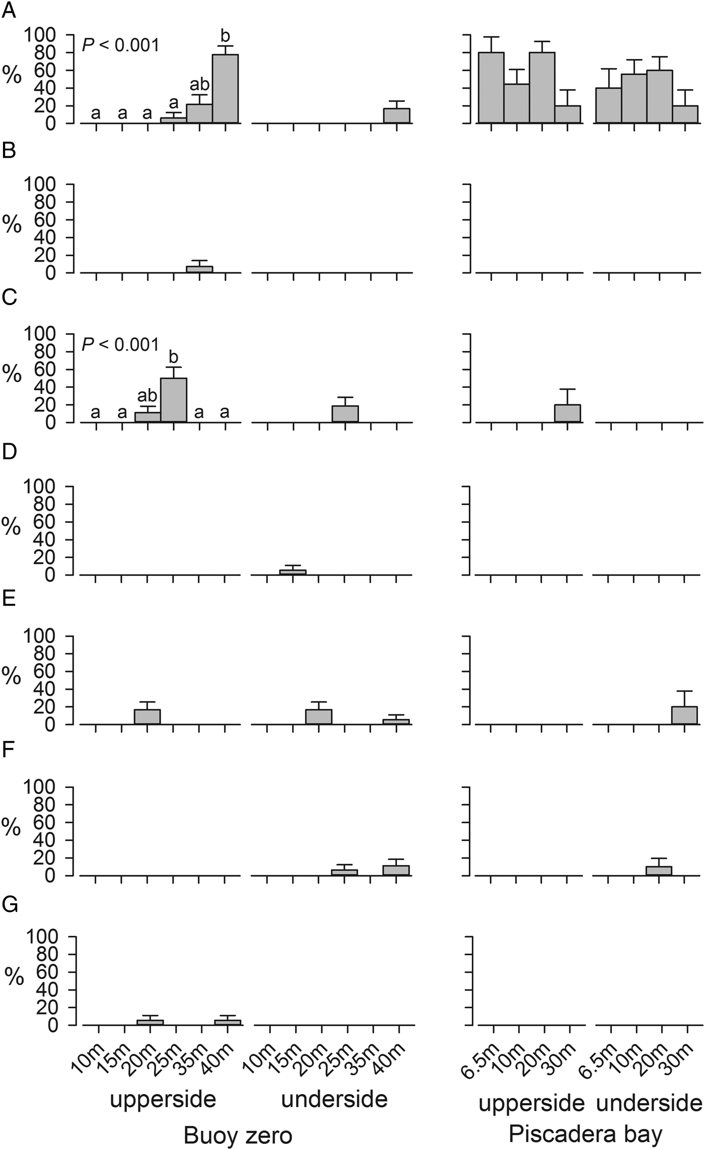
Although most epiphytic species were observed on the underside of the Lobophora variegata blades (Fricke et al., Reference Fricke, Titlyanova, Nugues and Bischof2011b), Neosiphonia howei was frequently found on the exposed upper, but hardly on the underside of L. variegata at B0 (Figure 1A). The percentage occurrence of N. howei on the upperside of L. variegata varied significantly among depths (heterogeneity G-test, GH = 48.52, df = 5, P < 0.001), with no occurrence at 10, 15 and 20 m and a maximum occurrence of 77.8% at 40 m. STP tests showed that 2 depth groups (10 m–35 m and 35 m–40 m) formed homogeneous sets. At PISC, N. howei was common on both upper- and under-sides of the blades across the entire depth gradient and its occurrence did not differ among depths (heterogeneity G-test, P > 0.05).
Percentage occurrence of: (A) Neosiphonia howei (Hollenberg) Skelton & G.R. Southand; (B) Polysiphonia binneyi Harvey; (C) Polysiphonia denudata (Dillwyn) Greville ex Harvey; (D) Polysiphonia sertularioides (Grateloup) J. Agardh; (E) Polysiphonia scopulorum var. villum (J. Agardh) Hollenberg; (F) Polysiphonia pseudovillum Hollenberg; (G) Polysiphonia subtilissima Montagne growing on the upper and undersides of Lobophora variegata blades for each site × depth combination. Analysed by heterogeneity G-tests, followed by simultaneous test procedures. Significance and homogeneous groups are indicated only when P < 0.05. Number of blades were for B0: 5 (10 m), 18 (15 m), 18 (20 m), 16 (25 m), 14 (35 m) and 18 (40 m) and for PISC: 5 (6.5 m), 9 (10 m), 10 (20 m) and 5 (30 m). Values are means +1 standard error.

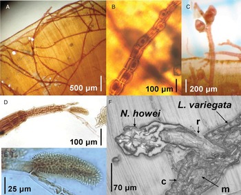
At present, N. howei is known as a typical epiphyte in shallow waters, intertidal to upper subtidal (Hollenberg, Reference Hollenberg1968; Abbott, Reference Abbott1999; Littler & Littler, Reference Littler and Littler2000). In benthic algal surveys by Van den Hoek et al. (Reference Van den Hoek, Colijn, Cortel-Breeman and Wanders1972) at one of our study sites (Buoy Zero) and on nearby sites, N. howei was reported to grow exclusively in the shallows, on mangrove roots. Due to its observed restricted appearance in the intertidal area, its ability to grow continuously submerged was even questioned by these authors. Nearly 40 years later, we can positively answer this question, as the alga was shown on the reef slope in all fertile life stages. Reproductive features and stages (i.e. tetraspongia, male gametophytes with spermatangial branches and female gametophytes with carpogonium) were commonly observed (Figure 2; personal observation). Interestingly, N. howei was absent on settlement tiles exposed for up to 333 days at depths ranging from 5 to 25 m depth at Buoy Zero during the same period (Fricke et al., Reference Fricke, Teichberg, Beilfuss and Bischof2011a). Whilst further information is needed on the distribution of N. howei on other macroalgae and dead coral substrates, these findings suggest that L. variegata may be a particularly well-suited substrate for N. howei, also that the spread of the former may have increased the depth distribution of the latter.
Neosiphonia howei (Hollenberg) Skelton & G.R. South 2007, growing epiphytically on Lobophora variegata (Lamouroux) Womersley ex E.C. Oliveira 1977 at 6.5 m depth, showing different life stages: (A) thallus covering the surface with branching prostrate axis; (B) erect axis with tetrasporangia; (C) female gametophyte with carposporangia; (D&E) male gametophyte with spermatangial branch; (F) microtome cut, showing attachment of N. howei on L. variegata blade in cross-section. Neosiphonia howei rhizoids (r) are attached to the cortex (c) and penetrate deeply into the medullary cells (m).

Neosiphonia howei was observed to be spreading over the L. variegata blade with its creeping filamentous thallus, growing over margin (Figure 2). As opposed to epiphytes which are loosely attached on the outer layer of their host (‘holo-epiphytes', Ducker & Knox (Reference Ducker and Knox1984)), N. howei was observed to anchor deeply in the host's medulla (‘amphi-epiphytes'), which was shown by microtome cuts (Figure 2F). Such firm attachment may indicate hemiparasitic behaviour, as it has been reported for Vertebrata lanosa, an obligate epiphyte of the brown alga Ascophyllum nodosum. This rhodophyte draws organic carbon from its host with its penetrating rhizoids (Garbary & Deckert, Reference Garbary and Deckert2004). Such firm attachment can cause lesions through which potential pathogens enter the host. As an example, a sibling species Neosiphonia savatierii promotes an opening for bacterial infection in the carrageenophyte Kappaphycus alvarezii, causing ‘goose bumps swellings' and ‘ice-ice disease' (Vairappan et al., Reference Vairappan, Chung, Hurtado, Soya, Lhonneur and Critchley2008; Loureiro et al., Reference Loureiro, Reis and Critchley2010). So far such disease symptoms have not been reported in L. variegata. The surface of L. variegata blade is commonly colonized by mutualistic gram-positive actinomycetes (Jiang et al., Reference Jiang, Jensen and Fenical1999). These filamentous bacteria may inhibit bacterial and fungal growth (Holmström & Kjelleberg, Reference Holmström, Kjelleberg and Evans2000; Rao et al., Reference Rao, Webb and Kjelleberg2006) due to production of macrolide antibiotics and prevent infections following epiphytic penetration.
Other species of the genera Neosiphonia and Polysiphonia are common epiphytes on L. variegata. Polysiphonia binneyi Harvey, P. denudata (Dillwyn) Greville ex Harvey, P. sertularioides (Grateloup) J. Agardh, P. scopulorum var. villum (J. Agardh) Hollenberg and P. subtilissima Montagne were occasionally found in our study (Figure 1B-G). Polysiphonia dotyi and P. exilis were reported by Hollenberg (Reference Hollenberg1968), and P. havenensis by Ballantine (Reference Ballantine1979). Despite their epiphytic occurrence, Polysiphonia and Neosiphonia species, including N. howei, are probably not restricted to L. variegata, as they were also observed growing on other hard substrata within Caribbean coral reefs (Hollenberg, Reference Hollenberg1968; Abbott, Reference Abbott1999; Littler & Littler, Reference Littler and Littler2000; Dawes & Mathieson, Reference Dawes and Mathieson2008). In the face of increasing macroalgal abundance on coral reefs, further research on epiphyte–host relations and potential substrate preferences are needed.
SYSTEMATICS
Class FLORIDEOPHYCEAE Cronquist, 1960
Subclass RHODYMENIOPHYCIDAE Saunders and Hommersand, 2004
Order CERAMIALES Oltmanns, 1904
Family RHODOMELACEAE Areschoug, 1847
Genus NEOSIPHONIA Kim and Lee, 1999
Neosiphonia howei (Hollenberg) Skelton and South, 2007 (Figure 1)
= Polysiphonia howei Hollenberg, 1945
DIAGNOSIS
Thallus filamentous prostrate, dark brown red coloured; prostrate axis 50–150 μm in diameter, segments mostly less than 1 diameter to 100–146 µm long, prostrate branches giving rise ventro-laterally to lateral branches in alternating positions; rhizoids 1(–2) cellular, with simple or digitate ends, cut off across-wall from the centre or distal end of the pericentral cells; erect axes formed at intervals of 2–6–8 segments, unbranched, 1.8–2.4 mm height, 70–150 μm in diameter and segments 85–120 µm long formed of 8–10(–12) pericentral cells, which form secondary pit connections to adjacent segments at either end; trichoblasts 24–28 µm in diameter at the base, to 353–500 µm long, with 3–4 dichotomies, diminishing towards cone-shaped tips, 15–17.5 µm in diameter, scar cells present, one per segment in no regular intervals; tetrasporangia roundish, tetrahedrally divided, 70–85 µm in diameter as swollen spiral series in outer branchlets; cystocarps ovoid 230–350 µm in diameter, celled stalk; spermatangial branches near branch tips 113 µm long and 37 µm wide on a sturdy 1-celled stalk, without a sterile tip, arising from primary branch of the trichoblast.
Comment
One additional characteristic defining Neosiphonia howei by Hollenberg (Reference Hollenberg1968), is the offset-position of pericentral cells. This characteristic is not mentioned in the descriptions of Taylor (Reference Taylor1945), Littler & Littler (Reference Littler and Littler2000), or Dawes & Mathieson (Reference Dawes and Mathieson2008) and was not clearly definable in the present species.
ACKNOWLEDGEMENTS
For kind assistance with diving and fieldwork we thank Svenja Beilfuss. For support and assistance with the microtome cutting procedure we acknowledge the working group on cell biology (Professor Stick, University of Bremen), especially Ute Helmboldt-Cesar. We are grateful to the staff of the CARMABI foundation: Dr Mark J.A. Vermeij, Dr Adolphe Debrot, Carlos Winterdaal, Oscar Frans and Sislin Rosalia for friendly support. For statistical support we thank Professor Clarke. This study was supported by the Bremen International Graduate School for Marine Sciences (GLOMAR) that is funded by the German Research Foundation (DFG) within the frame of the Excellence Initiative by the German federal and state governments to promote science and research at German universities.