The coronary artery revascularisation operation is necessary for all patients with congenital left main coronary artery atresia, because of a high sudden death rate. Reference Musiani, Cernigliaro, Sansa, Maselli and De Gasperis1 Surgical treatment involved surgical ostial angioplasty and paediatric coronary artery bypass surgery with internal thoracic artery grafting, depending on the length of the atretic or stenotic lesion from the left coronary ostium to the left coronary bifurcation. Kitamura Reference Kitamura2 reported that long-term survival and internal thoracic artery graft function are excellent in growing children. However, there are no reports of paediatric coronary artery bypass surgery patients who needed reintervention in childhood due to subsequent obstruction. We report a case with left main coronary artery atresia and obstruction of the bypass graft. Left main coronary artery reconstruction was performed with a good outcome.
Case report
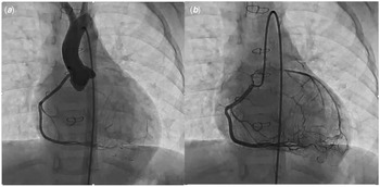
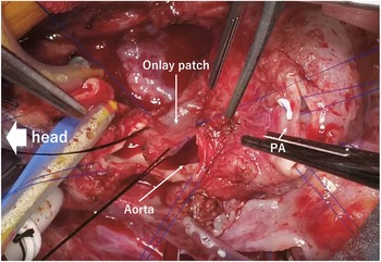
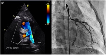
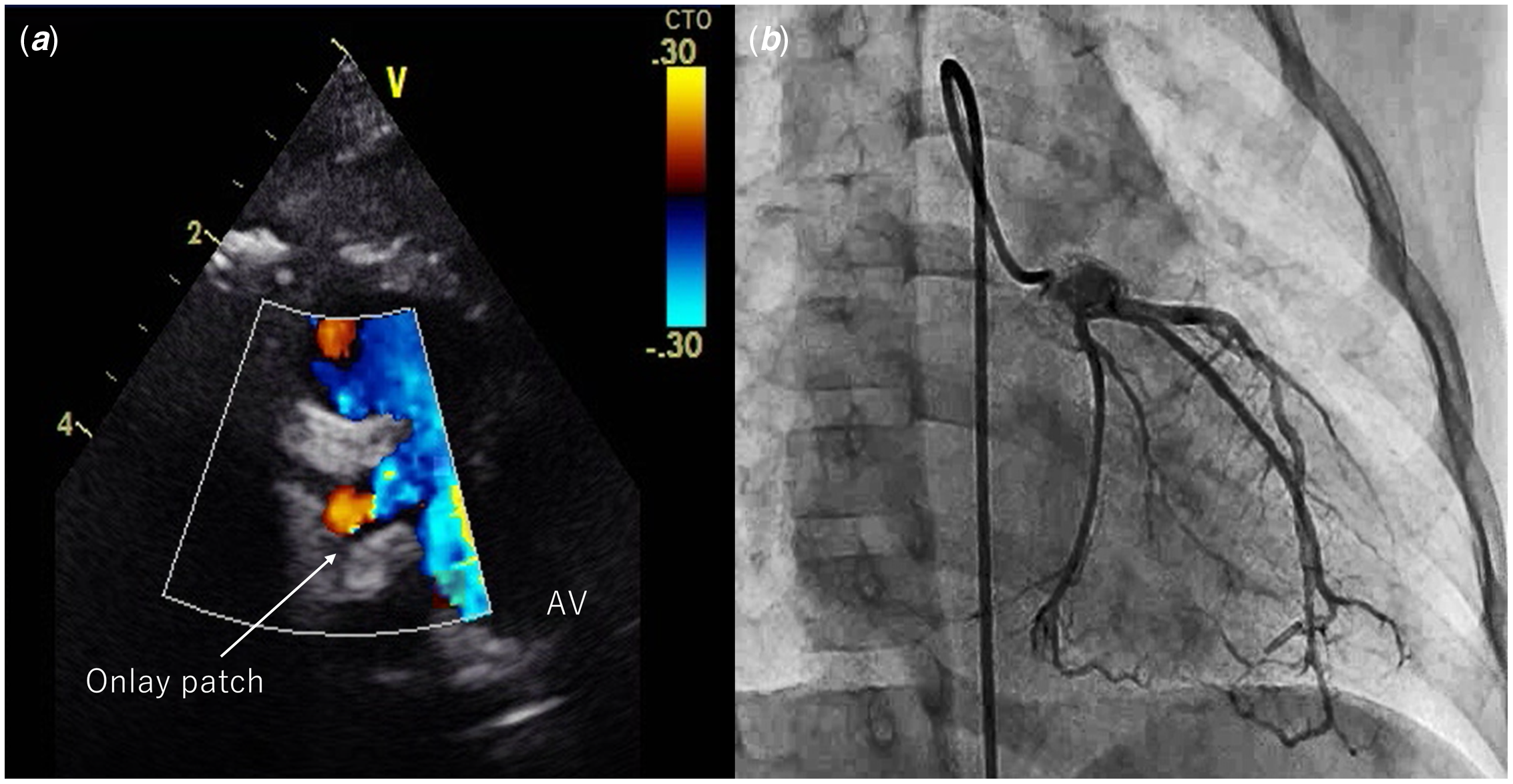
A 2-year and 3-month-old girl (10.5 kg) presented with a past medical history of left main coronary artery atresia; she had undergone paediatric coronary artery bypass surgery with the left internal thoracic artery at the age of one and a half years (9 kg). The arteriotomy was made at the proximal left anterior descending artery. The end-to-side anastomoses were performed by running suture with 8-0 monofilament suture. A follow-up angiogram showed obstruction of the internal thoracic artery graft, and right coronary artery angiography showed a collateral branch from the proximal right coronary artery coursing anteriorly and anastomosing with the proximal left anterior descending artery (Fig 1). Myocardial perfusion scintigraphy detected ischaemia; hence, we decided to perform left main coronary artery reconstruction. The operation was performed with standard cardiopulmonary bypass. Myocardial preservation was achieved through antegrade cold blood cardioplegia with additional retrograde blood cardioplegia. The main pulmonary trunk was transected to allow adequate exposure of the aortic root, left coronary ostium, and left main stem. After aortic cross-clamping, the incision was started on the anterior aspect of the aortic root and was extended laterally toward the involved coronary ostium. The incision was extended across the atretic area until a normal coronary arterial wall was reached at the circumflex bifurcation and left anterior descending artery branches. Glutaraldehyde-treated autologous pericardium was utilised as an onlay patch to enlarge the main coronary stem and the adjacent aortic incision, so as to give the new coronary ostium a funnel shape. The patch was sutured with 8-0 monofilament suture to the peripheral portion of the left main coronary artery and with 7-0 monofilament suture to the aortic wall continuously. We sutured the patch material to the visceral pericardium around the left main coronary ostium, in order to prevent post-operative left main coronary ostiumobstruction (Fig 2). The patient had an uneventful post-operative course and was extubated on post-operative day 1 and discharged without complications on post-operative day 15. She was administered low-dose aspirin and warfarin immediately post-operatively, and the administration will be maintained continuously. The post-operative echocardiogram demonstrated a wide-open orifice (Fig 3a). Coronary angiography performed 1 year after the procedure revealed perfect patency of the left main stem and no aneurismal change (Fig 3b).
Preoperative imaging. ( a ) Aortography shows left main coronary artery atresia. ( b ) Right coronary artery angiography shows a collateral branch from the proximal right coronary artery coursing anteriorly and anastomosing with the proximal left anterior descending artery.

Surgical technique. The main pulmonary trunk was transected to allow adequate exposure of the aortic root, left coronary ostium and left main stem. The left main stem beyond the obstruction is opened and the incision extended through the left main coronary artery to the bifurcation. Glutaraldehyde-treated autologous pericardium was utilised for an onlay patch.

Post-operative imaging. ( a ) The post-operative echocardiogram demonstrates a wide-open orifice. ( b ) The post-operative angiogram showed perfect patency of the left main stem.
Comment
Herein, we reported a case of successful reconstruction in a patient with bypass graft obstruction. Paediatric coronary artery bypass surgery with internal thoracic artery grafting is the most common treatment for left main coronary artery atresia and is associated with excellent short- and long-term outcomes. Reference Kitamura2 However; it can result in competitive flow and consumes bypass material. Surgical reconstruction of the left main coronary artery avoids these potential drawbacks, and in addition, does not preclude percutaneous coronary intervention for the distal coronary tree if necessary at a later stage. Reference Raanani, Kogan, Shapira, Sagie, Kornowsky and Vidne3 This is certainly a reasonable concept, and the surgical decision partly depends on the length of the atretic coronary trunk. In this case, we suppose that the bypass graft failed at an early stage by the flow competition with left anterior descending artery due to the well-developed collateral blood flow from the right coronary artery. We agree that paediatric coronary artery bypass surgery with internal thoracic artery grafting is the first choice for left main coronary artery atresia, but in this case with graft obstruction, rePCABS with the right internal thoracic artery would also result in obstruction, so we chose the surgical angioplasty. Surgical reconstruction of the left main coronary artery is a good palliative alternative for left main coronary artery atresia with internal thoracic artery–left anterior descending artery obstruction which can save the internal thoracic artery for future use.
Acknowledgements
We thank Editage (www.editage.jp) for English language editing.
Financial support
None.
Conflict of interest
None declared.
Ethical standards
Papers reporting the results of human and/or animal experimentation that do not contain an Ethical Standards statement will not be reviewed.