Introduction
Monogeneans of Ligophorus Euzet et Suriano, Reference Euzet and Suriano1977 are specific gill parasites of fish from the family Mugilidae Jarocki, 1822. The genus currently includes 66 nominal species (Euzet & Suriano, Reference Euzet and Suriano1977; Dmitrieva et al., Reference Dmitrieva, Gerasev and Pron'kina2007, Reference Dmitrieva, Gerasev, Gibson, Pronkina and Galli2012, Reference Dmitrieva, Gerasev and Gibson2013a; Abdallah et al., Reference Abdallah, De Azevedo and Luque2009; Soo & Lim, Reference Soo and Lim2012, Reference Soo and Lim2013; El Hafidi et al., Reference El Hafidi, Rkhami, De Buron, Durand and Pariselle2013a, Reference El Hafidi, Diamanka, Rkhami and Pariselleb; Kritsky et al., Reference Kritsky, Khamees and Ali2013; Sarabeev et al., Reference Sarabeev, Rubtsova, Yang and Balbuena2013; Marchiori et al., Reference Marchiori, Pariselle, Pereira, Agnèse, Durand and Vanhove2015; Rodríguez-González et al., Reference Rodríguez-González, Míguez-Lozano, Llopis-Belenguer and Balbuena2015a, Reference Rodríguez-González, Miguez-Lozano, Llopis-Belenguer and Balbuena2015b; Khang et al., Reference Khang, Soo, Tan and Lim2016; Pakdee et al., Reference Pakdee, Ogawa, Pornruseetriratn, Thaenkham and Yeemin2018). Identification of Ligophorus species is based mainly on the morphology of hard structures of the haptor and the distal parts of the female and male reproductive systems (Euzet & Suriano, Reference Euzet and Suriano1977; Sarabeev et al., Reference Sarabeev, Rubtsova, Yang and Balbuena2013). Many species are very morphologically similar to each other, creating difficulties for delimitation of species (Euzet & Suriano, Reference Euzet and Suriano1977; Dmitrieva et al., Reference Dmitrieva, Gerasev and Pron'kina2007, Reference Dmitrieva, Gerasev and Gibson2013a). Some of them were distinguished on the basis of DNA sequence data (Marchiori et al., Reference Marchiori, Pariselle, Pereira, Agnèse, Durand and Vanhove2015; Pakdee et al., Reference Pakdee, Ogawa, Pornruseetriratn, Thaenkham and Yeemin2018). However, these data are discrete or insufficient, representing 127 sequences of the different parts of the nuclear DNA ribosomal cluster for only 32 species (https://www.ncbi.nlm.nih.gov/nuccore), including 12 species from the Mediterranean Sea and two species from the Azov Sea (Mollaret et al., Reference Mollaret, Jamieson and Justine2000; Plaisance et al., Reference Plaisance, Littlewood, Olson and Morand2005; Blasco-Costa et al., Reference Blasco-Costa, Míguez-Lozano, Sarabeev and Balbuena2012; Rodríguez-González et al., Reference Rodríguez-González, Míguez-Lozano, Llopis-Belenguer and Balbuena2015a), two species from the West Atlantic Ocean off Brasilia (Marchiori et al., Reference Marchiori, Pariselle, Pereira, Agnèse, Durand and Vanhove2015), 14 species from the East Indian Ocean off Malaysia (Soo et al., Reference Soo, Tan and Lim2015; Khang et al., Reference Khang, Soo, Tan and Lim2016) and for three species from the South China Sea (Wu et al., Reference Wu, Zhu, Xie and Li2006, Reference Wu, Zhu, Xie and Li2007; Pakdee et al., Reference Pakdee, Ogawa, Pornruseetriratn, Thaenkham and Yeemin2018). Data on DNA sequences for Ligophorus species from the Black Sea and the Sea of Japan are still lacking.
Ligophorus kaohsianghsieni (Gusev, Reference Gusev and Bychovsky1962) Gusev, Reference Gusev and Bauer1985 was described from the so-iuy mullet P. haematocheilus (Temminck & Schlegel, 1845) from the Tumen-Ula River flowing into the Sea of Japan and the Liao River flowing into the Yellow Sea (Gusev, Reference Gusev and Bychovsky1962, Reference Gusev and Bauer1985), but its native range includes the Sea of Japan as such (Sarabeev et al., Reference Sarabeev, Rubtsova, Yang and Balbuena2013), as well as the East China and South China Seas (Zhang et al., Reference Zhang, Yang, Liu and Xuejuan2003; Dmitrieva et al., Reference Dmitrieva, Gerasev, Kolpakov, Nguen and Ha2013b). In the Black Sea, this monogenean was first found on P. haematocheilus off the coast of Crimea (Dmitrieva, Reference Dmitrieva1996). Subsequently, this parasite was repeatedly registered on the same fish species in the Black Sea, off Bulgaria, and in the Sea of Azov (Pankov, Reference Pankov2011; Sarabeev et al., Reference Sarabeev, Rubtsova, Yang and Balbuena2013), where it was introduced from the Sea of Japan. Morphological descriptions of L. kaohsianghsieni have been published based on specimens from the Tumen-Ula and Liao rivers (Gusev, Reference Gusev and Bauer1985) and from the Black and Azov seas (Dmitrieva, Reference Dmitrieva1996; Sarabeev et al., Reference Sarabeev, Rubtsova, Yang and Balbuena2013), but with no data on its morphology from the Sea of Japan, the region from which the host was introduced. This study presents the molecular characterization of L. kaohsianghsieni using 28S, 18S and internal transcribed spacer 1 (ITS1) (rDNA) gene clusters and provides new morphological data for this species across its native and introduced distribution.
Materials and methods
Sampling
Monogeneans were collected from the gills of P. haematocheilus, caught in the Tavrichan Bay of the Sea of Japan, near the mouth of the River Razdolnaya (43°19′48″N, 131°46′19″E) and mouth of the River Kievka (42°51′27.8″N 133°38′39.3″E), and off the coast of Crimea near Sevastopol (44°36′58.4″N, 33°30′14″E) and Karadag (44°54′41″N, 35°12′07″E), and in the Kerch Strait (45°07′52.0″N 36°25′31.1″E), Northern Black Sea (table 1). All monogeneans were collected alive, some of them were immediately mounted in glycerine jelly (prepared with 0.5 g carbolic acid) after Gusev (Reference Gusev1983), and parts of others were stored in absolute ethanol and kept at 5°C for DNA analysis. Additional materials of 15 specimens of L. kaohsianghsieni collected in the Black Sea near Crimea from the Marine Parasites Collection of the A. O. Kovalevsky Institute of Biology of the Southern Seas, Sevastopol, Russia (IBSS collection, http://marineparasites.org) were reinvestigated for morphometry.
Sampling data, sequenced material, voucher and GenBank accession numbers of Ligophorus kaohsianghsieni.

Morphology analyses
Measurements and light micrographs were made with Olympus CX41 microscopes (Olympus Corporation, Tokyo, Japan), at magnifications of ×800–1000, using phase-contrast optics and CellSense digital image analysis software (Olympus Corporation, Tokyo, Japan). The measuring scheme mainly followed that suggested for the Dactylogyridae by Gusev (Reference Gusev and Bauer1985) with some configurations according to Dmitrieva et al. (Reference Dmitrieva, Gerasev, Kolpakov, Nguen and Ha2013b). Abbreviations of the linear measurements are presented in table 2. All dimensions are given in micrometres. The mean, standard deviation and range were used to describe the linear measurements. Morphological analysis of 41 specimens was carried out using principal component analysis based on the correlation matrix (30 measurements of hamulus and bars were lоg10-transformed) using the Statistica 6 for Windows software package.
Primers used for amplification.

DNA extraction
Prior to DNA analysis, the voucher slides from the haptor of the specimens used for sequencing were prepared and deposited in the IBSS collection, then identified based on the haptoral structures (Gusev, Reference Gusev and Bauer1985; Dmitrieva, Reference Dmitrieva1996; Sarabeev et al., Reference Sarabeev, Rubtsova, Yang and Balbuena2013). DNA extraction was carried out using DNK-EXTRAN Kit (Syntol, Moscow, Russia). Single animals were incubated in 100 μl of lysis buffer (Syntol, Moscow, Russia) with 5 μl of Syntol Proteinase K and 1 μl of 2-mercaptoethanol at 56°C overnight. After lysing, animals were vortexed for 20 s and DNA extraction was carried out according to the DNK-EXTRAN Kit protocol. The elution volume was 30 μl. The DNA was stored at −20°C.
Polymerase chain reaction (PCR) amplification and sequencing
The PCR was performed in a total volume 20 μLmix, consisting of 5xPCR ScreenMix with magnesium chloride (Evrogen, Moscow, Russia), 0.5 μM of each primer and 2 μL template DNA. The primers for amplification of 28S, ITS1 and 18S of ribosomal DNA are presented in table 2.
The 28S, ITS1 and 18S were amplified using the same following conditions: initial denaturation at 95°C for 3 min, followed by 38 cycles of denaturation at 94°C for 40 s, annealing at 56°C for 30 s and extension at 72°C for 45 s, the final extension at 72°C for 4 min. Amplicons were separated with horizontal electrophoresis on 1% agarose/Tris-Borate-Ethylenediaminetetraacetic acid buffer gel with ethidium bromide and visualized using an ultraviolet transilluminator. PCR products were directly sequenced using an ABI Big Dye Terminator v.3.1 Cycle Sequencing Kit (Applied Biosystems, Waltham, USA), as recommended by the manufacturer, with the internal sequencing primers described by Tkach et al. (Reference Tkach, Littlewood, Olson, Kinsella and Swiderski2003) for 28S rDNA. PCR product sequences were analysed using an ABI 3500 Genetic Analyzer (Thermo Fisher Scientific, Waltham, USA) at the Federal Scientific Center of the East Asia Terrestrial Biodiversity Far Eastern Branch of the Russian Academy of Sciences. Molecular analyses were performed on a total of 14 samples. All nucleotide sequences obtained during this study were deposited in the international National Center for Biotechnology Information GenBank database (table 1).
Molecular taxonomy analyses
Ribosomal DNA sequences were assembled with SeqScape v.2.6 software (Applied Biosystems, Waltham, USA). The obtained fragments of rDNA were aligned in the BioEdit software program (Hall, Reference Hall1999) and then the alignment was manually refined. The multiple alignment was run by ClustalW (Thompson et al., Reference Thompson, Higgins and Gibson1994) in the MEGAX software (Kumar et al., Reference Kumar, Stecher, Li, Knyaz and Tamura2018). Sequence datasets for phylogenetic analysis include original data and all available rDNA sequences in the GenBank database (table 3). As Ergenstrema mugilis Paperna, 1964 occurred as the sister group to Ligophorus spp. within the marine Ancyrocephalinae (Blasco-Costa et al., Reference Blasco-Costa, Míguez-Lozano, Sarabeev and Balbuena2012), it was chosen as the outgroup (GenBank accession number JN996800). Phylogenetic analysis was performed on the basis of each rDNA fragment separately with the Bayesian and the maximum likelihood (ML) algorithms using MrBayes v. 3.1.2 (Huelsenbeck et al., Reference Huelsenbeck, Ronquist, Nielsen and Bollback2001) and PhyML v. 3.1 software (Guindon & Gascuel, Reference Guindon and Gascuel2003), respectively. The best nucleotide substitution models, the GTR + G, TIM3ef + I + G and TPM2uf + G (Posada, Reference Posada2003) were estimated with jModeltest v. 2.1.5 software (Darriba et al., Reference Darriba, Taboada, Doallo and Posada2012) for ribosomal 28S rDNA, 18S rDNA and ITS1 rDNA fragments data set, respectively, using Bayesian information criterion for Bayesian inference (BI). For ML analysis, the best nucleotide substitutions, GTR + I + G, TIM3 + I + G and GTR + G (Posada, Reference Posada2003), were chosen for ribosomal 28S rDNA, 18S rDNA and ITS1 rDNA, respectively, using Akaike's information criterion (Akaike, Reference Akaike1974). Bayesian analyses were performed using 10,000,000 generations with two independent runs. Summary parameters and the phylogenetic tree were calculated with a burn-in of 25% of generations. The significance of the phylogenetic relationships was estimated using posterior probabilities (Huelsenbeck et al., Reference Huelsenbeck, Ronquist, Nielsen and Bollback2001). Estimation of ML phylogenetic relationships’ significance was performed with the help of the approximate likelihood ratio test with eBayes support (Anisimova & Gascuel, Reference Anisimova and Gascuel2006). Estimates of average evolutionary divergence over sequence pairs within groups and between groups were conducted in MEGAX (Kumar et al., Reference Kumar, Stecher, Li, Knyaz and Tamura2018). All ambiguous positions were removed for each sequence pair (pairwise deletion option).
GenBank accession numbers of 28S rRNA, 18S rRNA and internal transcribed spacer 1 (ITS1) sequences of the Ligophorus species used in the phylogenetic analyses.

Results
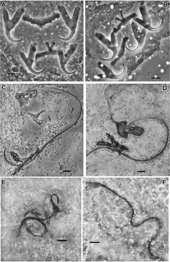
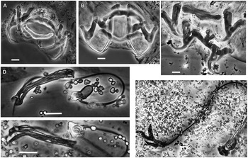
A comparison of the shape of dorsal and ventral anchors, dorsal and ventral bars, the male copulatory organ and the vagina of L. kaohsianghsieni specimens collected in different seas showed no obvious differences (fig. 1).
Haptoral structures (A, B), male copulatory organ (C, D) and vagina (E, F) of Ligophorus kaohsianghsieni ex Planiliza haematocheila from the Black Sea (A, C, E) and the Sea of Japan (B, D, F). Scale bar = 10 μm.

A comparative analysis of 45 newly obtained measurements of L. kaohsianghsieni from the Black Sea and Sea of Japan revealed no significant differences; the ranges of all corresponding measurements overlapped between samples from different seas (table 4). A small difference in the total length of the marginal hook was observed between specimens from the rivers of the Russian Far East and the Black Sea and Sea of Japan. In addition, two dimensions of the ventral bar anterior processes in the present study were smaller than in the previous studies (table 4). The latter is most likely due to some differences in the method of measurement Thirty measurements describing the main parameters of the anchors and bars were reduced to three principal components (Factors) describing 62.5% of their overall variance, and there was no clear distinction between specimens from different seas at these plots (fig. 2).
Plots of 41 specimens of Ligophorus kaohsianghsieni from the Black Sea and the Sea of Japan according to their scores in the first (A) and second (B) principal component analysis planes, run on metric data for log-transformed 30 characters of haptor structures.

Comparison of the dimensions of the body, haptoral and copulatory hard-parts of Ligophorus kaohsianghsieni from Planiliza haematocheila from the Black Sea and the Sea of Japan as the range followed by mean ± standard deviation; number of measurements in parentheses.

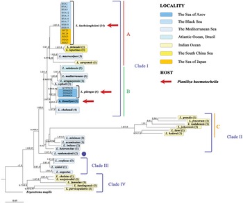
No intraspecific differences for L. kaohsianghsieni for each DNA marker were revealed. Maximum ML and BI showed identical topologies regarding major lineages based on each molecular marker (figs 3–5). Due to the discrete sequence data for Ligophorus species we were unable to reconstruct the representative phylogeny for these worms. Data on the 28S rRNA gene are available for most of the analysed species, so phylogenetic reconstruction based on this molecular marker is considered in detail. Additionally, a matrix of genetic distances between species was counted (online Supplementary 1). Four well-supported clades of Ligophorus containing different species, without agreement with geographical regions or hosts, were identified (fig. 3). Clade I consisted of two subclades (A and B), each with high nodal support and 5% average evolutionary divergence. Ligophorus kaohsianghsieni belonged to subclade A, whereas other species infecting P. haematocheilus (Ligophorus pilengas and Ligophorus llewellyni) were within subclade B. Clade II was poorly supported in general, but included well-supported subclade C and several species from the Mediterranean Sea, namely the closely related Ligophorus minimus, Ligophorus acuminatus,. Ligophorus imitans and Ligophorus heteronchus, which appeared as separate lineages. Ligophorus vanbenedeni represented a sister lineage to clades I and II with high support (fig. 3). Clade III was also highly supported and encompassed three species, namely Ligophorus confusus, Ligophorus szidati and Ligophorus angustus. Clade IV was poorly supported with the ML algorithm and highly supported with BI and consisted of five Ligophorus species from mullets from the Indian Ocean.
Phylogenetic tree derived from the 28S rRNA gene sequences using Bayesian analysis. The alignment length was 719 positions. Nodal numbers – posterior probabilities for Bayesian inference/maximum likelihood phylogenetic algorithms (only significant values (0.9–1.0) are provided). The number of available nucleotide sequences in GenBank is noted in parentheses next to each species. The branch length is drawn to scale, with the scale bar indicating the number of nucleotide substitutions. The species with a different position in phylogeny based on different genes are marked with the dark blue spot.

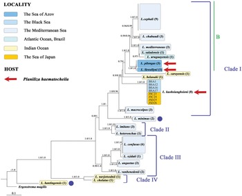
Phylogenetic tree derived from the internal transcribed spacer 1 rDNA sequences using Bayesian analysis. The alignment length was 667 positions. Nodal numbers – posterior probabilities for Bayesian inference/maximum likelihood phylogenetic algorithms (only significant values (0.9–1.0) are provided). The number of available nucleotide sequences in GenBank is noted in parentheses next to each species. The branch length is drawn to scale, with the scale bar indicating the number of nucleotide substitutions. The species with a different position in phylogeny based on different genes are marked with the dark blue spot.

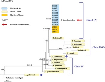
Phylogenetic tree derived from the 18S rRNA gene sequences using Bayesian analysis. The alignment length was 758 positions. Nodal numbers – posterior probabilities for Bayesian inference/maximum likelihood phylogenetic algorithms (only significant values (0.9–1.0) are provided). The number of available nucleotide sequences in GenBank is noted in parentheses next to each species. The branch length is drawn to scale, with the scale bar indicating the number of nucleotide substitutions. The species with a different position in phylogeny based on different genes are marked with the dark blue spot.

Intra-group and inter-group genetic divergence for each clade is presented in table 5. The highest intra-group sequences divergence was observed for clade IV (10%) and subclade C (12%). These two patterns consist of species from the Indian Ocean. Intra-group sequences divergences of subclades A and B, as well as for clade III ranged from 2% to 5%.
Estimates of average evolutionary divergence over sequence pairs within groups (boldface type, in the diagonal) and between groups (above the diagonal) based on 28S variability.

The topologies of the phylogenetic trees based on 18S and ITS1 rDNA were similar to that based on 28S in respect of mean clades, except some species, which were out of their clades. Probably the lack of ITS1 sequence data for species from the Indian Ocean, which formed clade C in the tree based on 28S, led to the exclusion of L. minimus from clade II (fig. 4). The reduction in the number of species in this analysis also resulted in a lack of support for subclade A. A similar situation is observed for phylogeny based on 18S (fig. 5). Ligophorus careyensis dropped out of clade IA, although this species is closer to clade I than to clade II in genetic distances. Nevertheless, both phylogenetic trees based on 18S and ITS1 rDNA keep a tendency of species clustering on the 28S rDNA-based tree, indicating the basal position of Ligophorus species from mullets of the Indian Ocean and terminal position of species ex hosts from the Black Sea and the Sea of Japan, including the position of L. kaohsianghsieni in subclade B. Obviously, 18S rDNA and ITS1 rDNA have good potential for more active use for phylogenetic studies of Ligophorus species in the future.
Discussion
Based on the present data and taking into account the previously published information (Gusev, Reference Gusev and Bychovsky1962; Sarabeev et al., Reference Sarabeev, Rubtsova, Yang and Balbuena2013), morphometric characters that allow to clearly distinguish specimens of L. kaohsianghsieni from the Black-Azov Sea region compared to rivers of the Russian Far East and the Sea of Japan has not been found.
The obtained sequences of three fragments of the ribosomal cluster of nuclear DNA (18S, ITS1 and 28S) from 14 individuals of L. kaohsianghsieni from the different regions are identical. This is consistent with the previously obtained data, since no mutations were observed in the 28S rRNA gene fragment between four individuals of L. pilengas, five individuals of L. confusus, four individuals of L. chabaudi, and in ITS1 between nine individuals of L. cephali and six individuals of L. confusus (Blasco-Costa et al., Reference Blasco-Costa, Míguez-Lozano, Sarabeev and Balbuena2012, figs 3 and 4). This confirms that this DNA region is highly conserved for Ligophorus at the intraspecific level.
At the same time, high interspecific genetic divergence is observed between the analysed Ligophorus species (figs 3–5). Even those species which formed monophyletic groups on the phylogenetic tree based on 28S rRNA gene sequences and parasitizing the same host species in the same region (fig. 3: subclade C and clade IV) are genetically significantly different from each other. The question arises, what contributes to this deep divergence between sympatric and synxenic species?
Specimens of L. kaohsianghsieni cluster with species parasitizing fish of the genera Planiliza and Chelon from the Atlantic and Pacific Oceans (clade I) in both phylogenetic reconstructions based on 28S and ITS1 rRNA (figs 3 and 4). Whereas L. kaohsianghsieni is significantly distant from L. pilengas and L. llewellyni occur on the same host (P. haematocheilus) in the same seas. Moreover, the latter two species have merged into a monophyletic group with species of Ligophorus infecting fish of the genus Mugil in the Atlantic and Pacific Oceans (fig. 3: subclade A vs. subclade B), and L. chabaudi, found in both oceans, occupies a basal position in this clade.
Previously, Blasco-Costa et al. (Reference Blasco-Costa, Míguez-Lozano, Sarabeev and Balbuena2012) obtained a similar result in reconstructing the phylogenetic relationships between 14 species of Ligophorus from the Mediterranean and Azov Seas based on 28S and ITS1, where two species (L. pilengas and L. llewellyni) from P. haematocheilus of north-western Pacific Ocean origin and Ligophorus spp. from widespread Mugil cephalus formed one group, distancing themselves from species parasitizing only hosts with Mediterranean Sea and north-east Atlantic Ocean distribution.
However, the addition of more representatives of Ligophorus from the Pacific Ocean and the north-west Atlantic Ocean into the phylogenetic analysis (fig. 3) revealed that some species from different host species and oceans were closer and included in one monophyletic lineage than species from the same host and region, for example, Ligophorus belanaki and L. careyensis entered clade I, and Ligophorus chelatus, Ligophorus navjotsodhii, Ligophorus parvicopulatrix, Ligophorus funnelus and Ligophorus bantingensis into clade IV (fig. 3), even though they all infected Planiliza subviridis from Malaysia. While Ligophorus macrocolpos, which parasitizes Chelon saliens in the Mediterranean and Black Seas, clustered with species distributed in the north-west Pacific Ocean (fig. 3: subclade A), and was significantly separated from other species occurring on the same host and in the same region, namely L. minimus, L. acuminatus, L. imitans, L. heteronchus, L. szidati (fig. 3: clades II and III), and L. vanbenedeni. Thus, there is no correspondence between the phylogenetic and geographical proximity of hosts and relation of Ligophorus species parasitizing them. Previously, the absence of relatedness between about half of the Ligophorus species infecting the same host species was suggested based on the analysis of morphological similarity (Sarabeev & Desdevises, Reference Sarabeev and Desdevises2014).
As a whole, the results of our study demonstrate the main vector of Ligophorus phylogeny, showing a constant basal position for certain species from Indian Ocean mullets, a middle position of other certain species from Mediterranean mullets and terminal position of Ligophorus species from mullets of different seas, including the Indo-West Pacific Ocean, Mediterranean Sea and Atlantic Ocen fauna. It cannot be excluded that the fauna of the Mediterranean Sea and Indo-Pacific Ocean Ligophorus species from terminal clades has secondary origin in these regions, occurring through possible host-switching processes using different mullet fish species after long-term spatial isolation. We suppose that some representatives of the ancestral form of the studied monogeneans, inhabiting in the Indian Ocean, could have migrated to other regions, for example, to the Mediterranean Sea (according to the results of phylogenetic analysis), using host-switching, where deep divergence and speciation occurred. Later, these new species could secondarily settle Indian Ocean territories using different mullet fish species. This hypothesis partially explains the deep genetic diversity of sympatric species; it should be studied in more detail in the future. Additionally, the widespread species complex M. cephalus can be considered as a key host species in the secondary distribution of monogeneans throughout different zoogeographical areas.
It was previously shown that differences in the morphology of attachment (haptoral) structures between Dactylogyrus species (related Ligophorus to taxon) occurring on the same host contribute to niche segregation and increased reproductive isolation of related species to prevent hybridization, just as monogeneans occupying the same niche differ greatly in shape and size of copulatory organ (Šimková et al., Reference Šimková, Ondračková, Gelnar and Morand2002). Thus, the morphology of both of these structures is of great evolutionary importance. Khang et al. (Reference Khang, Soo, Tan and Lim2016), analysing the haptoral morphology of 13 species from Malaysia, which formed two different clusters on a phylogenetic tree constructed based on 28S, ITS1, 18S rRNA sequences, obtained good agreement with the clustering of these species by these morphological characters. Similarly, Sarabeev & Desdevises (Reference Sarabeev and Desdevises2014), comparing reconstructions of the relatedness of 14 species from the Mediterranean and Black Seas based on 28S and ITS1 rRNA sequences with the results of morphological analysis, mainly related to the characters of haptoral structures and copulatory organs, concluded that morphological and molecular phylogenetic trees are congruent.
However, it should be noted that the species analysed in both studies (Sarabeev & Desdevises, Reference Sarabeev and Desdevises2014; Khang et al., Reference Khang, Soo, Tan and Lim2016) were from the same region, respectively, the Mediterranean and Black Seas in the first article and Malaysia in the second. The closest species to L. kaohsianghsieni according to all phylogenetic reconstructions (based on 28S, ITS1 and 18S) in the present study is L. belanaki, also parasitizing mullet of the genus Planiliza in the coastal seas of the western Pacific Ocean.
At the same time, the two species differ greatly in the morphology of their haptoral structures and copulatory organ: in L. kaohsianghsieni anchors have a relatively short distal part (blade) compared to their proximal part, ventral bar with closely spaced anterior processes, copulatory organ with a long tube and distally bifurcated accessory part (fig. 1), whereas L. belanaki anchors with a slender blade that is much longer than their proximal part, the ventral bar has rather widely spaced anterior processes, and the copulatory organ tube is rather short and its accessory part is not bifurcated distally (Soo & Lim, Reference Soo and Lim2013). It can be said that these species are opposite in most morphological characters of haptor and male copulatory organ, while in molecular data they are closely related species. Similarly, species infecting P. subviridis, which form clade IV, are quite diverse in size and shape of haptoral structures and copulatory organ (Soo & Lim, Reference Soo and Lim2012). Thus, it should be taken into account that closely related species may differ significantly in morphology.
Morphological differences in haptor structures of phylogenetically closely related species may be the result of adaptation to different hosts or to a specific attachment site on the gills. On the other hand, the marked differences in morphology of both haptor and copulatory organ between L. kaohsianghsieni and two other species from P. haematocheilus (L. pilengas and L. llewellyni) (fig. 6) are consistent with significant genetic divergence between them.
Comparison of the morphology of haptoral structures and the copulatory organ of Ligophorus kaohsianghsieni (C, F), Ligophorus llewellyni (A, D) and Ligophorus pilengas (B, E) ex Planiliza haematocheila from the Black Sea: A–C, haptoral structures; and D–F, male copulatory organ. Scale bar = 10 μm.

Overall, at least two groups of species of different origin parasitize P. haematocheilus in its natural range, the Sea of Japan, as well as in the region of introduction, the Black Sea.
Supplementary material
To view supplementary material for this article, please visit http://doi.org/10.1017/S0022149X22000724.
Financial support
This work is funded by a scientific theme of the A. O. Kovalevsky Institute of Biology of the Southern Seas, Sevastopol, Russia number 121030100028-0, Russian Foundation for Basic Research number 20-44-920004 and a scientific theme of Federal Scientific Center of the East Asia Terrestrial Biodiversity Far Eastern Branch of the Russian Academy of Sciences, project number 0207-2021-0008.
Conflicts of interest
None.
Author contributions
Vodiasova E. A.: conceptualization, writing – original draft, review and editing, visualization, funding acquisition, genetic data curation. Atopkin D.: investigation, genetic analysis, software, writing – original draft, review and editing. Plaksina M.: sampling, investigation, morphology analysis, visualization. Chelebieva E.: investigation, genetic analysis, writing – review and editing. Dmitrieva E.: conceptualization, morphology data curation, writing – original draft, review and editing, project administration.
Ethical standards
All applicable institutional, national and international guidelines for the care and use of animals were followed. All studied fishes are listed as a ‘Least Concern’ species by the International Union for Conservation of Nature.