Introduction
Pre-operative cross-sectional imaging is a fundamental component of evaluation prior to cochlear implantation. Assessment should fulfil two roles: firstly, to confirm cochlear implant candidacy (e.g. the presence of a cochlea and cochlear nerve); and, secondly, to guide surgical feasibility. This ‘feasibility’ includes prediction of aberrant anatomy both in the approach to the cochlea and in the cochlea itself. It influences surgical planning, laterality of implantation and electrode choice. Imaging may also inform discussion of risks and outcomes with the patient and family.
An evolution from single modality computed tomography (CT) scanning alone to dual modality (CT and magnetic resonance imaging (MRI)) assessment has occurred, alongside development in imaging quality.Reference Young, Ryan and Young1 Many institutions now apply a protocol recommending dual modality imaging in selected cases.Reference Tamplen, Schwalje, Lustig, Alemi and Miller2–Reference Sennaroglu, Saatci, Aralasmak, Gursel and Turan4 Many view MRI scanning to be mandatory in pre-operative cochlear implant assessment, given its superior ability to assess the crucial factor of cochlear nerve presence coupled with excellent evaluation of intracranial pathology.Reference Bamiou, Worth, Phelps, Sirimanna and Rajput5 There is, as yet, a lack of consensus as to whether both CT and MRI are routinely required.
Pre-operative evaluation of a paediatric cochlear implant candidate represents a markedly different situation to that of the adult patient. Congenital abnormalities of the inner ear can be found in up to 20 per cent of patients with congenital hearing loss.Reference Jackler, Luxford and House6–Reference Sennaroglu8 Furthermore, sedation or general anaesthesia may be required to obtain the scan; this is associated with practical, clinical and cost implications.Reference Trimble, Blaser, James and Papsin3 Unfortunately, because of commercial reasoning, the exact cost of CT and MRI cannot be disclosed by our institution. However, based on available tariff data in the private setting, these figures are estimated to be around £500 each for both CT and MRI respectively.9 We agree that it is ideal for both CT and MRI to be performed under the same episode of sedation or anaesthesia; however, logistical factors frequently limit this possibility. Computed tomography scanning also has a shorter acquisition time, but is associated with radiation exposure.
Cochlear implant candidates currently undergo both CT and MRI assessments at our institution, a tertiary paediatric centre. We aimed to summarise this topic, specifically in the paediatric setting. We conducted a retrospective pilot study to evaluate the utility of the imaging scans currently obtained.
Materials and methods
A retrospective review of 25 patients who underwent paediatric cochlear implant assessment between July 2015 and April 2016 was conducted.
According to our imaging protocol, all patients underwent high-resolution CT (without contrast), which includes 0.6 mm slices with axial and coronal reconstruction. An MRI assessment of the inner ear was performed using constructive interference in steady state, and our standard brain sequence composed of axial, T2-weighted, coronal fluid-attenuated inversion recovery (‘FLAIR’), and coronal and sagittal T1-weighted images for assessment of the internal acoustic meatus and brain. For patients under two years of age, the T2-weighted sequence is replaced by a dual-echo, axial, short-tau inversion recovery (‘STIR’) sequence.Reference Saunders, Thompson, Gunny, Jones, Cox and Chong10
‘Control scores’ describing the expected visualisation of specific features by CT and MRI imaging were established by a consultant paediatric neuroradiologist. Scores of 2 (optimal), 1 (adequate), −1 (suboptimal) or −2 (inadequate) were given for each investigation (Table I).
Allocated ‘control scores’ for specific features on CT and MRI

Scoring: 2 = optimal, 1 = adequate, −1 = suboptimal and −2 = inadequate. CT = computed tomography; MRI = magnetic resonance imaging; n/a = not applicable; SCC = semicircular canal; IAM = internal auditory meatus
An independent review of all CT and MRI scan features was then compared to the control scores. Primary outcome measures included the detection of radiological abnormalities and agreement of radiological appearances with control scores. Co-morbidities and the aetiology of hearing loss were also recorded.
Institutional approval was granted, with the study considered a service evaluation project.
Results
Breakdown of imaging
Fifty consecutive examinations (25 CT and 25 MRI scans) of prospective cochlear implant candidates were reviewed. Scans of 15 males and 10 females, aged between 6 months and 19 years at the time of assessment, were evaluated.
One CT and two MRI scans imported from other institutions were excluded, as the images did not comply with our institutional protocols. Of the remaining 47 examinations, 21 (45 per cent) were performed under general anaesthesia, 15 (32 per cent) without sedation and 9 (19 per cent) with sedation; 2 patients (4 per cent) underwent feed and wrap. In total, 17 per cent of examinations (four CT and four MRI scans) were performed under the same episode of sedation. No scans were repeated because of movement artefacts.
Scoring agreement and abnormalities
Agreement with control scores occurred in 20 out of 24 CT scans (83 per cent) and in 21 out of 23 MRI scans (91 per cent) (Figure 1). A specific feature within three of four CT scans, and two of two MRI scans was ‘under-scored’, because of artefacts or abnormal anatomy obscuring underlying structures. These features were less adequately visualised in these particular scans than expected from the imaging modality. ‘Under-scored’ features on CT included the facial nerve course, vestibular aqueduct, and internal acoustic meatus communications and size. On MRI, ‘under-scored’ features included the endolymphatic sac and cochlear nerve.
Flow diagram illustrating correlation with control scores for computed tomography (CT) and magnetic resonance imaging (MRI).

In total, radiological abnormalities of the brain were demonstrated in 16 per cent of scans, whilst radiological abnormalities of the temporal bone were seen in 18 per cent of investigations.
Hearing loss aetiology and co-morbidities
Seventy-four per cent of patients had an underlying diagnosis or co-morbidity associated with hearing loss. Within our cohort, 14 patients had congenital hearing loss, 6 patients had progressive hearing loss and 5 patients had sudden-onset sensorineural hearing loss.
Underlying diagnoses included: kernicterus (n = 2); coloboma, heart defects, atresia choanae, growth retardation, genital abnormalities, and ear abnormalities (‘CHARGE’) association (n = 2); cytomegalovirus (n = 2); connexin 26 mutation (n = 2); and Jervell and Lange-Nielsen syndrome (n = 1). Of the remaining five patients with an unknown aetiology, all had associated co-morbidities, including three premature babies and two term babies born with perinatal complications.
Amongst patients with progressive hearing loss, an underlying diagnosis of auditory neuropathy spectrum disorder was found in one patient. Of the remaining five patients with an unknown aetiology, three had associated co-morbidities: bulbar palsy, neonatal jaundice and prematurity.
Amongst patients with sudden-onset hearing loss, diagnoses included meningitis (n = 2), critical illness associated hearing loss (n = 2) and chemotherapy-related ototoxicity (n = 1).
Discussion
Our results show a high similarity with control scores, demonstrating consistency across all independently reviewed images. A relatively high yield of radiological abnormalities (16 per cent and 18 per cent for brain and temporal bone respectively), and a 74 per cent incidence of underlying diagnoses and associated co-morbidities, illustrates the complexity of patients within our paediatric population. In a previous larger evaluation of brain abnormalities on MRI, in cochlear implant candidates at our centre, abnormalities were found in 30 per cent of cases.Reference Jonas, Ahmed, Grainger, Jephson, Wyatt and Hartley11 Although this was not a specific focus, most brain abnormalities (e.g. cytomegalovirus, kernicterus) correlated with hearing loss aetiology. This was less clear for temporal bone abnormalities, where some patients with unknown underlying diagnoses were found to have inner-ear malformations (e.g. incomplete partition type 2, cochlear aplasia, semicircular canal aplasia).
The relative and individual strengths of CT and MRI have become increasingly well-defined; however, the superior modality for cochlear implant pre-operative assessment remains open to debate, particularly in the paediatric setting.
Bettman and colleagues have described a CT protocol designed to give optimal information, and proposed that CT alone gives sufficient temporal bone information for cochlear implant assessment.Reference Bettman, Van Olphen, Zonneveld and Huizing12 It was also suggested that the sensitivity and specificity of CT in the assessment of cochlear patency are comparable to MRI; omission of MRI from the pre-operative evaluation of candidates known to have a cause of deafness outside of the central acoustic pathway was proposed. Conversely, several authors have asserted that MRI adds greater total information than CT, principally greater accuracy regarding the presence and size of the cochlear nerve and soft tissue abnormalities within the inner ear.Reference Ellul, Shelton, Davidson and Harnsberger13, Reference Parry, Booth and Roland14 Other studies, such as that of Gleeson and colleagues, have suggested that a combination of CT and MRI is not superior to either modality alone.Reference Gleeson, Lacy, Bresnihan, Gaffney, Brennan and Viani15
Our novel scoring system has reflected the advantages of both imaging modalities as described within the literature. We agree with the conclusion of Trimble et al. that a policy of dual modality imaging can detect abnormalities related to hearing loss that would otherwise not have been found using either modality alone.Reference Trimble, Blaser, James and Papsin3
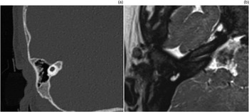
We advocate the use of dual modality imaging; together, the modalities are both diagnostic and prognostic in paediatric patients undergoing cochlear implant assessment. Information provided by imaging can improve pre-operative patient counselling, for example, in cases where previously unknown brain abnormalities are diagnosed, influencing the success of cochlear implantation. In addition, complementary information provided by both CT and MRI can help to minimise missed radiological abnormalities (Figure 2), and drive forward the investigation of inner-ear malformations such as the new classification proposed by SennarogluReference Sennaroglu8 (Figure 3).
A case of cochlear aplasia with associated dysplastic vestibule. This is easily detected on computed tomography (a); however, on magnetic resonance imaging (b), it may be confused with a fluid-containing mastoid air cell and misdiagnosed as complete labyrinthine aplasia.

Computed tomography (a) and magnetic resonance imaging (MRI) (b) scans demonstrating incomplete partition type 3. The MRI scan better demonstrates that internal cochlear structure is present.

The aetiology of hearing loss within the paediatric population varies widely compared to that of the adult population. Paediatric patients often present with complex medical conditions or syndromes that may be associated with a variety of radiological abnormalities; this further supports the use of both imaging modalities in this subset. We also recommend the use of a ‘dataset’ to both standardise and optimise the evaluation of CT and MRI.
This retrospective pilot study samples a small population. A larger study including several independent neuroradiological and otological evaluations would reduce bias (e.g. minimise the level of subjectivity in the allocation of control scores) and yield greater evidence. This study focused on information gained from cross-sectional imaging, and thus does not evaluate important associated factors including cost, logistics and radiation dosage.
• Pre-operative cross-sectional imaging is fundamental in evaluation prior to cochlear implantation
• Assessment should confirm cochlear implant candidacy (e.g. presence of cochlea and cochlear nerve) and guide surgical feasibility
• Consensus is lacking as to whether both computed tomography (CT) and magnetic resonance imaging (MRI) are routinely required
• Pre-operative evaluation of a paediatric cochlear implant candidate represents a markedly different situation to adult patients
• Both CT and MRI contribute valuable information and remain necessary in paediatric cochlear implant pre-operative assessment
In addition, cone beam CT could be considered for pre-operative paediatric cochlear implant assessment, alongside MRI. It has high spatial resolution and low cost when compared to conventional CT, and is associated with a smaller radiation dose. However, drawbacks include successful imaging in older children only, as imaging under general anaesthesia is only suitable in supine machines, which are less readily available, and cone beam CT takes a longer time to complete compared to conventional high-resolution CT.
Conclusion
Dual modality imaging is an optimal investigative approach for paediatric cochlear implant assessment in our patient population in terms of the radiological knowledge gained. Despite some duplication of information, CT and MRI may individually contribute valuable information to the cochlear implantation team and better inform patient care.
Acknowledgement
We would like to thank the Department of Radiology at Great Ormond Street Hospital for Children for their contribution to this study.
Competing interests
None declared.