Significant outcomes
Glutamate level in serum was positively correlated with severity of depression in depressed post-stroke patients.
Rehabilitation, with or without rTMS, improved depressive symptoms in post-stroke patients.
Rehabilitation in combination with rTMS decreased d-serine in post-stroke patients with depression, but rehabilitation without rTMS increased d-serine.
Limitations
This study was not a randomised controlled trial.
The present study considered only post-stroke patients with mild to moderate symptoms of depression.
This study did not include post-stroke patients without upper limb hemiparesis.
Introduction
Stroke is the leading cause of physical disability. However, it has been reported that a stroke can also cause mental disabilities such as depression (Burvill et al., Reference Burvill, Johnson, Jamrozik, Anderson, Stewart-Wynne and Chakera1995). Therefore, post-stroke patients often suffer from multiple symptoms consisting of physical and mental disorders (Ellis et al., Reference Ellis, Grubaugh and Egede2013).
Recently, many studies have reported that repetitive transcranial magnetic stimulation (rTMS) is beneficial to post-stroke patients (Lefaucheur et al., Reference Lefaucheur, André-Obadia, Antal, Ayache, Baeken, Benninger, Cantello, Cincotta, de Carvalho, De Ridder, Devanne, Di Lazzaro, Filipović, Hummel, Jääskeläinen, Kimiskidis, Koch, Langguth, Nyffeler, Oliviero, Padberg, Poulet, Rossi, Rossini, Rothwell, Schönfeldt-Lecuona, Siebner, Slotema, Stagg, Valls-Sole, Ziemann, Paulus and Garcia-Larrea2014). The rTMS treatment can be applied to a wide variety of post-stroke symptoms such as upper limb hemiparesis (Niimi et al., Reference Niimi, Sasaki, Kimura, Hara, Yamada and Abo2019) and depression (Duan et al., Reference Duan, Yao, Liu, Cui and Yang2018).
In addition to stroke effects, it has been reported that rTMS is beneficial in the treatment of various brain illnesses such as depression, schizophrenia, anxiety disorders, Alzheimer’s disease, and Parkinson’s disease (Lefaucheur et al., Reference Lefaucheur, André-Obadia, Antal, Ayache, Baeken, Benninger, Cantello, Cincotta, de Carvalho, De Ridder, Devanne, Di Lazzaro, Filipović, Hummel, Jääskeläinen, Kimiskidis, Koch, Langguth, Nyffeler, Oliviero, Padberg, Poulet, Rossi, Rossini, Rothwell, Schönfeldt-Lecuona, Siebner, Slotema, Stagg, Valls-Sole, Ziemann, Paulus and Garcia-Larrea2014). The rTMS treatment is considered to modulate neuronal plasticity by altering cortical excitability. Some studies have shown that the rTMS modulates the release of N-methyl-D-aspartic acid receptor (NMDAR)-related amino acids in rat brains (Keck et al., Reference Keck, Sillaber, Ebner, Welt, Toschi, Kaehler, Singewald, Philippu, Elbel, Wotjak, Holsboer, Landgraf and Engelmann2000). A few studies have reported that rTMS treatment can potentiate glutamatergic neurotransmission in depressed patients. One study showed that glutamate levels in the left dorsolateral prefrontal cortex increased after high-frequency rTMS treatment in depressed patients (Yang et al., Reference Yang, Kirton, Wilkes, Pradhan, Liu, Jaworska, Damji, Keess, Langevin, Rajapakse, Lebel, Sembo, Fife and MacMaster2014). Another study showed that high-frequency rTMS treatment increased glutamine/glutamate ratios in the left dorsolateral prefrontal cortex and anterior cingulate cortex in patients with treatment-refractory major depressive disorder (Croarkin et al., Reference Croarkin, Nakonezny, Wall, Murphy, Sampson, Frye and Port2016). In addition, it has been reported that rTMS increased NMDAR expression in the hippocampus of a rat model of vascular dementia (Zhang et al., Reference Zhang, Xing, Wang, Tao and Cheng2015). To our knowledge, however, there have been no studies investigating the influence of rTMS on NMDAR-related amino acids in the blood of post-stroke patients.
Aims of the study
The present study was intended to determine the effects of rTMS on NMDAR-related amino acid levels (e.g., glutamine, glutamate, glycine, l-serine, and d-serine) in the serum of post-stroke patients. In addition, we investigated the relationship between the depressive parameters and NMDAR-related biomarkers in the serum of post-stroke patients.
Materials and methods
Subjects
The subjects were inpatients admitted to Tokyo General Hospital for rehabilitation therapy for hemiparesis following a stroke during the period between February 2012 and March 2014. The inclusion criteria were as follows: (1) Age 30–90 years at intervention. (2) Time after onset of stroke > 1 month. (3) Mild or moderate upper limb hemiparesis (able to flex fingers of the affected upper limb). (4) At a probable plateau state with regard to recovery of upper limb hemiparesis as determined by serial evaluation after onset of stroke [no increase in the Fugl–Meyer Assessment (FMA) score during the last two weeks]. (5) No cognitive deficits [Mini Mental State Examination (MMSE) > 23 and if suffering from aphasia, Raven’s colored progressive matrices (RCPM) > average score for the same age – 2sd]. (6) No history of convulsions. (7) No intracranial metal or cardiac pacemaker. The exclusion criteria were as follows: (1) Unacceptable quality of patient’s blood samples (hemolysis, insufficient volume or melting of frozen samples) and (2) missing values of clinical evaluation before and/or after intervention. These subjects were the same as the subjects in a previous study (Niimi et al., Reference Niimi, Hashimoto, Kakuda, Miyano, Momosaki, Ishima and Abo2016). We conducted additional analysis.
The study was approved by the ethics committees of The Jikei University School of Medicine and Tokyo General Hospital and the Biomedical Research Ethics Committee of the Graduate School of Medicine at Chiba University. A signed informed consent about participation in this study and rTMS treatment was obtained from each patient. The authors assert that all procedures contributing to this work comply with the ethical standards of the relevant national and institutional committees on human experimentation and with the Helsinki Declaration of 1975, as revised in 2008. This clinical trial was registered with http://www.umin.ac.jp/(UMIN000034303).
Application of rTMS
Low-frequency rTMS inhibits cortical excitability in the stimulated site, while high-frequency rTMS facilitates cortical excitability (Lefaucheur et al., Reference Lefaucheur, André-Obadia, Antal, Ayache, Baeken, Benninger, Cantello, Cincotta, de Carvalho, De Ridder, Devanne, Di Lazzaro, Filipović, Hummel, Jääskeläinen, Kimiskidis, Koch, Langguth, Nyffeler, Oliviero, Padberg, Poulet, Rossi, Rossini, Rothwell, Schönfeldt-Lecuona, Siebner, Slotema, Stagg, Valls-Sole, Ziemann, Paulus and Garcia-Larrea2014). Low-frequency rTMS has often been applied to the primary motor area of the non-lesional hemisphere in order to enhance cortical excitability of the lesional hemisphere through a reduction of interhemispheric inhibition from the non-lesional hemisphere to the lesional hemisphere (Lefaucheur et al., Reference Lefaucheur, André-Obadia, Antal, Ayache, Baeken, Benninger, Cantello, Cincotta, de Carvalho, De Ridder, Devanne, Di Lazzaro, Filipović, Hummel, Jääskeläinen, Kimiskidis, Koch, Langguth, Nyffeler, Oliviero, Padberg, Poulet, Rossi, Rossini, Rothwell, Schönfeldt-Lecuona, Siebner, Slotema, Stagg, Valls-Sole, Ziemann, Paulus and Garcia-Larrea2014). A 70-mm figure-8 coil and MagPro R30 stimulator (MagVenture Company, Farum, Denmark) were used for application of rTMS. According to safety recommendations and hospital protocol (Lefaucheur et al., Reference Lefaucheur, André-Obadia, Antal, Ayache, Baeken, Benninger, Cantello, Cincotta, de Carvalho, De Ridder, Devanne, Di Lazzaro, Filipović, Hummel, Jääskeläinen, Kimiskidis, Koch, Langguth, Nyffeler, Oliviero, Padberg, Poulet, Rossi, Rossini, Rothwell, Schönfeldt-Lecuona, Siebner, Slotema, Stagg, Valls-Sole, Ziemann, Paulus and Garcia-Larrea2014), 1-Hz rTMS was applied over the motor area responsible for the first dorsal interosseous (FDI) muscle movement in the non-lesional hemisphere. The stimulated region was defined as the location where the largest motor-evoked potentials (MEPs) in the FDI muscle of the unaffected upper limb were provoked by electromyography. The resting motor threshold (RMT) of the non-lesional hemisphere was defined as the lowest stimulation intensity that produced MEPs of the FDI muscle of the unaffected upper limb (about 50 μV in five out of 10 trials) at rest. The intensity of the rTMS application was subsequently set at 90% of the measured RMT of the FDI muscle. Each session consisted of 1200 pulses and two sessions were conducted per day. Each patient underwent a total of 22 treatment sessions delivered on a daily basis except for holidays.
Rehabilitation content
In both groups, patients received rehabilitation comprised of 60-min training in the morning and 60-min training in the afternoon, provided by a physiotherapist, every day over a period of 2 weeks. Rehabilitation mainly consisted of shaping techniques and repetitive task practice designed to intensively use the affected upper limb. The shaping techniques included reaching forward to move a cup from one place to another, wiping the surface of a table with a towel, picking up a hairbrush and combing hair, writing letters with a pencil, drawing pictures with a pen, handling chopsticks to pick up small objects, folding an umbrella, and other activities of daily living. The repetitive task practice typically included turning over cards, squeezing clay, gripping a small ball, and pinching small coins. Although each 60-min training time usually included 30 min of shaping techniques and 30 min of repetitive task practice, the proportion of training time was modified if necessary depending on improvement in motor function of the affected upper limb. In patients who received rTMS, each 60-min training was scheduled to start soon after application of rTMS.
Clinical evaluation
The MMSE was carried out to measure cognitive function before treatment. RCPM was conducted instead of MMSE in patients with aphasia. BDI was used to evaluate the depressive state before and after treatment. The MMSE consists of various questions classified into various categories, assessing orientation to time, orientation to place, registration, attention and calculation, recall, language, repetition, and complex commands (Folstein et al., Reference Folstein, Folstein and McHugh1975). With a maximum score of 30 points, a total score of 23 or less suggests cognitive impairment. The RCPM consists of 36 visual multiple-choice tests and does not require verbal responses, so it is used to evaluate cognition in an aphasic patient (Villardita et al., Reference Villardita1985). The BDI consists of 21 questions about mood in the past week. Each question has a four-point ordinal scale (0–3) with a maximum score of 63 points. Total score of 10 or more indicates depression and a higher score suggests more severe depression (Beck et al., Reference Beck, Ward, Mendelson, Mock and Erbaugh1961).
Blood collection
Blood samples were taken between 8:30 a.m. and 9:00 a.m. after a 7:00 a.m. breakfast at the Tokyo General Hospital (Tokyo, Japan). Blood samples were collected twice; before and after treatment. Blood samples before treatment were collected on the day after admission. Fourteen days after collection of the first blood samples, blood samples were again collected after treatment. The obtained samples were anonymised and immediately stored at −20°C. Within 1 week of collection, the stored samples were transported under freezing condition to the laboratory of Chiba University Center for Forensic Mental Health and stored at −80°C until subjected to analysis. Some of blood samples were used for measurement of other biomarkers in a previous study (Niimi et al., Reference Niimi, Hashimoto, Kakuda, Miyano, Momosaki, Ishima and Abo2016).
Measurement of serum amino acid levels
Measurement of total, d-, and l-serine levels in plasma was carried out using a column-switching high-performance liquid chromatography (HPLC) system (Shimadzu Corporation, Kyoto, Japan), as previously reported (Hashimoto et al., Reference Hashimoto, Yoshida, Ishikawa, Fujita, Niitsu, Nakazato, Watanabe, Sasaki, Shiina, Hashimoto, Kanahara, Hasegawa, Enohara, Kimura and Iyo2016). The measurement of glycine, glutamine, and glutamate was carried out using an HPLC system with fluorescence detection, as previously reported (Hashimoto et al., Reference Hashimoto, Yoshida, Ishikawa, Fujita, Niitsu, Nakazato, Watanabe, Sasaki, Shiina, Hashimoto, Kanahara, Hasegawa, Enohara, Kimura and Iyo2016). Briefly, serum (20 µL) was homogenised in 180 µL of methanol (HPLC grade) on ice. The homogenates were centrifuged at 3000 × g for 6 min at 4°C, and 20 µL of supernatant was evaporated to dryness at 40°C. To the residue, 20 µl of H2O (HPLC grade), 20 μL of 0.1 M borate buffer (pH 8.0), and 60 μL of 50 mM 4-fluoro-7-nitro-2,1,3-benzoxadiazole (NBD-F; Tokyo Kasei Kogyo Co., Ltd., Tokyo, Japan) in CH3CN (HPLC grade) were added. The reaction mixture was heated at 60°C for 2 min, then quenched immediately with 100 μL of H2O/CH3CN (90/10) containing 0.1% trifluoroacetic acid to stop the reaction.
A 20 µL aliquot of the resultant solution was injected into the HPLC system. A reversed-phase ODS column [TSKgel ODS-80 Ts (Tosoh Corporation, Tokyo, Japan) as Column 1] was used for the separation and quantification of total (d- and l-) serine, and the gradient elution of the mobile phase was maintained at a constant flow rate of 0.8 mL/min. Mobile phase 1a consisted of H2O/CH3CN (90/10) containing 0.1% TFA, and phases 1b and 1c, of H2O/CH3CN (10/90) containing 0.1% TFA and CH3CN, respectively. The time program for gradient elution was as follows: 0–25 min 1a : 1b : 1c = 92 : 8 : 0, 25–25.1 min liner gradient from 8% 1b to 100% 1b, 25.1–35 min 1a : 1b : 1c = 0 : 100 : 0, 35–35.1 min liner gradient from 8% 1b to 100% 1c, 35.1–40 min 1a : 1b : 1c = 0 : 0 : 100, and 40.1–60 min 1a : 1b : 1c = 92 : 8 : 0. The chiral column (Column 2) used for the separation and quantification of d- and l-serine with NBD-F was comprised of two Sumichiral OA-2500 columns (S) (Sumika Chemical Analysis Service Ltd., Osaka, Japan), connected in tandem. The mobile phase was 15 mM citric acid in MeOH. The flow rate for isocratic pumping was 1.0 mL/min. Column temperatures were maintained at 35°C. Fluorescence detection was performed at 530 nm with an excitation wavelength of 470 nm.
For determination of glycine, glutamine, and glutamate, a reversed-phase ODS column (TSKgel ODS-80 Ts, Tosoh Corporation, Tokyo, Japan) was used. The gradient elution of the mobile phase was kept at a constant flow rate of 0.8 mL/min. The time program for gradient elution was programmed as follows: 0–50.5 min 1a: 1b : 1c = 95 : 5 : 0, 50.5–55.5 min 1a : 1b : 1c = 0 : 100 : 0, and 55.5–57 min, 1a : 1b : 1c = 0 : 0 : 100. The temperature of all columns was maintained at 35°C. Fluorescence detection was performed at 530 nm with an excitation wavelength of 470 nm.
Statistical analysis
All statistical analyses were performed using SPSS version 21.0 (IBM, Somers, NY). The Student’s t-test was used for comparison of normally distributed parameters and the Mann–Whitney test was used for comparison of parameters that showed a skewed distribution pattern. The chi-squared test was used for comparison of categorical data. Data were analyzed using two-way analysis of variance (ANOVA). Correlations between BDI and serum biomarkers were carried out using Spearman’s correlation. A p value of less than 0.05 was considered to denote statistical significance.
Results
The median of BDI score was 6.0 [2.0–11.0] in 95 patients. In 27 patients, the BDI score was 10 or higher.
Comparison between depressed patients and non-depressed patients
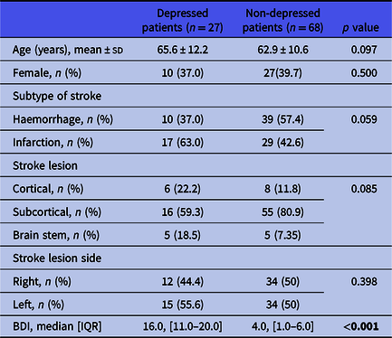
The baseline clinical characteristics of 95 subjects are summarised in Table 1. There were no differences in the clinical characteristics between depressed patients and non-depressed patients.
Comparison of clinical characteristics at baseline
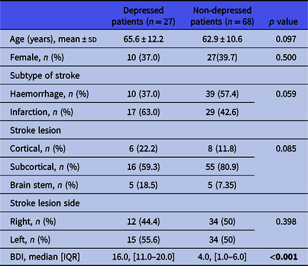
BDI, Beck Depression Inventory
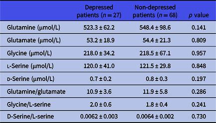
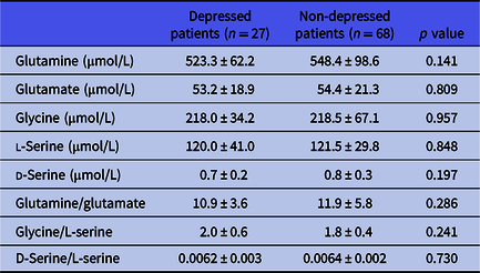
There were no significant differences in the levels of five amino acids in serum and the ratio of amino acids between depressed patients and non-depressed patients before treatment (Table 2).
Levels of amino acids in serum and ratio of amino acids in subjects at baseline
Comparison between depressed patients in the rehabilitation with rTMS group and without rTMS group
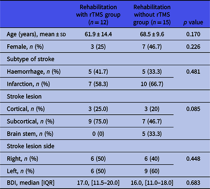
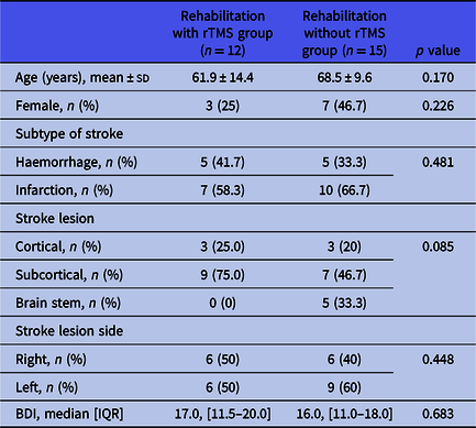
Among the 27 depressed patients, 12 patients received rTMS with rehabilitation, while 15 patients received rehabilitation only without rTMS. The baseline clinical characteristics of the depressed patients are summarised in Table 3. There were no differences in the clinical features between the depressed patients in the rehabilitation with rTMS group and without rTMS group.
Comparison of clinical characteristics at baseline between rehabilitation with rTMS group and rehabilitation without rTMS group
BDI, Beck Depression Inventory
Correlations between levels of glutamate-related amino acids in serum and depressive parameters
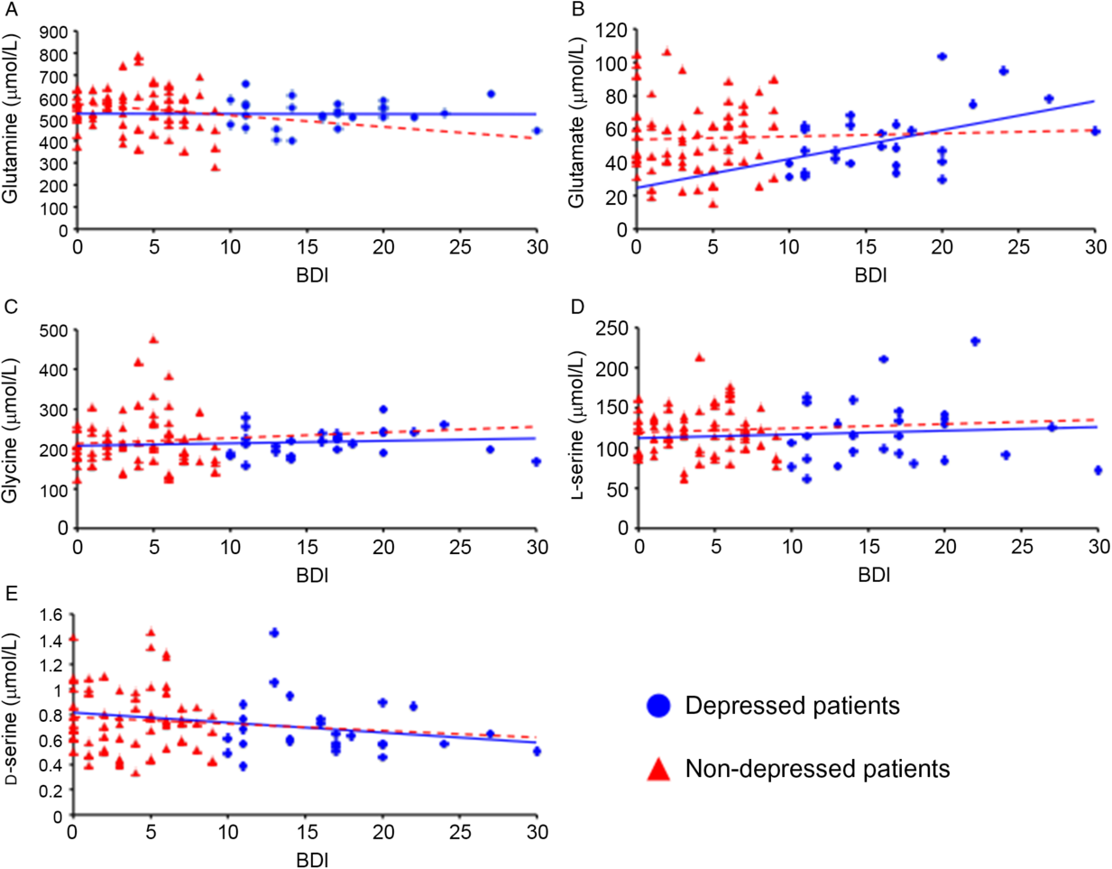
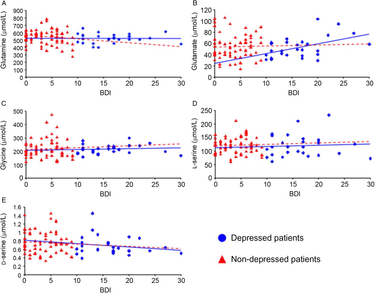
There was a significant correlation between serum glutamate levels and BDI in depressed patients (n = 27, ρ = 0.428, p = 0.026) (Fig. 1). There was no significant correlation between levels of other NMDAR-related amino acids in serum and BDI in depressed patients. There was no significant correlation between the five amino acids and BDI in non-depressed patients.
(A) There was no significant correlation between serum levels of glutamine and BDI in depressed patients (n = 27, ρ = −0.021, p = 0.919) and non-depressed patients (n = 68, ρ = −0.107, p = 0.387). (B) There was a significant positive correlation between serum levels of glutamate and BDI in depressed patients (ρ = 0.428, p = 0.026). There was no significant correlation between serum levels of glutamate and BDI in non-depressed patients (ρ = 0.092, p = 0.454). (C) There was no significant correlation between serum levels of glycine and BDI in depressed patients (ρ = 0.254, p = 0.202) and non-depressed patients (ρ = 0.013, p = 0.919). (D) There was no significant correlation between serum levels of l-serine and BDI in depressed patients (ρ = 0.090, p = 0.656) and non-depressed patients (ρ = 0.046, p = 0.711). (E) There was no significant correlation between serum levels of d-serine and BDI in depressed patients (ρ = −0.159, p = 0.427) and non-depressed patients (ρ = −0.040, p = 0.745).
Effect of treatment on BDI score in depressed patients
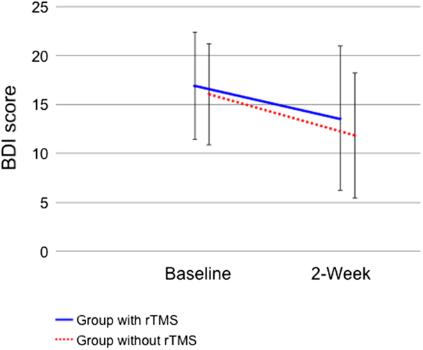
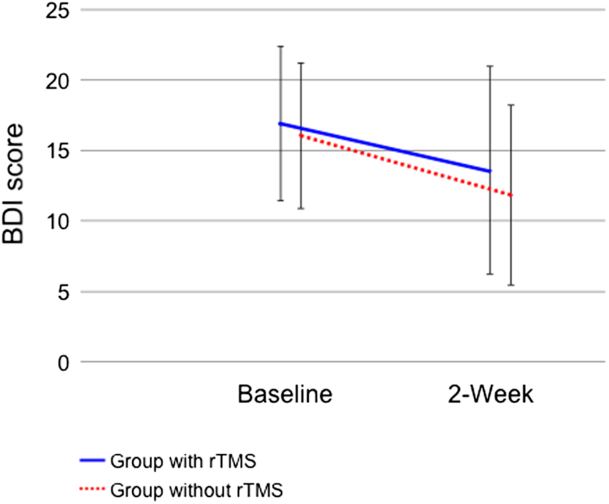
The BDI score significantly decreased in both the rehabilitation with rTMS group and rehabilitation without rTMS group after treatment (Fig. 2).
There was a significant decrease of BDI score in both rehabilitation with rTMS group and rehabilitation without rTMS group (group: F = 0.5, df = 1,25, p = 0.489, time: F = 6.2, df = 1,25, p = 0.020, interaction (group × time): F = 0.1, df = 1,25, p = 0.777).
Effect of treatment on levels of glutamate-related amino acids in serum
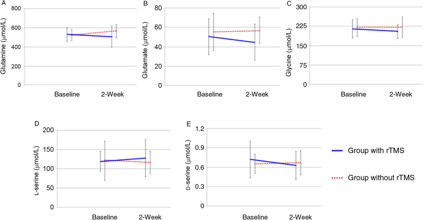
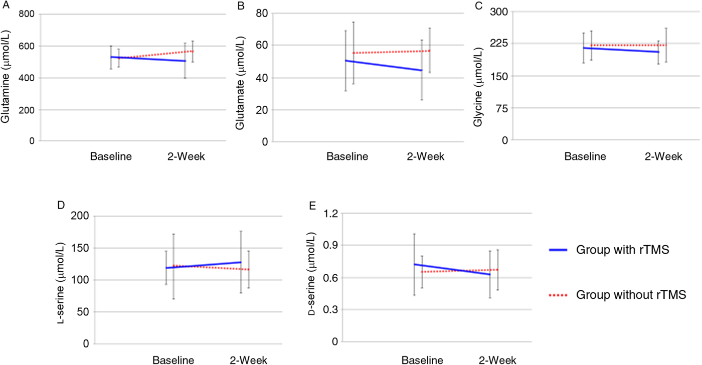
There was a significant relation between group and time in the levels of d-serine in serum. However, there were no significant effects and group-and-time relation for other NMDAR-related amino acids (Fig. 3).
(A) Two-way repeated measures ANOVA of glutamine data showed no significant effects and group-and-time interaction (group: F = 1.1, df = 1,25, p = 0.306, time: F = 0.4, df = 1,25, p = 0.531, interaction (group × time): F = 4.1, df = 1,25, p = 0.055). (B) Two-way repeated measures ANOVA of glutamate data showed no significant effects and group-and-time interaction (group: F = 2.0, df = 1,25, p = 0.169, time: F = 0.4, df = 1,25, p = 0.520, interaction (group × time): F = 1.2, df = 1,25, p = 0.278). (C) Two-way repeated measures ANOVA of glycine data showed no significant effects and group-and-time interaction (group: F = 0.9, df = 1,25, p = 0.346, time: F = 0.4, df = 1,25, p = 0.479, interaction (group × time): F = 0.8, df = 1,25, p = 0.387). (D) Two-way repeated measures ANOVA of l-serine data showed no significant effects and group-and-time interaction (group: F = 0.1, df = 1,25, p = 0.738, time: F = 0.1, df = 1,25, p = 0.798, interaction (group × time): F = 0.8, df = 1,25, p = 0.391). (E) Two-way repeated measures ANOVA of d-serine data showed a significant group-and-time interaction (group: F = 0.03, df = 1,25, p = 0.858, time: F = 1.5, df = 1,25, p = 0.232, interaction (group × time): F = 5.0, df = 1,25, p = 0.034).
Effect of treatment on glutamate-related amino acids ratio in serum
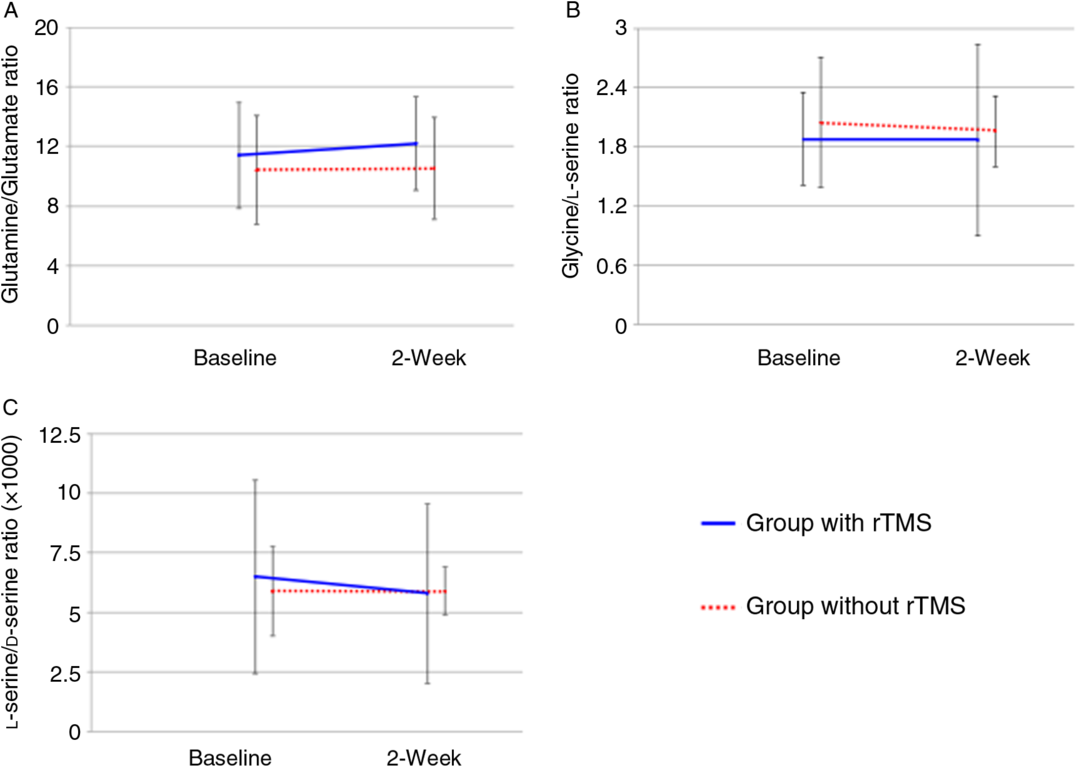
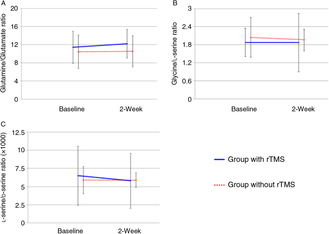
There were no significant effects and group-and-time relation in NMDAR-related amino acids ratio in serum (Fig. 4).
(A) Two-way repeated measures ANOVA of the ratio of glutamine to glutamate showed no significant effects and group-and-time interaction (group: F = 1.5, df = 1,25, p = 0.240, time: F = 0.4, df = 1,25, p = 0.546, interaction (group × time): F = 0.2, df = 1,25, p = 0.655). (B) Two-way repeated measures ANOVA of the ratio of glycine to l-serine showed no significant effects and group-and-time interaction (group: F = 0.3, df = 1,25, p = 0.566, time: F = 0.2, df = 1,25, p = 0.628, interaction (group × time): F = 0.1, df = 1,25, p = 0.709). (C) Two-way repeated measures ANOVA of the ratio of l-serine to d-serine showed no significant effects and group-and-time interaction (group: F = 0.1, df = 1,25, p = 0817, time: F = 2.7, df = 1,25, p = 0.114, interaction (group × time): F = 1.8, df = 1,25, p = 0.195).
Discussion
The present study reported the effect of rTMS on levels in serum of five NMDAR-related amino acids. Especially, this study demonstrated for the first time that 2-week low-frequency rTMS decreases levels of d-serine in serum in mild to moderate depressed stroke patients.
A previous study showed that levels of d-serine in serum of depressed patients were significantly higher than in control subjects (Hashimoto et al., Reference Hashimoto, Yoshida, Ishikawa, Fujita, Niitsu, Nakazato, Watanabe, Sasaki, Shiina, Hashimoto, Kanahara, Hasegawa, Enohara, Kimura and Iyo2016). Another study demonstrated that d-serine levels in cerebrospinal fluid (CSF) were also significantly higher in depressed patients than in healthy controls (Madeira et al., Reference Madeira, Lourenco, Vargas-Lopes, Suemoto, Brandão, Reis, Leite, Laks, Jacob-Filho, Pasqualucci, Grinberg, Ferreira and Panizzutti2015). In the present study, however, there was no significant difference in the levels of five amino acids in serum and ratio of amino acids. This may be because the present study used as subjects mild to moderate depressed stroke patients with a BDI score ranging from 10 to 27.
Some studies have shown that levels of glutamate in plasma are higher in depressed patients than in healthy controls (Mauri et al., Reference Mauri, Ferrara, Boscati, Bravin, Zamberlan, Alecci and Invernizzi1998, Mitani et al., Reference Mitani, Shirayama, Yamada, Maeda, Ashby and Kawahara2006). One study demonstrated that increased levels of glutamate were observed in the frontal cortex of postmortem brains from depressed patients (Hashimoto et al., Reference Hashimoto, Sawa and Iyo2007). In addition, Mitani et al. have shown that levels of glutamate in serum are correlated positively with severity of depression (Mitani et al., Reference Mitani, Shirayama, Yamada, Maeda, Ashby and Kawahara2006). In the present study, there was no difference in levels of glutamate between non-depressed and depressed patients. However, the levels of glutamate were correlated with severity of depression in depressed patients in the present study. It suggests that abnormalities of glutamatergic neurotransmission would be related to post-stroke depression.
Some meta-analyses have shown that physical exercise has a significant antidepressant effect in people with depression (Schuch et al., Reference Schuch, Vancampfort, Richards, Rosenbaum, Ward and Stubbs2016). In addition, other meta-analyses have shown that rTMS also has a significant antidepressant effect in depressed patients (Slotema et al., Reference Slotema, Blom, Hoek and Sommer2010). In the present study, there was a significant decrease in the BDI score of depressed patients in both rehabilitation with rTMS group and rehabilitation without rTMS group. However, levels of d-serine in serum decreased in depressed patients receiving rTMS, but levels of d-serine in serum increased in those not receiving rTMS. This result may suggest that physical exercise and rTMS treatment have different effects on NMDAR-related amino acid neurotransmission.
A few studies have reported on the relationship between rTMS and NMDAR-related amino acids. Keck et al. examined the critical effect of high-frequency rTMS over the left frontal cortex upon release of NMDAR-related amino acids in the right dorsal hippocampus of an anesthetised rat (Keck et al., Reference Keck, Sillaber, Ebner, Welt, Toschi, Kaehler, Singewald, Philippu, Elbel, Wotjak, Holsboer, Landgraf and Engelmann2000). They showed high-frequency rTMS increases the release of serine, but there were no significant changes in the release of glutamine and glutamate. Croarkin et al. demonstrated that glutamine/glutamate ratio increased in conjunction with depressive symptom improvement after high-frequency rTMS application to depressed patients by the use of magnetic resonance spectroscopy (MRS) (Croarkin et al., Reference Croarkin, Nakonezny, Wall, Murphy, Sampson, Frye and Port2016). This may suggest that rTMS can modulate NMDAR-related neurotransmission. In addition, Seewoo et al. have reported the effects of high and low-frequency rTMS on cortical levels of glutamine and glutamate in rats using MRS (Seewoo et al., Reference Seewoo, Feindel, Etherington and Rodger2019). They showed high-frequency rTMS with 10 Hz significantly increased glutamine levels and non-significantly increased glutamate levels in the sensorimotor cortex, whereas low-frequency rTMS with 1 Hz slightly decreased glutamine and glutamate levels. This accords well with our results.
The present study has several limitations. First, this study was not a randomised controlled trial and the sample size was too small. Further randomised controlled studies of larger populations are needed in order to allow for firm conclusions. Second, the present study subjected patients with no or mild to moderate symptoms of depression. Therefore, the results obtained by this study should not be applied to patients with severe symptoms of depression. Third, post-stroke patients with upper hemiparesis were consecutively recruited in the present study. This means that the present study did not engage post-stroke patients without upper limb hemiparesis. Therefore, the results obtained by this study should not be applied in understanding depression severity in post-stroke patients without upper limb hemiparesis.
In conclusion, the present study showed that rTMS decreases levels of d-serine in serum of post-stroke patients with depression. The result suggests that rTMS can modulate neurotransmission by the NMDAR-related amino acids.
Acknowledgements
The authors gratefully acknowledge the support and the participation of the patients in this study.
Author contributions
MN, KH, and MA designed the study protocol. MN, YF, and TI obtained the data. MN, NS, TH, and NY interpreted data. MN wrote the manuscript. MN, KH, and MA reviewed the manuscript.
Financial support
This work was supported by JSPS KAKENHI Grant Number 17K13070.
Conflict of interest
None.