The coronary artery fistula is a strange anomaly characterised by an abnormal communication between a coronary artery and a cardiac chamber, the pulmonary artery, the coronary sinus, or the pulmonary veins. A great part of the coronary fistulas are congenital. Congenital coronary anomalies are described in 0.5–2% of the population. Of them, the coronary fistula constitutes 15–30%. They originate from the right coronary and the left coronary artery, being the drainage to the pulmonary circulation in 90% of the occasions.Reference Barriales-Villa, Morís and López-Muñiz 1 Owing to its low prevalence, diagnosis becomes a challenge. It must be suspected in those symptomatic or asymptomatic patients who present with constant cardiac murmur on the left esternal edge or in the low edge of the breastbone.
We report the case of a 26-year-old woman who presented for consultation after the detection of a cardiac murmur in a medical routine recognition. She denied having dyspnoea, thoracic pain, or palpitations. The patient had an active life, without any constraint. Physical exploration confirmed the presence of a mesosystolic heart murmur on the left esternal edge with a normal behaviour of the second sound that was interpreted as innocent. The rest of the physical examination, the chest X-ray, and the electrocardiogram results were normal.
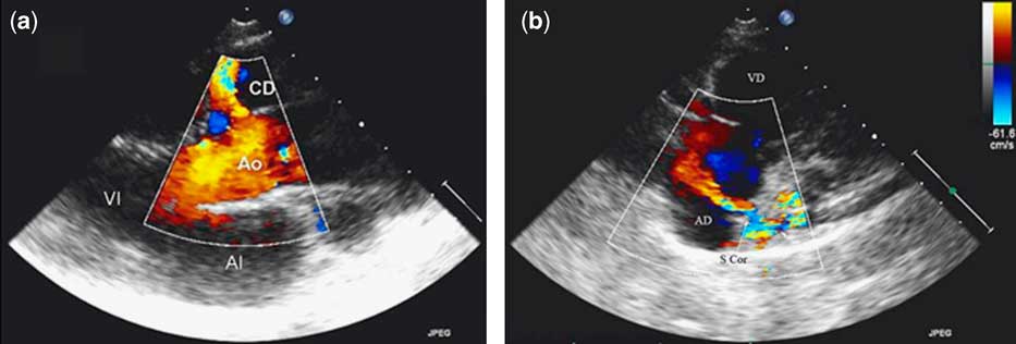
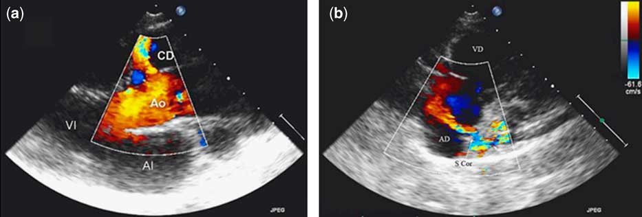
Doppler echocardiography showed the origin of the right coronary artery extremely dilated and an expanded tortuous vascular structure in the right auriculoventricular sulcus. In this zone, the existence of a constant turbulent flow was notable, in addition to the expansion and increase of the flow of the coronary sinus suspicious of drainage to this level (Fig 1).
Transthoracic echocardiography: (a) Left paraesternal long axis: it is observed from the aortic root (Ao) the origin of a great coronary right artery (CD) with turbulent flow. (b) Apical four-chamber plane: the doppler colour shows a fistulous connection to the coronary sinus (S.cor), which is expanded. The arrow indicates the turbulent flow from the coronary sinus to the right auricle (AD). (VI: Left ventricle; VD: Right ventricle; AI: Left auricle).
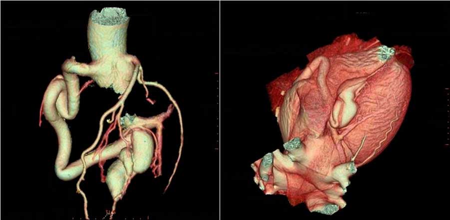
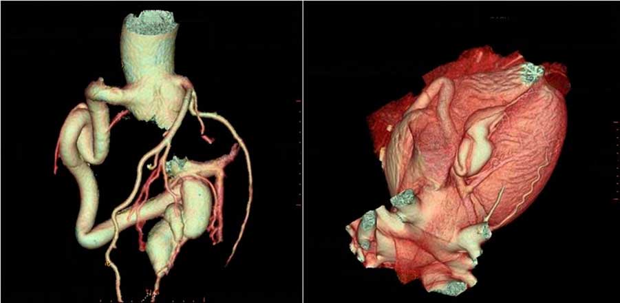
The 64-row multidetector computed tomography confirmed the presence of a significant enlargement of the right coronary artery winding in the contour of the right ventricle and its fistulosa connection to the coronary sinus. The left coronary artery was normal (Fig 2).
Volumetric reconstruction by multidetector computed tomography that allows to observe the whole path of the right coronary artery from its origin to its fistulisation in the coronary sinus. The left coronary artery is normal.
The coronary fistulas generally arise as a congenital isolated anomaly. The expansion of the coronary arteries is frequent, although the degree of expansion not always depends on the size of the short circuit. The clinical presentation of a coronary congenital fistula can vary considerably, depending on its anatomy and the size of the fistulosa connection. Many subjects are asymptomatic, as our patient, especially those in whom the fistula has a small caliber. In general, symptoms are infrequent before the third decade of life. Over the fifth decade, common symptoms such as dyspnoea and angina, signs of congestive heart failure, supraventricular arrhythmias, and, in exceptional cases, pulmonary hypertension or infective endocarditis are present. A progressive dilation of the right or left ventricle and consequently heart failure can also occur.
Traditionally, coronary fistulas have been diagnosed using coronary angiography. Recently, other non-invasive assessment procedures such as multidetector computed tomography and nuclear magnetic resonance are being used. The coronary angiography with multidetector computed tomography has turned into a valuable method for the evaluation of the malformations of the coronary arteries.Reference Lim, Tsai and Ko 2 The three-dimensional reconstruction with multidetector computed tomography allows an exact analysis of the coronary anomalous arteries, with this being a tool of first choice in the diagnosis.Reference Budoff, Achenbach and Blumenthal 3 The natural history of the disease partly depends on the size, as well as on the short circuit that the fistula originates from, which is directly proportional to the risk of thrombosis, endocarditis, or rupture. There is a general agreement in recommending that large fistulas should be closed surgically or by means of embolisation in symptomatic patients or in patients with an important short circuit. On the other hand, small fistulas are generally well tolerated, and they do not usually cause symptoms. The treatment of asymptomatic adults with non-significant short circuits is still controversial. Some authors recommend, because of the potential complications, treatment by surgery or embolisation. Although patients under non-invasive treatment are asyntomatic, they must be followed-up in order to look for appearance of new symptoms.
Although the coronary fistula in our patient had a considerable size, there was no ventricular dilation. Therefore, we chose, according to the desire of the patient, not to perform any other imaging stress test and not to intervene, as well as to evaluate the patient regularly. The plan of follow-up was to review her in consultation and perform an echocardiography every 12–18 months, and realise an imaging stress test as soon as possible.
After 1 year of follow-up, she continued to be asymptomatic and no ventricular dilation was detected on echocardiography. No imaging stress test was done, although it was planned.
Acknowledgement
Special thanks to S. Romero Arenas for his contribution in preparing the figures.