Introduction
The vast majority (95 per cent) of malignant melanoma arises from melanocytes, the pigment-producing cells located mainly in the basal layer of the epidermis. However, this tumour can also arise from melanocytes residing in non-cutaneous tissues, as is the case for mucosal melanoma.Reference Markovic, Erickson, Rao, Weenig, Pockaj and Bardia1
The treatment of mucosal melanoma is challenging, and exact pre-operative staging is crucial. 18F-fluoro-2-deoxy-d-glucose positron emission tomography/computed tomography (FDG-PET/CT) enables evaluation of the primary tumour extent as well as the detection of lymph node and distant metastases, all in one imaging procedure. The successful use of this imaging procedure in cases of mucosal melanoma of the head and neck has been previously reported.Reference Goerres, Stoeckli, von Schulthess and Steinert2
We report the first case of a middle-ear melanoma imaged with FDG-PET/CT.
Case report
An 88-year-old woman presented with intermittent otorrhoea of the left ear for several months. Otoscopy revealed a livid protrusion of the tympanic membrane. No suspicious neck lymph nodes were palpable.
Audiometry showed severe combined hearing loss of the left ear.
The differential diagnosis at this stage was middle-ear adenoma, paraganglioma, tympanic meningioma, malignant tumour and infectious diseases such as tuberculosis.
A transmembranous biopsy was undertaken and the diagnosis of melanoma was confirmed histologically.
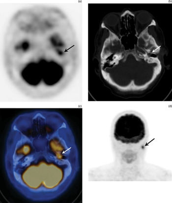
Computed tomography was performed to evaluate the local extent of the lesion. This showed a mass lesion within the left middle ear with obliteration of the mastoid cells and erosion of the tegmen and skull base. The auricular ossicles were intact and there was no infiltration of the inner ear. Fluoro-2-deoxy-d-glucose positron emission tomography/computed tomography was performed for further staging. This demonstrated increased FDG uptake (maximum standardized uptake value (SUVmax) = 5.1) of the mass lesion in the left tympanic cavity (Figure 1a to 1c). Additionally, there was focal FDG accumulation in the ipsilateral parotid gland (SUVmax = 9.1) (Figure 1d), suspicious for intraparotid lymph node metastasis. No distant metastases were visible on FDG-PET/CT imaging.
(a)–(c) Axial Fluoro-2-deoxy-d-glucose positron emission tomography/computed tomography (FDG-PET/CT) scans showing FDG-active mass lesion in the left tympanic cavity (arrows). (d) Coronal FDG-PET/CT SCAN showing FDG accumulation in the ipsilateral parotid gland (arrow).

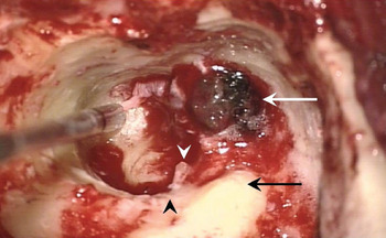
The patient underwent subtotal petrosectomy and melanoma resection as well as lateral left-sided parotidectomy and a modified left-sided neck dissection. Intra-operatively, the lesion presented as a dark, brittle tumour at the tympanic orifice of the eustachian tube (Figure 2).
Intra-operative view showing a brittle, black and purple tumour at the tympanic orifice of the Eustachian tube (white arrow). The stapes (white arrowhead), lateral semicircular canal (black arrow) and facial nerve (black arrowhead) are also visible.

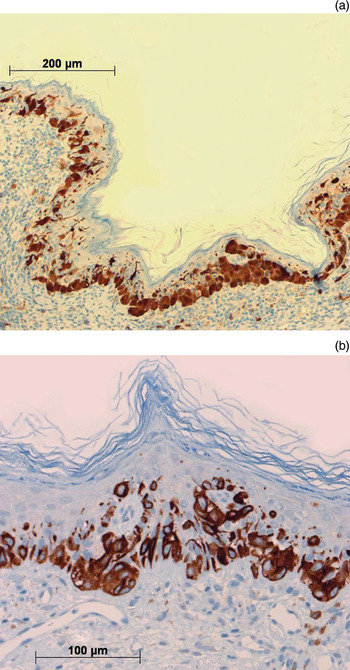
Immunohistological examination of the resected middle-ear tumour confirmed a mucosal melanoma by immunopathological positivity for S-100 (Figure 3a) and Melan-A (Figure 3b). The lesion in the parotid gland was confirmed as a lymph node metastasis.
Photomicrographs showing tympanic membrane with in situ malignant melanoma, with immunopathological positivity for (a) S-100 and (b) Melan-A.

The patient recovered quickly from surgery.
Four months later, an enlarged, left-sided, level II cervical lymph node was palpated. Subsequent fine needle aspiration cytology revealed a melanoma metastasis. Neck dissection (level II and III) was performed, but revealed no evidence of additional lymph node metastases.
Six months after initial presentation, the patient developed multiple distant metastases in the liver and abdominal lymph nodes and died.
Discussion
A few case reports of malignant melanoma of the middle ear have previously been published.Reference McKenna, Holmes and Harwick3–Reference Uchida and Matsunami6 Approximately 1.3 per cent of all melanomas arise within the mucosal surfaces of the body, and 55 per cent of these occur in the head and neck.Reference Patel, Prasad, Escrig, Singh, Shaha and Kraus7
The treatment of mucosal melanomas is challenging. The only curative treatment is complete resection, but clear surgical margins are often difficult to obtain and this is considered to contribute to the poor prognosis.Reference Patel, Prasad, Escrig, Singh, Shaha and Kraus7, Reference Bradley8 Some studies suggest that improved local control could be achieved by post-operative radiotherapy.Reference Bradley8
Malignant melanoma can metastasise to any organ, and the location of metastases is often unpredictable. Metastasising melanoma is refractory to chemotherapy in most cases, although several clinical trials of vaccines and cytokines are in progress.Reference Thompson, Scolyer and Kefford9 Recent studies suggest a therapeutic option involving inhibiting signalling pathways in tumours with the v-Raf murine sarcoma viral oncogene homolog B1 gene (BRAF) mutation.Reference Dhomen and Marais10 If distant metastasis of melanoma occurs then the outcome is poor, with an average survival of only four to six months.Reference Markovic, Erickson, Rao, Weenig, Pockaj and Bardia1, Reference Balch, Soong, Atkins, Buzaid, Cascinelli and Coit11
Of the above-mentioned patients with middle-ear melanoma, only half survived longer than eight months after diagnosis.
Clinical evaluation and conventional imaging modalities generally underestimate the true extent of malignant melanoma. Autopsies of patients with known cutaneous malignant melanoma have revealed a higher prevalence of metastases than is generally clinically documented.Reference Amer, Al-Sarraf and Vaitkevicius12–Reference Budman, Camacho and Wittes14
• This paper reports a rare case of primary mucosal melanoma of the middle ear, imaged with fluoro-deoxyglucose positron emission tomography/computed tomography (FDG-PET/CT)
• In melanoma cases, pre-operative FDG-PET/CT is a valuable aid to surgical planning and oncological treatment
• The presented patient underwent subtotal petrosectomy and melanoma resection plus lateral left-sided parotidectomy and a modified left-sided neck dissection
Precise staging of malignant melanoma is essential for early detection of metastases and initiation of appropriate treatment.Reference Balch, Soong, Atkins, Buzaid, Cascinelli and Coit11, Reference Balch, Buzaid, Soong, Atkins, Cascinelli and Coit15 The majority of the available literature suggests that FDG-PET/CT is the most accurate imaging modality for the detection of metastases in melanoma patients, due to the high avidity of FDG for melanoma tumour cells.Reference Friedman and Wahl16–Reference Krug, Crott, Lonneux, Baurain, Pirson and Vander Borght18
Conclusion
Malignant melanoma of the middle ear is a very rare entity. However, it should be considered in the differential diagnosis of mucosal tumours. It may mimic other conditions found in the middle ear, and may produce similar symptoms such as hearing loss and otorrhoea. Whenever melanoma is considered, the patient should undergo FDG-PET/CT to search for local and distant metastases, which if found may modify surgical and oncological treatment.