Introduction
Type one tympanoplasties are surgical procedures designed to deal with perforated or diseased tympanic membranes. They have traditionally been classified as overlay or underlay procedures, depending on whether the graft is inserted lateral or medial to the remnants of the tympanic membrane respectively. Both techniques can be effective. Their aim is not only to repair the tympanic membrane,Reference Sheehy and Glasscock1 but also to obtain anatomically normal ears. This is because post-operative eardrum alterations, such as retraction pockets, scars, thinned areas, tympanosclerosis, lateralisation or blunting, may lead to impaired functional results.Reference Glasscock2–Reference Gersdorf, Garin, Decat and Juantegui4
When dealing with large perforations or extended areas of unhealthy membrane, we prefer to remove the tympanic membrane remnants and graft the entire surface to prevent anatomical defects. The graft must be stable, and care must be taken to save the areas from which re-epithelialisation of the neo-tympanum should begin. A key point is the vibrating anterior tympanomeatal angle, where scarring or blunting are frequent complications.Reference Farrior5 To overcome these problems, we propose the Felix tympanoplasty, a modification of the annular wedge tympanoplasty developed by Pappas and Sympson in 1984 and published in 1992.Reference Pappas and Sympson6
Surgical technique
The posterior ear canal is infiltrated with normal saline solution and 1:5000 adrenaline, and then temporalis fascia is harvested via a post-auricular approach. The posterior external auditory meatus skin is incised as close as possible to the posterior annulus, and lifted medially exposing the posterior bony canal wall which is usually calibrated with a burr. Incisions are then made at the 6 and 12 o'clock positions of the Gerlach annulus, and its posterior half is removed and discarded.
All remnants of the tympanic membrane are removed, leaving in place only the anterior half of the annulus which is carefully freed from its squamous epithelium (Figure 1). Only the squamous epithelium of the distal part of the malleus handle is spared as this is useful to re-epithelise the graft; leaving it in place also avoids unnecessary surgical trauma to the ossicles. The anterior part of the external auditory meatus is left untouched unless it is so protruding as to prevent access to the anterior tympanomeatal angle; in this case only, a lateral-based flap exposing the bony canal bulge is elevated and the anterior canal bone is calibrated with a burr.
The skin incision is marked with black dashes, the annulus is shown in blue, and the part of the annulus and the remnants of the tympanic membrane to be removed are shown in red.

In annular wedge tympanoplasty, a slit is created in the graft which is inserted medial to the malleus handle and pulled superiorly by the slit edges. However, we believe that such a slit is unnecessary. Instead, we insert the fascia medial to the handle of the malleus, avoiding cuts which may provoke migration of the skin under the graft during epithelialisation. We shape the anterior part of the fascia with two little triangular ‘cat's ears’; these must be inserted in two pockets obtained by lifting the anterior annulus from the anterior bony canal wall (Figures 2 and 3). Unlike annular wedge tympanoplasty, we do not separate the entire anterior annulus from the canal wall, to prevent lack of graft support if the annulus is thin and fragile.
The anterior part of the annulus (shown in blue) is spared and lifted from the anterior bony external auditory canal in two little spots. The graft (shown in orange) is shaped with two ‘cat's ears’.
The fascia (shown in orange) is placed medial to the malleus handle, and the anterior annulus is overlaid with the two ‘cat's ears’ (marked by thin black dashes) tucked between the annulus and the anterior bony auditory meatus. The skin incisions are marked with thick black dashes.
The graft is stabilised anteriorly by the two ears jammed between the annulus and the canal wall, and posteriorly up the posterior canal wall, covered by the skin of the posterior auditory meatus. Lateralisation of the graft is prevented by the handle of the malleus being lateral to the fascia. The meatus can then be packed with Gelfoam® and expandable sponges.
The post-auricular skin incision can be sutured with 3-0 absorbable sutures. The sutures pass through both the subcutaneous tissue and the dermis, making external suturing and subsequent stitch removal unnecessary.
Materials and methods
This study comprised 64 patients (age range = 10–70 years, with a mean age of 40 years), on whom we performed the Felix tympanoplasty from January 2001 through to January 2011 to obtain adequate follow-up data. Patients were operated on at the Otorhinolaryngology Department of Azienda Sanitaria Locale CN1 in Savigliano. Tympanoplasties that included ossiculoplasty or surgery for cholesteatoma were not taken into account. Felix tympanoplasty was chosen only for those patients who had a perforation of the eardrum greater than 40 per cent of the surface of the tympanic membrane, or for those who had a perforated, unhealthy, thin and scarred or tympanosclerotic membrane.
Hearing results were evaluated according to the Committee on Hearing and Equilibrium Guidelines for the evaluation of results of treatment of conductive hearing loss.7 Pre-operative, short-term post-operative (6-months post-operative), and long-term post-operative (2–10-years post-operative) hearing results were evaluated. Analyses were performed according to the availability of hearing data at each time point. In order to account for the potential attrition bias deriving from a selective evaluation of patients at each time point, two sensitivity analyses were performed: one on patients in whom all audiological data were available at each follow up (early and late post-operative data available: n = 18), and another one on all patients, imputing missing hearing data with the last observation carried forward (n = 64).
The mean of the thresholds at the frequencies 0.5, 1, 2 and 3 kHz were used to form a four-tone pure tone average. At each time point, the air–bone gaps (ABG) were reported (with 95 per cent confidence intervals (CI)) as the four-tone pure tone average for air conduction minus the same average for bone conduction using values determined at the same time.
Changes from baseline of ABGs were evaluated by applying linear regression models, comparing levels of short-term and long-term post-operative ABG hearing results with the ABG levels recorded during the pre-operative examination. The standard errors of regression coefficients were adjusted in this analysis (for clustering, as a result of repeated measurements taken in the same patient) with the Huber–White sandwich estimator.Reference White8
Analyses were performed using Stata, version 11.2 (StataCorp, College Station, Texas, USA).
Results
Anatomical results
Two of the 64 patients had a re-perforation in the first year after the operation. A further perforation occurred five years post-operatively, caused by an upper airway infection.
No patients showed any retraction pockets, areas of atrophy or anterior tympanic membrane blunting. Three patients (4.68 per cent of cases) developed a little pearl of cholesteatoma on the anterior annulus: this could easily be removed under local anaesthesia without hospitalisation, and none recurred.
Ten patients were operated on whilst having a discharging ear; graft take was achieved in all of these patients. In the first two months after surgery, three cases (4.68 per cent) had a discharging ear, which we treated successfully with antibiotic and steroidal drops. Sixty-one patients (95.31 per cent) showed a non-discharging intact tympanic membrane at late follow up (2–10 years post-operatively).
No patients dropped out of the follow-up cohort earlier than two years. Overall, 61 of the 64 patients had good anatomical results, with a repaired tympanic membrane at late follow up.
Functional results
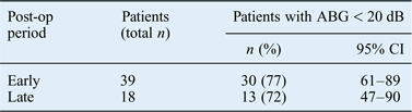
The pre-operative, and early and late post-operative hearing results, and results for patients showing ABGs inferior to 20 dB at early and late post-operative follow up, are presented in Tables I and II.
Pre-operative, and early and late post-operative hearing results*
* Mean thresholds at 0.5, 1, 2 and 3 kHz were used to form a four-tone pure tone average. †With respect to pre-operative mean air–bone gap. Pts = patients; ABG = air–bone gap; CI = confidence interval; pre-op = pre-operative; post-op = post-operative
Patients showing abg inferior to 20 db early and late post-operation
ABG = air–bone gap; post-op = post-operative; CI = confidence interval
The pre-operative mean air conduction pure tone average was 46.0 dB (standard deviation (SD) = 15.7; range = 22.5–82.5 dB). The pre-operative mean bone conduction pure tone average was 24.4 dB (SD = 11.4; range = 10–56.3 dB). The mean pre-operative ABG was 21.7 dB (95 per cent CI = 19.2–24.1).
Thirty-nine of the 64 patients underwent post-operative audiological evaluation: we could not obtain post-operative audiological data on 25 patients, even if they agreed to be followed for a minimum of 2 years. The average length of audiological follow up was 29 months. At the short-term follow up, 33 patients (84.6 per cent) showed ABG improvements; ABG remained unchanged in 5 patients (12.8 per cent) and the ABG was closed in 6 patients (15.3 per cent). One patient had worsening of the ABG post-operatively. The mean post-operative short-term ABG was 11.3 ± 9.6 dB (range = 0–32.5 dB); the mean post-operative long-term ABG was 15 ± 9.7 dB (range = 0–40 dB). Eight patients (29.5 per cent) showed a post-operative high-frequency sensorineural hearing loss, with bone conduction worsening between 10 and 60 dB at 4 kHz. There were no cases of ‘dead ears’.
Discussion
In all cases, the Felix tympanoplasty resulted in a healthy, intact tympanic membrane at late follow up, with the exception of three patients. In 1 patient, a micro-perforation was detected at 15 months after surgery. This remained stable and no further surgery was required as the patient was happy with the hearing results. Bathing was even possible as the perforation was of such a small diameter that water could not penetrate into the middle ear. One patient had stenosis of the ear canal due to exuberant scarring, and developed iatrogenic cholesteatoma which needed revision surgery. This patient had an extremely narrow auditory meatus and her anterior ear canal was drilled with accidental minimal penetration into the temporomandibular joint. This poor result is therefore regarded as a technical failure. Another patient had an intact neo-tympanum for five years; this eventually re-perforated as a result of recurrent episodes of upper airways infection.
A pearl of cholesteatoma, developing early, anteriorly near the annulus, is a possible complication. It is important to recognise this before it enlarges in size. It can easily be excised under local anaesthesia (conducted as an out-patient procedure) whilst still small. In our series, the pearls of cholesteatoma were probably caused by remnants of the squamous epithelium that were not completely removed from the anterior part of the annulus. Three patients (4.68 per cent) had such a complication; after removal, there was no recurrence at late follow up.
• Myringoplasties are used to repair perforated tympanic membranes whilst retaining anatomically normal ears for optimal functional results
• The avoidance of anterior tympanomeatal angle blunting and good graft stability are key for optimal results
• The Felix tympanoplasty, developed in our institution, is a reliable technique, with good anatomical and functional results
We believe that the lack of retraction pockets, areas of atrophy or anterior tympanic membrane blunting account for the good hearing results. In particular, the avoidance of blunting is likely attributed to the minimal dissection at the anterior annulus with the Felix technique. Careful packing of the anterior meatal skin is necessary only in the few cases when this is elevated to reduce the bony canal bulge.
Conclusion
Felix tympanoplasty proved to be a reliable procedure to repair tympanic membranes with large perforations, or unhealthy, scarred and tympanosclerotic tympanic membrane remnants. The avoidance of blunting at the anterior tympanomeatal angle or of other eardrum deformities allowed for good functional results.
Acknowledgement
We wish to thank Dr Dong Ching Chiu for advice and revision.