Introduction
Human embryonic stem cells (hESCs) extracted from the blastocyst are pluripotent primordial cells. Stable ESCs have important significance for gene detection and selection, genetic engineering, and the construction of human disease models (Bogliotti et al., Reference Bogliotti, Wu, Vilarino, Okamura, Soto, Zhong, Sakurai, Sampaio, Suzuki, Izpisua Belmonte and Ross2018). ESC self-renewal and pluripotency characteristics enable them to differentiate into germ cells in vivo and into the cells that originate in tridermic layers, such as brain cells, skin, muscle, blood, bone, lung and liver cells. Researchers have successfully induced pluripotent stem cells to differentiate in vitro into neuroepithelial cells (Jeon et al., Reference Jeon, Kumar, Conway, Park, Jothi and Jetten2019), pancreatic cells, cardiomyocytes, vascular endothelial cells (Hofsteen et al., Reference Hofsteen, Robitaille, Strash, Palpant, Moon, Pabon and Murry2018), and smooth muscle cells using suitable conditions and induction factors.
The differentiation of ESCs requires the participation of multiple signalling pathways. The combination treatment of FLI1 overexpression and VEGF-1 enhanced the ratio of ESC differentiation into endothelial cell through the PKC-ε/η pathway (Zhao et al., Reference Zhao, Li, Ouyang, Lin and Hu2020). ERK1/2 and GSK3β inhibitors were used to maintain the pluripotency of mESCs (Tabata et al., Reference Tabata, Hara and Kitajima2019). Laminin promoted ESC differentiation by affecting the integrin/FAK/PI3K p85 pathway (Wang et al., Reference Wang, Wang, Liu, Tong, Ying, Sachinidis, Li and Peng2019). Moreover, the different expression of lncRNAs and microRNAs is a promising biomarker for cell differentiation (Zhu et al., Reference Zhu, Wang, Yu, Xia, Huang, Wang, Liu, Tao, Liang and Li2019; Divisato et al., Reference Divisato, Passaro, Russo and Parisi2020).
PGCs originate from the ectoderm and first appear in the hindgut before migrating to the gonadal ridge containing unlimited proliferation and multidirectional differentiation capabilities (Chen et al., Reference Chen, Liu, Lukianchikov, Hancock, Zimmerman, Lowe, Kim, Galic, Irie, Surani, Jacobsen and Clark2017; Hackett et al., Reference Hackett, Huang, Günesdogan, Gretarsson, Kobayashi and Surani2018). This process is accompanied by complex molecular regulation mechanisms, DNA methylation and reprogramming, histone modification, and other epigenetic processes. During development, the determination of germ cell fate marks the beginning of important processes in biology and medicine (Kojima et al., Reference Kojima, Sasaki, Yokobayashi, Sakai, Nakamura, Yabuta, Nakaki, Nagaoka, Woltjen, Hotta, Yamamoto and Saitou2017). Researchers have found that the fate of mouse germ cells was regulated by bone morphogenetic protein 4 (BMP4) and Wnt3a at the ectoderm (Oestrup et al., Reference Oestrup, Hall, Petkov, Wolf, Hyldig and Hyttel2009; Hackett et al., Reference Hackett, Huang, Günesdogan, Gretarsson, Kobayashi and Surani2018). In contrast, the formation of trophoblast-like cells was decreased when the Activin/Nodal signal was inhibited (Fei et al., Reference Fei, Zhu, Xia, Zhang, Li, Han and Chen2010; Sudheer et al., Reference Sudheer, Bhushan, Fauler, Lehrach and Adjaye2012). Germ cell differentiation from stem cells can aid in treating adult infertility (Mansouri et al., Reference Mansouri, Salehi, Omrani, Niknam and Ardeshirylajimi2017).
Wnt is a complex, highly conserved signalling system that regulates the development, proliferation, differentiation, and migration of cells (Zuccarini et al., Reference Zuccarini, Giuliani, Ziberi, Carluccio, Iorio, Caciagli and Ciccarelli2018), and is vital for various stages of life development. hESCs are extremely sensitive to Wnt ligands as they can rapidly induce differentiation. Wnt/β-catenin can regulate the self-renewal and differentiation of stem cells in a dose-dependent manner through its dual function. Therefore, the self-renewal of hESCs requires a strong mechanism to ensure that the Wnt pathway is not hyperactive (dysregulated) but is responsive in cells (Sierra et al., Reference Sierra, Hoverter, Ramirez, Vuong, Mortazavi, Merrill, Waterman and Donovan2018). Canonical Wnt/β-catenin signals play an indispensable role in gonad development and sexual differentiation in mammals. Another study found that Wnt5 could promote hESC differentiation through the non-canonical Wnt/JNK signalling pathway (Susman et al., Reference Susman, Karuna, Kunz, Gujral, Cantú, Choi, Jong, Okada, Scales, Hum, Hu, Kirschner, Nishinakamura, Yamada, Laird, Jao, Gygi, Greenberg and Ho2017; Velasco-Tirado et al., Reference Velasco-Tirado, Alonso-Sardón, Lopez-Bernus, Romero-Alegría, Burguillo, Muro, Carpio-Pérez, Muñoz Bellido, Pardo-Lledias, Cordero and Belhassen-García2018; Bothun and Woods, Reference Bothun and Woods2019).
The main aim of this study was to perform primordial germ cell-like cell (PGCLC) induction in in vitro experiments and regulate the Wnt signalling pathway to verify the role of this signalling pathway in PGCLC differentiation of ESCs.
Materials and Methods
Materials
SOX2 (ab97959), NANOG (ab109250), OCT4 (ab19857), SOX17(ab84990), GSK (ab75745), β-catenin (ab32572), Frizzled (ab83042), Wnt3α (ab28472), Wnt5α (ab174963), Mvh (ab13840), DAZL (ab34139), Blimp1 (ab13700), TFAP2C (ab218107), Gata4 (ab84593), Nodal (ab55676), Activin (ab109300), EOMES (ab23345), APC (ab193785), and goat anti-rabbit IgG (HRP) (ab205718) were purchased from Abcam. Recombinant Wnt3α protein (ab81484), BMP4 (ab87063), Activin (ProSpec, CYT-145), Matrigel (BD), the RNeasy Mini Kit (Qiagen, 74104), and the QuantiNova SYBR Green PCR Kit (Qiagen, 208054) were used.
Cell culture and differentiation
hESC lines were cultured on mouse embryonic fibroblasts in hESC medium following protocols described in previous studies. The medium was composed of 20% knockout serum replacement (GIBCO, 10828-028), 100 μM l-glutamine (GIBCO, 25030-081), 1× MEM non-essential amino acids (NEAA) (GIBCO, 11140-050), 55 μM 2-mercaptoethanol (GIBCO, 21985-023), 10 ng/ml recombinant human fibroblast growth factor (FGF) basic (R&D systems, 233-FB), 1× penicillin/streptomycin (GIBCO, 15140-122), and 50 ng/ml primocin (InvivoGen, ant-pm-2) in DMEM/F12 (GIBCO, 11330-032). Cells were placed in incipient mesoderm-like cell (iMeLC) medium.
The differentiation medium contained 15% KSR, 0.1 mM NEAA, 0.1 mM 2-mercaptoethanol, 100 U/ml penicillin, 0.1 mg/ml streptomycin, 2 mM l-glutamine, 1 mM sodium pyruvate, 500 ng/ml BMP4 (R&D Systems), 1 μg/ml human LIF (SCI), 100 ng/ml SCF (R&D Systems), 50 ng/ml EGF (R&D Systems), and 10 μM ROCK inhibitor in GMEM (Gibco, 11710-035); 2 µM IWP2 (Sigma), 10 µM SB (Sigma) and 100 ng CER1 (Abcam) were needed during differentiation (Chi et al., Reference Chi, Saarela, Railo, Prunskaite-Hyyryläinen, Skovorodkin, Anthony, Katsu, Liu, Shan, Salgueiro, Belo, Davies, Yokouchi and Vainio2011; Martyn et al., Reference Martyn, Kanno, Ruzo, Siggia and Brivanlou2018). The inhibitors were added to the differentiation medium at the initiation of differentiation (Koel et al., Reference Koel, Võsa, Krjutškov, Einarsdottir, Kere, Tapanainen, Katayama, Ingerpuu, Jaks, Stenman, Lundin, Tuuri and Salumets2017).
The experimental research programme was supervised by the Ethics Committee of Qinghai Provincial People’s Hospital. This research used commercial cell lines and did not involve any sample collection from humans.
Immunofluorescence
Immunofluorescence was used to identify the totipotency of hESCs and the differentiation marker of PGCLCs. Cells were washed in 6-well cell culture plates with phosphate-buffered saline (PBS), fixed with 4% paraformaldehyde, and washed again. Then 0.25% Triton X-100 was added to permeabilize the membrane for 15 min. The plate was blocked with PBS containing 5% BSA. Cells were incubated with specific primary antibodies at 4°C overnight. After washing with PBS, the secondary antibody was added for 1 h, then cells were incubated with 4′,6-diamidino-2-phenylindole for nuclear staining. Cells were washed with PBS, then cells were observed under a fluorescence microscope. Image-plus software (Media cybernetics, Rockville, MD, USA) was used to analyze fluorescence intensity.
Flow cytometry
Here, 0.25% trypsin/EDTA was used to detach the differentiated cells, and the supernatant was discarded after centrifugation at 300 g for 5min. Cells were washed twice with PBS containing 0.5% BSA, then SOX17 antibody (Abcam, ab84990) was added and cells were incubated at 4°C for 40 min in the dark. At the same time, negative control cells were treated using the same method. The experimental method has been described in previous research (Cheng et al., Reference Cheng, Zhai, Chang, Yao, He, Wang, Kong, Xin, Wang, Jin, Gong, Gu, Yang, Wu, Ji and Sun2017).
Western blot
Cell samples were lysed using cell lysis buffer for 30 min on ice. Following that, the lysates were centrifuged. Next, 60 µl of supernatant was collected, 20 µl of 4× loading buffer was added, and the mixture was vortexed. The samples were placed in a 100°C water bath and then cooled on ice. SDS-PAGE was performed, then proteins were transferred to a polyvinylidene fluoride membrane. The membrane was blocked using 5% skimmed milk and washed three times with PBS containing 0.05% Tween-20. Membranes were incubated with primary antibodies at 4°C overnight, membranes were washed as described previously before incubation with secondary antibodies at room temperature for 1 h, before washing with phosphate-buffered saline Tween. The ECL method was used to develop the obtained images. ImageJ Pro software was used for grey scale analysis of the image.
qRT-PCR
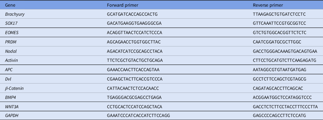
Total RNA was extracted using a commercial kit. RNA concentration was determined using a NanoDrop 2000 spectrophotometer. Reverse transcription and cDNA synthesis were performed. qPCR was performed using a 2× QuantiNova SYBR Green PCR Master Mix. The experimental steps were as follows: Step 1: 95°C, 2 min, 1 cycle; Step 2: 95°C, 5 s, 60°C, 10 s, 40 cycles. The credibility of the amplification curve and dissolution curve for real-time PCR was confirmed after the reaction. mRNA relative levels were calculated using the equation F = 2–ΔΔCt. Each sample was prepared in triplicate. The primer sequences are listed in Table 1.
PCR primer sequences

Statistical analysis
Statistical analysis was carried out using SPSS v.20.0 software. One-way analysis of variance was used for comparison of multiple groups, and inter-group pairwise comparison was carried out using the least significant difference t-test. A difference of P < 0.05 indicated statistical significance. *P < 0.05; **P < 0.01; n.s. means no significant difference.
Results
Effect of the Wnt signalling pathway on pluripotency of embryonic stem cells
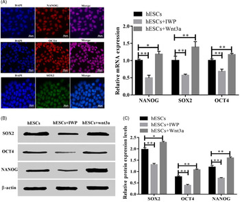
Octamer-binding transcription factor (OCT4), sex determining region Y-box (SOX2), and NANOG were identified by immunofluorescence in hESCs (Fig. 1A). hESCs were subjected to the inhibitor and activator of Wnt signalling. The results showed that the expression levels of pluripotency-associated factors were decreased significantly after IWP was added (Fig. 1B) (P < 0.05). However, the action of Wnt3a reversed this phenomenon. After Wnt3a induction, protein levels of NANOG, SOX2 and OCT4 were increased. This result showed that Wnt signalling activation could maintain and enhance the totipotency of ESCs; inhibitors of the Wnt signalling pathway promoted differentiation of cells (Price et al., Reference Price, Yin, Jones, van Ijcken, Grosveld and Rudnicki2013; Osei-Sarfo and Gudas, Reference Osei-Sarfo and Gudas2014; Chen et al., Reference Chen, Nefzger, Rossello, Sun, Lim, Liu, de Boer, Knaupp, Li, Davidson, Polo and Barberi2018).
Activation of Wnt maintains the pluripotency of ESCs. (A) Pluripotency factors were detected by immunofluorescence assay. (B) Changes in pluripotency factors induced by IWP and Wnt3A. (C) Protein levels of SOX2, OCT4 and NANOG were upregulated by recombination protein Wnt3A. Means compared with hESCs. *P < 0.05, **P < 0.01.

Effects of the Wnt signalling pathway on differentiation of primordial germ cell-like cells
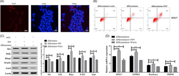
In flow cytometry and immunofluorescence experiments, we observed that ESCs could differentiate and produce PGCLC-like markers. DAZL was measured by immunofluorescence and SOX17 was marked by fluorescence activated cell sorting (Fig. 2A,B). From the flow cytometry results, we also saw that PGCLCs accounted for 10% of the cells during induction. However, expression of SOX17 was affected by the use of Wnt3a and IWP. Treatment with Wnt3a decreased the ratio of SOX17-positive cells sorted. Conversely, the ratio SOX17-positive cells was enhanced by IWP (Fig. 2B). At the same time, the effect of regulation on the Wnt signal on differentiation of PGCLCs was investigated at the protein and mRNA levels. The expression changes of Mvh, DAZL, Blimp1, TFAP2C, Gata4 showed the similar trends. The use of IWP enhanced protein expression, but Wnt3a reduced the expression levels (Fig. 2C). There were significant differences among groups. Similarly, the mRNA expression levels of SOX17, EOMES, Brachyury and PRDM1 were upregulated by the use of IWP, and downregulated by Wnt3a (Fig. 2D).
Dual roles for Wnt/β-catenin in regulating the PGCLC differentiation process. (A) Expression of DAZL during differentiation and detected by immunofluorescence assay. (B) Changes in SOX17 induced by WNT3A and IWP during differentiation. (C) Effect of Wnt3A and IWP on PGCLCs markers during differentiation. (D) Wnt3A and IWP affect mRNA levels of differentiation markers. Means compared with differentiation. *P < 0.05, **P < 0.01.

BMP–Wnt–Activin/Nodal mediated embryonic stem cell differentiation
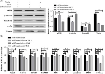
The effect of IWP on the Wnt pathway was significant. Use of IWP upregulated the expression of pGSK, and downregulated the protein levels of β-catenin, Frizzled and WNT5A (Fig. 3A).
The signalling pathway involved in embryonic stem cell differentiation. (A) Wnt signal is regulated by Wnt3A and IWP during differentiation. (B) BMP and Nodal signals participate in differentiation with the form of BMP4–WNT–Nodal. Means compared with differentiation. *P < 0.05, **P < 0.01, n.s, means no significant difference.

To investigate the signalling pathway, the inhibitors CER1 and SB were used to regulate the BMP and Nodal signals, respectively. Results showed that both these inhibitors affected differentiation. The effect of CER1 on the WNT signal was significant in the differentiation process, APC, Dvl, Wnt3a and β-catenin mRNA levels were decreased (Fig. 3B). But there was no difference with SB and the WNT pathway for the duration of differentiation. Both inhibitors could regulate differentiation, but with different inhibition effects.
Discussion
The successful differentiation and culture of PGCLCs in vitro provide a route for treating infertility (Mucksová et al., Reference Mucksová, Reinišová, Kalina, Lejčková, Hejnar and Trefil2019). Infertility is a global public health problem due to errors in stages in gametogenesis (Eskandari et al., Reference Eskandari, Hassani Moghaddam, Atlasi, Amini Mahabadi, Taherian and Nikzad2018). Most of our knowledge on mammalian PGC development has been obtained through the study of laboratory mice. However, regulation of the Wnt signalling pathway can effectively control ESCs to directionally form PGCs.
Wnt signalling begins with the secretion of Wnt proteins, Wnt ligands participate in PGCLC specification and are essential for the formation of primitive streak (PS), mesoderm, and Nodal. The Wnt signalling pathway is an important target of p53 to regulate mouse ESC differentiation (Lee et al., Reference Lee, Li, Michalowski, Zhang, Liao, Chen, Xu, Wu and Huang2010). During retinoic acid (RA)-induced differentiation, it is the main effector mechanism that can activate meiosis in germ cells (Suzuki et al., Reference Suzuki, Kanai-Azuma and Kanai2015) and is regulated through the Wnt signalling pathway. Wnt5a and its receptor Ror2 participate in PGC migration; Wnt4 participates in female sex differentiation; and sex establishment and differentiation is β-catenin dependent (Cantú et al., Reference Cantú, Altshuler-Keylin and Laird2016).
Dynamic changes in BMP, Wnt and Nodal signalling components are considered as the underlying germ-layer specification conserved across mammals. The initiation of the gastrulation signal is BMP signalling that activates WNT signalling, the activation of WNT signalling leads to the activation of the NODAL signal, and the NODAL signal maintains BMP signalling. The signalling cascade eventually leads to the formation of PS (Chhabra et al., Reference Chhabra, Liu, Goh, Kong and Warmflash2019). Wnt/BMP and Activin/Nodal participate in most stages of differentiation, such as neuronization of ESCs, and the regulation of signals could promote differentiation (Bertacchi et al., Reference Bertacchi, Pandolfini, D’Onofrio, Brandi and Cremisi2015; Morizane et al., Reference Morizane, Doi, Kikuchi, Nishimura and Takahashi2011). The signal of Activin/Nodal was identified as the major component in self-renewal of hESCs. Inhibition of Activin/Nodal affected BMP4-mediated differentiation activation, having the opposite effects as found for FGF2 inhibition (Koel et al., Reference Koel, Võsa, Krjutškov, Einarsdottir, Kere, Tapanainen, Katayama, Ingerpuu, Jaks, Stenman, Lundin, Tuuri and Salumets2017). The development of the early posterior epiblast into germ or somatic cells was regulated by Nodal/Bmp signalling (Senft et al., Reference Senft, Bikoff, Robertson and Costello2019).
Throughout our experiment, we found that the presence of WNT3A in the hESC culture could significantly promote the expression of totipotent genes and decrease PGCLC marker gene levels. The effects of BMP inhibition were the reverse of Wnt inhibition: Wnt inhibition promoted cell differentiation, while BMP inhibition promoted maintenance of pluripotency. IWP can inhibit self-renewal in ESCs and block Porcupine (an essential enzyme for acylation of Wnt proteins) to inhibit cells from producing active Wnt proteins. Addition of WNT3A alleviates this effect. Therefore, ESCs require Wnt to block differentiation (ten Berge et al., Reference ten Berge, Kurek, Blauwkamp, Koole, Maas, Eroglu, Siu and Nusse2011). Wnt and Nodal/Activin have crucial roles in the de-differentiation of different vertebrate models, and this is mediated by the bi-directional regulation of the Wnt signalling pathway (Zhong et al., Reference Zhong, Lai, Yu, Xia, Yuan, Ouyang, Shan and Chen2017). Therefore, we speculated whether the PGCLC differentiation conditions could be improved by adding an inhibitor of Wnt signalling, making ESC differentiation into PGCs more easy in the future. Moreover, the realization of this process and whether it can solve a series of safety problems due to immune rejection and autoimmune reactions, as well as ethical and legal conditions, were considered. In the near future, if we are able to induce ESCs to differentiate into PGCs, this research domain will be truly used to solve patients’ needs and become a milestone in human history.
Acknowledgements
Thanks to Science and Technology project of Qinghai (2017-ZJ-740) for their support and help.
Financial support
This study was supported by Science and Technology project of Qinghai (2017-ZJ-740).
Conflict of interest
Not applicable.
Ethical standards
All experiments meet the standards of stem cell application.