INTRODUCTION
There are numerous species of Sarcocystis named from the avian hosts worldwide (Dubey et al. Reference Dubey, Speer and Fayer1989). Little is known of the avian Sarcocystis species from Egypt. Recently we described a species of Sarcocystis in the common moorhen (Gallinula chloropus) from Egypt (El-Morsey et al. Reference El-Morsey, El-Seify, Desouky, Abdel-Aziz, Sakai and Yanai2014). We have now molecularly characterized this parasite and propose a new name Sarcocystis chloropusae.
MATERIALS AND METHODS
Same material described by El-Morsey et al. (Reference El-Morsey, El-Seify, Desouky, Abdel-Aziz, Sakai and Yanai2014) was used here for morphological description. For molecular characterization sarcocysts had been preserved in 70% ethyl alcohol and stored frozen at −20 °C. Genomic DNA was extracted from two infected samples (MR9 and MR11) using QIAGEN DNeasy Tissue kit® according to the manufacturer protocol. For targeting the internal transcribed spacer-1 region (ITS-1); P-ITSF (5′ATTGAGTGTTCCGGTGAATTA3′) and P-ITSR (5′ GCCATTTGCGTTCAGAAATC 3′) primer pair (Kutkiene et al. Reference Kutkiene, Prakas, Sruoga and Butkauskas2010), was used in PCR amplification.
PCR reactions were carried out in the final 50 μL volume consisting of 10X Ex Taq PCR buffer 5 μL, Takara Ex Taq (DNA polymerase) (5 units μL−1) 0·25 μL, dNTP Mixture (2·5 mm each) 4 μL, forward and reverse primers 1 μL each, DNA template 5 μL and finally sterilized distilled water up to 50 μL. Reaction conditions required for amplification of the (ITS-1) region were adjusted according to Kutkiene et al. (Reference Kutkiene, Prakas, Sruoga and Butkauskas2010).
Amplification products were analysed on 2% agarose gel. DNA bands were purified using Qiaquick gel extraction kits® according to the Manufacturer recommendations. Purified PCR products for the target gene were sequenced directly with Applied Biosystems 3730xL DNA Analyzer using the same primer pairs as for the PCR amplification. Multiple sequence alignments were performed using the MUSCLE algorithm (Edgar, Reference Edgar2004).
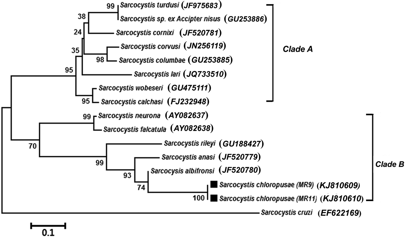
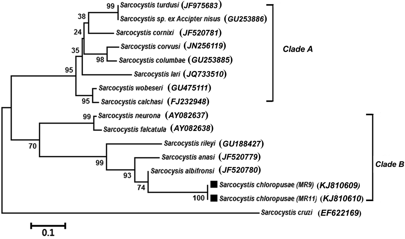
Phylogenic status of the newly identified S. chloropusae was reconstructed within the Sarcocystidae family, through a distance-based Neighbour-Joining analysis, as implemented by MEGA 6 version (Tamura et al. Reference Tamura, Stecher, Peterson, Filipski and Kumar2013). Analysis represented 15 taxa (16 nucleotide sequences including 2 sequences KJ810609 and KJ810610 those belonged to the MR9 and MR11 isolates of S. chloropusae, respectively). It was constructed for (ITS-1) region sequences and based on Sarcocystis cruzi as outgroup. The phylogenetic tree was constructed using Neighbour-Joining method (Saitou and Nei, Reference Saitou and Nei1987). The Kimura-2 parameter distance model was used for Neighbour-Joining analysis (Kimura, Reference Kimura1980).
RESULTS
Neighbour-Joining analysis of ITS-1 region sequences showed that two well-supported clades were recognized; the first clade (Clade A) contained (Sarcocystis turdusi, Sarcocystis sp. ex Accipiter nisus, Sarcocystis cornixi, Sarcocystis corvusi, Sarcocystis columbae, Sarcocystis lari, Sarcocystis wobeseri and Sarcocystis calchasi). Two well-supported subgroups were distinguished within the second clade (Clade B). The two isolates (MR9 and MR11) of the Sarcocystis species under investigation; S. chloropusae, was phylogenetically closely related to (Sarcocystis albifronsi, Sarcocystis anasi, Sarcocystis rileyi) those formed together a separate subgroup. Sarcocystis falcatula and Sarcocystis neurona were linked in another subgroup (Fig. 1).
Phylogenetic tree of species of the genus Sarcocystis based on ITS-1 region sequences. Note the distinctive position of S. chloropusae and its grouping with S. rileyi, S. anasi and S. albifronsi.
DESCRIPTION
Sarcocystis chloropusae n. sp.
(Fig. 2a, b and c)
Morphology of S. chloropusae n. sp. (a) Section of sarcocyst. Note vp (opposing arrows) on the cyst wall giving it striated appearance, and banana-shaped bradyzoites (b); (b)TEM micrograph of the sarcocyst wall. Note pvm is wavy and lined by an electron dense layer. The vp are irregular in size and shape, contain multiple electron dense granules (ed) or vesicle-like structures (v), and absence of microtubules. Also note relatively thin and smooth ground substance (gs), septa (s), electron lucent metrocytes (met) and Bradyzoites (b) with many amylopectin granules (vacuolated areas); (c) TEM micrograph of a longitudinally cut bradyzoite with a conoid (c), a centrally located nucleus (n) and several dense granules (dg) that ranged from 2 to 12 and scattered within the cytoplasm of the bradyzoites.

Sarcocysts were microscopic, up to 650 μm long and up to 185 μm wide. By light microscopy, sarcocyst wall appeared striated (Fig. 2a). By Transmission Electron Microscopy (TEM) cyst wall measured 2–4·5 μm thick. The parasitophorous vacuolar membrane (pvm) was wavy, underlined with the electron dense layer 50–100 nm thick with irregularly shaped, crowded 1·5–4 μm long and up to 2 μm wide villar protrusions (vp) filled with multiple vesicle-like structures but no microtubules. The ground substance layer measured 200–750 nm thick without vesicles (Fig. 2b). Banana-shaped bradyzoites were 6–12 × 1–2 μm in size (Fig. 2c).
Taxonomic summary
Type intermediate host: the Common Moorhen (Gallinula chloropus).
Definitive host: unknown.
Locality: Brolos Lake, Northern region of Nile Delta (coordinates: 31°06′42″N 30°56′45″E), Egypt.
GenBank accession numbers: KJ810604 (18S rRNA), KJ810605 (28S rRNA), ITS-1 region from 2 isolates (MR9; KJ810609) (MR11; KJ810610).
Specimens deposited: TEM materials under accession numbers (GF100-MR9; GF101-MR11) and DNA samples under numbers (MR9 and MR11) were deposited at the Laboratory of Veterinary Pathology, Department of Veterinary Medicine, Faculty of Applied Biological Sciences, Gifu, Japan.
Etymology: Latin name of the common moorhen is used for the species name.
DISCUSSION
Sarcocystis chloropusae proposed here is morphologically and molecularly distinct from other species of Sarcocystis in birds. Morphologically, the sarcocyst wall of S. chloropusae is distinctive from other Sarcocystis species, and it vaguely resembled type 10 cyst wall classified by Dubey et al. (Reference Dubey, Speer and Fayer1989) and Dubey and Odening (Reference Dubey, Odening, Samuel, Pybus and Kocan2001). However, unlike other species with type 10 cyst wall, the vp are not uniform in shape and size, are crowded, microtubules are absent and vesicles are discrete, whereas the vp on cyst walls of S. albifronsi, and anasi are type 9 with microtubules. The cyst wall of S. rileyi is completely different than in any other Sarcocystis species; the vp are highly branched (Dubey et al. Reference Dubey, Speer and Fayer1989; Kutkienė et al. Reference Kutkienė, Sruoga and Butkauskas2006, Reference Kutkienė, Sruoga and Butkauskas2008, Reference Kutkienė, Prakas, Sruoga and Butkauskas2012).
Molecularly the sequences of the 18S and 28S rRNA genes and the ITS-1 region of both isolates derived from the common moorhen are distinct from any previously reported sequences in GenBank. The life cycle of S. chloropusae is unknown but the red fox (Vulpes vulpes) appears to be most likely candidate as a definitive host.
ACKNOWLEDGEMENTS
The authors are grateful to the members and technicians of the Laboratory of Veterinary Pathology, Faculty of Applied Biological Sciences, Gifu University for their helpful assistance in the preservation and processing of the samples for the current molecular study.
FINANCIAL SUPPORT
Ahmed El-Morsey is a PhD researcher funded by the Missions Department of the Ministry of Higher Education and Scientific Research, Egypt (Grant No. MIS; 2007–2012).