Introduction
Malignant neoplasm of the external auditory canal (EAC) is a rare pathology with an incidence of between one and six per million people.Reference Oya, Takenaka, Takemura, Ashida, Shimizu and Kitamura1 The most common neoplasm is squamous cell carcinoma (SCC). Less common types include basal cell carcinoma (BCC), adenoid cystic carcinoma (ACC), ceruminous adenocarcinoma and middle ear adenocarcinoma.Reference Allanson, Low, Clark and Gupta2 Early diagnosis can prove challenging owing to the rarity of the disease together with overlapping symptoms between EAC neoplasm and more prevalent pathologies such as chronic/necrotising otitis externa. All three can present with otalgia and discharge that are refractory to antibiotics.
Cutaneous SCC (cSCC) has known risk factors of fair skin phenotype being exposed to high ultraviolet (UV) radiation and immunosuppression.Reference Allanson, Low, Clark and Gupta2 The carcinogenic risk factors for SCC of the EAC (EACSCC) are less clear with none of the above definitively shown to be causal. Chronic inflammation leading to metaplasia is the leading theory of pathogenesis, but the mechanism by which this occurs is unknown and may involve changes in pH of the EAC.Reference Yin, Ishikawa, Honda, Arakawa, Harabuchi and Nagabashi3, Reference Martinez Devesa, Willis and Capper4 Alternative theories of pathogenesis include the association of human papilloma virus (HPV) with EACSCC,Reference Tsai, Li, Jin, Chao and Su5 or previous radiation.Reference Lim, Goh and Chan6
The genetic basis for EACSCC remains largely unknown owing to its rarity, in contrast to the genetic mapping available for cSCC and HNSCC. Sato et al. have been the first to map genetic alterations using genome sequencing on 11 primary tumours of the EAC from 10 different patients with two-thirds of the tumours showing TP53 mutations.Reference Sato, Komune, Hongo, Koike, Niida and Uchi7
Initial management after biopsy proven diagnosis is staging by magnetic resonance imaging (MRI) and computerised tomography (CT) (Figure 1), with subsequent Multidisciplinary Team (MDT) discussion.Reference Lechner, Sutton, Murkin, Masterson, O’Flynn and Wareing8 The Pittsburgh Staging SystemReference Moody, Hirsch and Myers9 is an accepted staging system in reference to cancers of the EAC. The primary treatment modality is surgery with the aim of en-bloc resection (temporal bone and parotid) with post-operative radiotherapy (PORT) in advanced tumours.Reference Lechner, Sutton, Murkin, Masterson, O’Flynn and Wareing8 Late stage and high grade differentiation of SCC are associated with worse prognostic factors.Reference Lechner, Sutton, Murkin, Masterson, O’Flynn and Wareing8, Reference Yin, Ishikawa, Honda, Arakawa, Harabuchi and Nagabashi3
(a) MRI left internal acoustic meatus showing increased soft tissue in the left external auditory canal and in the surrounding regions. (b) CT left petrous bone demonstrating soft-tissue thickening in the left external auditory canal.

The aim of this correspondence is to highlight the increasing incidence of neoplasms affecting the EAC as documented in this regional case series with seven cases over an 18-month period with reference to the Hospital Episode Statistics (HES) database,10 and identify potential aetiological or mechanistic factors underlying this rise.
Methods
We analysed all (n = 7) cases of primary neoplasms of the EAC diagnosed at a regional National Health Service (NHS) trust over an 18-month period. All biopsy proven patients discussed at the MDT were included. Data were collected via an electronic database on patient demographics (age at diagnosis, sex); histological subtype and grade; treatment modality; marginal status (clear, involved, marginal); and recurrence. The Pittsburgh Staging SystemReference Moody, Hirsch and Myers9 was used to stage the cancers. Tumour specific analysis included histopathological characterisation using staining of tumour invasion, next generation sequencing (NGS) and HPV status using in-situ hybridisation.
Results
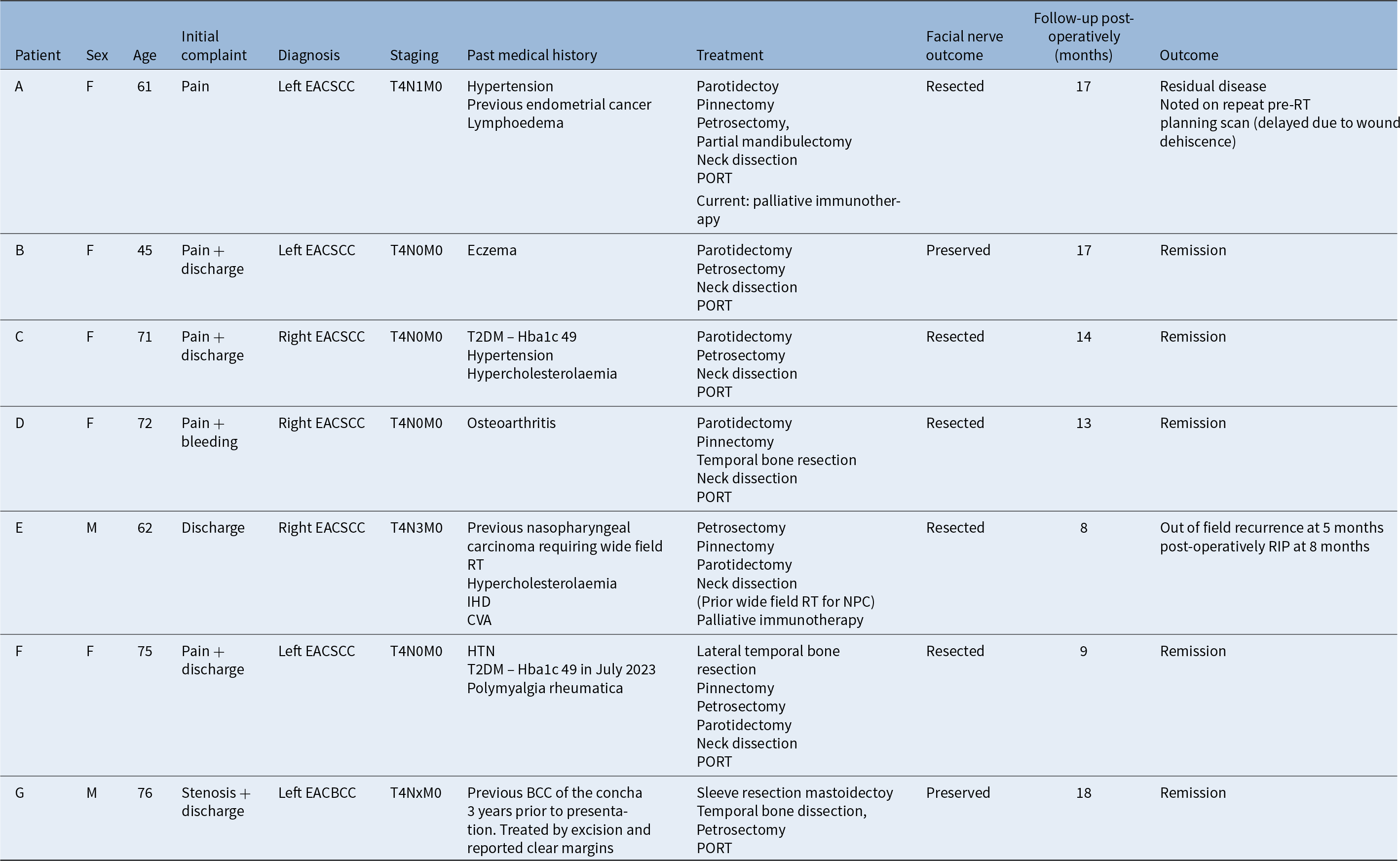
Table 1 summarises the main demographics and treatment pathways of each patient. Six were diagnosed with squamous cell carcinoma of the EAC and one with BCC of the EAC, with all cases graded as T4. The male to female ratio was 2:5, and the average age was 64.5years. Mean time (months) of follow up post-surgery (correct to May 2024) is 14.7 months with one mortality and one patient with residual disease. Patients A and E were the only reported case to have nodal disease (Table 2). No cases demonstrated distant metastases.
Summary of patients included in case series highlighting key demographics, pathology, treatment and outcomes

Note: Follow up correct to December 2023.
BCC = basal cell carcinoma; CVA = cerebrovascular accident; EACBCC = external auditory canal basal cell carcinoma; Hba1c = glycated haemaglobin A1C; HTN = hypertension; IHD = ischaemic heart disease; NPC = nasopharyngeal carcinoma; PORT = post-operative radiotherapy; RIP = rest in peace; RT = radiotherapy; T2DM = type 2 diabetes mellitus
Tumour characteristics

BCC = basal cell carcinoma; diff = differentiated; ECS = extracapsular spread; HPV = human papilloma virus; LN = lymph node; LVI = lymphovascular invasion; mod = moderately; NGS = next generation sequencing; SCC = squamous cell carcinoma
To test whether the rise in incidence seen locally was reflected nationally, we performed a search on the HES database,10 which highlighted no diagnostic code for cancers of the external auditory canal. As a control we used titles of “Carcinoma in situ: Skin of ear and external auricular canal” and “Malignant neoplasm: Middle ear”. From the data available it was not possible to comment on any increase in cases of EAC malignancy over a five year period for carcinoma of the external ear and EAC. There were 514 cases seen in 2022–202311 compared to that of 491 in 2016–2017.12 For carcinomas of the middle ear there were 26 cases in 2016–201712 rising to 50 in 2021–2022,13 and 39 from 2022–2023.11 We also performed a systematic literature review with broad search terms of “neoplasm OR carcinoma OR tumour OR malignancy OR cancer” AND “External auditory OR ear canal OR EAC OR ear” and did not find any reports of rising incidence.
With regards to potential underlying risk factors, three of the seven patients had well controlled type 2 diabetes with all of them having a haemoglobin A1c of less than 50 mmol. Patient B suffered with chronic eczema affecting her ears and swam regularly. Patient F had a previous BCC of the concha but had clear margins with excision three years prior to re-presentation. All SCC cases had prolonged antibiotic treatment in both primary and secondary care for greater than four weeks prior to diagnosis. This was either due to suspicion of necrotising externa or severe otitis externa. Of the four patients who had ear swabs sent for microbiology analysis, none showed any growth.
Table 2 summarises the tumour characteristics observed. All patients yielded negative P16/HPV analysis. All had bony involvement, with two cases having parotid involvement, and the BCC case was the only example to show peri-neural invasion. Sequencing showed all seven cases demonstrating TP53 mutation, with two cases over more than 50 per cent, with ERBB2 and CDKN2A mutations observed separately in one case each (Table 2). Patient F (Figure 2) displayed SCC infiltration of bone and patient D (Figure 3) demonstrated circumferential severe epithelial dysplasia of the external auditory canal epidermis with an exo-endophytic growth pattern.
High power image to show moderate to poorly differentiated squamous cell carcinoma infiltrating bone and associated with a fibromyxoid stromal reaction. Haematoxylin and eosin staining of tissue and cell sections.

(a) Low power image of squamous cell carcinoma arising from widespread circumferential severe epithelial dysplasia of the external auditory canal epidermis with an exo-endophytic growth pattern. (b) Severe epithelial dysplasia is discontinuous with polypoid tumour protruding into the canal lumen. Haematoxylin and eosin staining of tissue and cell sections.

All patients included in this report underwent en-bloc temporal bone resection. Six patients underwent PORT with two subsequently undergoing palliative immunotherapy (Cemiplimab) due to either recurrence (patient E) or residual tumour (patient A). Patient A was noted to have residual disease on her PORT planning imaging and underwent five weeks of PORT, but this was stopped after subsequent imaging showed further tumour progression.
Patient E is the only reported mortality to date of the series and had associated N3 staging. They had previous nasopharyngeal cancer (NPC) treated 20 years ago in China with wide field radiotherapy, such that adjuvant RT was not possible. Intra-operatively he had multiple frozen sections positive at the sinodural angle. His post-operative pathology showed cancer involving the dura, mastoid and parotid. His recurrence occurred five months after surgery, and he passed away 8 months after surgery.
Of the seven completed operations the facial nerve was preserved in two and sacrificed in five of the resections. Neck dissections were performed in all patients with EACSCC. In this series only two patients were seen to have nodal involvement with associated extracapsular spread (ECS) (Table 2).
Discussion
This report shows a rise in incidence of EACSCC with all cases showing TP53 mutations. Given the rarity in clinical practice, and by extension in the research literature, there is currently limited understanding of underlying aetiology.
Our findings are largely comparable with other published case series. The largest case series review in Australia was of 39 patient patients collating 20 years of data from 1974 to 1995.Reference Arriaga, Curtin, Takahashi, Hirsch and Kamerer14 They advocated for the use of a congruous staging system to allow comparison between studies. They comment on resection margin status as a key prognostic factor and a non-significant but important role of radiotherapy, where resection margins were positive. A notable feature of their series was recurrence in the EAC from previous pinna and peri-auricular sites. Our smaller series only noted one recurrent BCC with previous excision from the concha three years prior to the lesion in the EAC emerging.
A review of 95 patients with both middle ear carcinomas (MESCC) and EACSCC in Japan in 2006Reference Yin, Ishikawa, Honda, Arakawa, Harabuchi and Nagabashi3 came to similar conclusions from the Australian study. They both established early surgical treatment followed by chemo/radiotherapy provided the best survival rates. Staging, clear tumour margins, recurrence and metastasis had the greatest significance influence on survival. Of note only 12 per cent of patients in their study had a history of recurrent otitis externa and tended to present atypically. This highlights some of the challenges faced with early diagnosis of the EACSCC with patients often presenting with generic non-specific symptoms, overlapping with otitis externa resulting in prolonged antibiotic treatment. This report supports a lower threshold for biopsy and imaging after a period of ineffective antibiotic therapy.
A more recent literature reviewReference Lechner, Sutton, Murkin, Masterson, O’Flynn and Wareing8 focused on SCC of the temporal bone. The study echoed findings from previous papers noting a lack of universal treatment strategy, with late stage and differentiation of cancer giving the worst prognostic factors.Reference Lechner, Sutton, Murkin, Masterson, O’Flynn and Wareing8 The review also identifies the need for a more collaborative effort in producing high quality research to help overcome the rarity of this disease. The last review of EAC neoplasms based on UK practice was published in 1993, focusing on the efficacy of surgical treatments,Reference Prasad and Janecka15 highlighting the need for more up to date local guidance on the disease. Overall UK incidence is yet to be established and formalised as reflected by no specific category within HES database.
Sequencing shows TP53 mutation in all cases
The genetic analysis from our case series of patients demonstrated mutations in TP53 with one displaying an additional ERBB2 mutation. These findings are consistent with other genetic studies of EACSCC.Reference Sato, Komune, Hongo, Koike, Niida and Uchi7 Sato et al.Reference Sato, Komune, Hongo, Koike, Niida and Uchi7 found that in their 11 sample case series, the mutations in TP53 were the most common gene mutation found in 63.3 per cent of patients. The study also showed that the activation of apolipoprotein B mRNA‐editing enzyme catalytic polypeptide‐like (APOBEC) was associated with the mutational progression, similar to other SCC subtypes. This suggests a link between chronic inflammation and tissue damage and the mutational pathway in which APOBEC is dominant.Reference Sato, Komune, Hongo, Koike, Niida and Uchi7 APOBEC mutations have been shown to drive carcinogenesis in aggressive cSCC.Reference Cho, Alexandrov, den Breems, Atanasova, Farshchian and Purdom16
Chronic inflammation may underlie tumorigenesis
The implication of chronic inflammation in the metaplasia–dysplasia requires large scale cohort data collection referencing past medical history and duration of ear pathology to try and identify potential drivers of inflammation. Within our smaller sample size potential inflammatory drivers could have arisen from diabetes, skin conditions and chronic discharge. However, this sample size was too small to draw significant conclusions. Likewise whether the coronavirus disease 2019 (Covid-19) pandemic had any impact either in pathogenesis or clinical practice is unclear but HES data pre- and post-pandemic shows no obvious increase in incidence when applied to carcinomas on the ear skin and EAC.11–13 It has been suggested that higher pH of the skin in the auditory canal results in a more severe otitis externa lending to chronicity.Reference Martinez Devesa, Willis and Capper4 Although the pH of the EAC was not measured in this study, this could be investigated further with a hypothesis of alkaline pH being a local risk factor for progression to EACSCC.
The relevance of HPV as a risk factor for EACSCC and middle ear SCC has been shown to be less significant compared to its role in oropharyngeal SCC.Reference Sato, Komune, Hongo, Koike, Niida and Uchi7 However, HPV 16-18 have been found in MESCCReference Martinez Devesa, Willis and Capper4, Reference Jin, Tsai, Li, Chang, Yan and Chao17 with Masterson et al. also finding HPV-16 in EACSCC tumours.Reference Masterson, Winder, Marker, Sterling, Sudhoff and Moffat18 These findings warrant further investigation with larger data samples and standardised analysis to truly clarify the role of HPV in the pathogenesis of EACSCC.
Future work
The role of epigenetics could perhaps have the largest influence in the rise in incidence of EACSCC. While the genetic basis for cancer is well understood, it has been proposed that epigenetic changes can be the drivers of initiating and progression of cancer as a consequence of aberrant regulation of signalling pathways.Reference Sharma, Kelly and Jones19, Reference Feinberg, Ohlsson and Henikoff20 One such association is the disruption of progenitor cells resulting in reduced expression Wnt gene signalling and subsequent stem cell instability.Reference Feinberg, Ohlsson and Henikoff20 This is pertinent to colorectal cancers where 90 per cent of early progression are linked with altered Wnt signalling.Reference Fodde, Smits and Clevers21 In addition, epigenetic mutations in DNA methylation are widely detected in lung adenocarcinomas and are implicated in their pathogenesis, with links between environmental exposure to cigarette smoke and DNA methylation of the genome.22 The epigenetic mutations and environmental influences in EACSCC are yet to be elucidated but warrant further investigation given the documented rise in incidence. Whether pH changes, chronic inflammation or HPV are involved in the observed TP53 mutation remains to be seen but holds major therapeutic potential given the reversibility of epigenetic changes (unlike genetic mutations). Furthermore, immunotherapy is a promising new avenue of treatment and may potentially be used in the neoadjuvant setting, and not solely in the context of palliative or unresectable cases.Reference Boutros, Cecchi, Tanda, Croce, Gili and Arecco23
• Primary neoplasms of the external auditory canal are historically reported as rare, but local data in this report suggest a rise in incidence
• Squamous cell carcinomas of the external auditory canal can mimic the same presenting symptoms of otitis externa and necrotising otitis externa, which can lead to a delay in diagnosis and management
• Treatment is largely centred on resection and post-operative radiotherapy
• Little is known on the predictors of disease however higher staging and differentiation of cancer have been associated with the poor outcomes
• This case series highlights a significant rise in incidence of TP53 mutant carcinomas in primary squamous cell carcinomas in the external auditory canal
• Further work centred around larger cohorts are required to clarify clinical traits and molecular pathophysiology specific to squamous cell carcinomas of the external auditory canal, which will subsequently help guide prompt diagnosis and effective management
Conclusion
Primary SCC of the external auditory canal has historically been considered a rare disease. This case series highlights a significant rise in incidence of TP53 mutant carcinomas. This spike has highlighted the need for up-to-date guidance and pathways given the importance of early diagnosis in treatment. Further work centred around larger cohorts are required to clarify clinical traits and molecular pathophysiology specific to EACSCC. Developments can help guide local practice and also generate better insight into therapeutic interventions especially in high grade advanced disease where prognosis is particularly poor.
Acknowledgements
No further acknowledgements.
Author contribution
Aashish Pandya (ORCID ID: 0000-0003-1281-7483): data curation, formal analysis, roles/writing - original draft. Deepak Chandrasekran (ORCID ID: 0000-0002-3427-3926): data curation, formal analysis, roles/writing - original draft, writing - review and editing. Krishna Suchak (ORCID ID: 0009-0008-6541-0963): data curation, methodology, formal analysis writing - review and editing, data curation. Jagdeep S Virk (ORCID ID: 0000-0001-9065-8962): conceptualisation; data curation, supervision, formal analysis writing - review and editing.
Data availability statement
The data that support the findings of this study are available from the corresponding author (A.P.), upon reasonable request.
Funding
No funding contributed to this report. This research received no specific grant from any funding agency, commercial or not-for-profit sectors.
Competing interests
The authors declare none.
Ethical considerations
All data were part of routine clinical care. Patient information used in the article was anonymised to gender and age. Participants were not allocated to particular study arms and treatment was decided via Head and Neck MDT irrespective of this article. No experimental treatment was undertaken. Verbal consent was obtained from all patients during clinical follow up with the ENT team.