Non-technical Summary
Carcharodontosaurids were giant carnivorous dinosaurs that occupied the top of the food pyramid in the Early Cretaceous and were replaced by more advanced tyrannosaurids in the Late Cretaceous. Carcharodontosaurids are almost unknown from Asia. We publish a find of a carcharodontosaurid maxilla from Uzbekistan, confirming that they coexisted with tyrannosauroids at that time in the extreme west of what was then Asia. This find is a partially preserved maxilla from the lower Cenomanian Khodzhakul Formation, referred to Ulughbegsaurus sp. It is similar to the maxilla of Ulughbegsaurus uzbekistanensis previously described from the Turonian Bissekty Formation of Uzbekistan. The new maxilla shows the presence of tooth-root bulges that extend above the dorsal margin of the maxilla and a medial alveolar canal that supplied the alveoli medially, characters that are very rare or unique for theropod dinosaurs.
Introduction
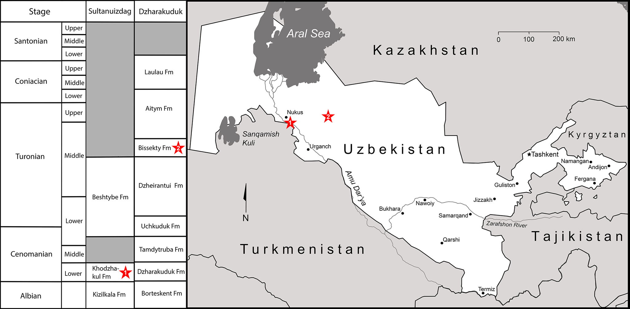
Carcharodontosauridae is a clade of allosauroid theropod dinosaurs that were dominant predators on most continents during the Early Cretaceous and attained their maximum sizes in the mid-Cretaceous (Aptian–Cenomanian). It reached its peak diversity shortly before its extinction in the early Late Cretaceous (Brusatte and Sereno, Reference Brusatte, Benson and Hutt2008; Brusatte et al., Reference Brusatte, Benson, Chure, Xu, Sullivan and Hone2009; Tanaka et al., Reference Tanaka, Anvarov, Zelenitsky, Ahmedshaev and Kobayashi2021; Canale et al., Reference Canale, Apesteguía, Gallina, Mitchell, Smith, Cullen, Shinya, Haluza, Gianechini and Makovicky2022). In Africa and South America, the stratigraphically youngest carcharodontosaurids are known from the Albian–Cenomanian interval, and a late-surviving carcharodontosaurid was previously reported from the Turonian of China (Brusatte et al., Reference Brusatte, Benson, Chure, Xu, Sullivan and Hone2009). Recently, Tanaka et al. (Reference Tanaka, Anvarov, Zelenitsky, Ahmedshaev and Kobayashi2021) described a new carcharodontosaurid from the Turonian Bissekty Formation of Uzbekistan, Ulughbegsaurus uzbekistanensis, which they considered the apex predator in its ecosystem. This new taxon was based on a fragment of a left maxilla (UzSGM 11-01-02), which was found at the Dzharakuduk locality, contrary to the information presented in Tanaka et al. (Reference Tanaka, Anvarov, Zelenitsky, Ahmedshaev and Kobayashi2021), by a member of the Uzbekistan/Russian/British/American/Canadian joint paleontological expeditions to the Kyzylkum Desert (URBAC) expeditions, the late Yuri A. Chikin (1951–2014), in 2006 and was originally housed at the Zoological Institute of the National Academy of Sciences of Uzbekistan. Aside from the holotype UzSGM 11-01-02, Tanaka et al. (Reference Tanaka, Anvarov, Zelenitsky, Ahmedshaev and Kobayashi2021) referred to U. uzbekistanensis two maxillary fragments from Dzharakuduk, CCMGE 600/12457 and ZIN PH 357/16, which had previously been referred to the dromaeosaurid Itemirus medullaris (Sues and Averianov, Reference Sues and Averianov2014). Sues et al. (Reference Sues, Averianov and Britt2023) questioned their attribution of Ulughbegsaurus uzbekistanensis and noted that the holotype lacks unambiguously diagnostic features of Carcharodontosauridae. These authors concluded that its purported carcharodontosaurid characters are either taphonomic artifacts or characters also shared by dromaeosaurids. However, an additional specimen, a fragment of a right maxilla (ZIN PH 2368/16) from the stratigraphically older Cenomanian Khodzhakul Formation, which outcrops in the southwest Kyzylkum Desert, Uzbekistan (Fig. 1), makes it now possible to reassess the phylogenetic position of Ulughbegsaurus and confirm its original assignment to Carcharodontosauridae.
Stratigraphic successions of Cretaceous deposits at the Sultanuizdag and Dzharakuduk sections in the southwest and central Kyzylkum Desert, Uzbekistan, respectively (left) and a map of Central Asia showing the positions of these sections (right; asterisks 1 and 2, respectively).

The Bissekty Formation at the Dzharakuduk locality complex produces the best-sampled and most diverse dinosaur assemblage currently known from the Cretaceous continental strata in the Kyzylkum Desert, Uzbekistan. It is intercalated between the marine or near-shore marine Dzheirantui and Aitym formations, which have yielded many specimens of chronostratigraphically useful invertebrates such as ammonoids, allowing precise dating of the Bissekty Formation as middle Turonian (Pyatkov et al., Reference Pyatkov, Pyanovskaya, Bukharin and Bykovskii1967; Feldmann et al., Reference Feldmann, Schweitzer, Redman, Morris and Ward2007; Redman and Leighton, Reference Redman and Leighton2009; Sues et al., Reference Sues, Averianov and Britt2023; Ward et al., Reference Ward, King, Morris and Kennedy2023). The assemblage of non-avian dinosaurs from the Bissekty Formation includes the rebbachisaurid Dzharatitanis kingi Averianov and Sues, Reference Averianov and Sues2021, titanosaurs (Sues et al., Reference Sues, Averianov, Ridgely and Witmer2015; Averianov and Sues, Reference Averianov and Sues2017a, Reference Averianov and Suesb), the carcharodontosaurid Ulughbegsaurus uzbekistanensis Tanaka et al., Reference Tanaka, Anvarov, Zelenitsky, Ahmedshaev and Kobayashi2021, the tyrannosauroid Timurlengia euotica Brusatte et al., Reference Brusatte, Averianov, Sues, Muir and Butler2016 (Averianov and Sues, Reference Averianov and Sues2012), the alvarezsaurid Dzharaonyx eski Averianov and Sues, Reference Averianov and Sues2022 (Averianov and Sues, Reference Averianov and Sues2017c), a new ornithomimid taxon (Sues and Averianov, Reference Sues and Averianov2016a), therizinosauroids (Sues and Averianov, Reference Sues and Averianov2016b), the caenagnathid Caenagnathasia martinsoni Currie et al., Reference Currie, Godfrey and Nessov1994 (Sues and Averianov, Reference Sues and Averianov2015), the troodontid Urbacodon sp. (Averianov and Sues, Reference Averianov and Sues2007; Averianov and Sues, Reference Averianov and Sues2016), the dromaeosaurid Itemirus medullaris Kurzanov, Reference Kurzanov1976 (Sues and Averianov, Reference Sues and Averianov2014), the enigmatic theropod tooth taxa Paronychodon asiaticus Nesov, Reference Nesov1995 and Richardoestesia asiatica Nesov, Reference Nesov1995 (Sues and Averianov, Reference Sues and Averianov2013; Averianov and Sues, Reference Averianov and Sues2019), the hadrosauroid Levnesovia transoxiana Sues and Averianov, Reference Sues and Averianov2009, the ceratopsoid Turanoceratops tardabilis Nesov et al., Reference Nesov, Kaznyshkina, Cherepanov, Bogdanova and Khozatsky1989 (Sues and Averianov, Reference Sues and Averianov2009), and the ankylosaurid Bissektipelta archibaldi (Averianov, Reference Averianov2002) (Kuzmin et al., Reference Kuzmin, Petrov, Averianov, Boitsova, Skutschas and Sues2020).
Materials and methods
The fragment of a right maxilla reported here, ZIN PH 2368/16, was found by L.A. Nesov during his first expedition to the southwest Kyzylkum Desert in 1974. It was one of the first of the many dinosaur bones he collected from the Upper Cretaceous deposits in the Kyzylkum Desert. Nesov mentioned this specimen in his monograph on Eurasian dinosaurs (Nesov, Reference Nesov1995, p. 98), observing that it was part of a large jaw with labiolingually moderately compressed alveoli and vertically oriented swellings along the dorsal margin of the bone containing the roots of the teeth (= tooth-root bulges in the present paper). He noted the presence of similar swellings along the medial surface of the dentary in Ceratosaurus nasicornis Marsh, Reference Marsh1884 (Gilmore, Reference Gilmore1920, pl. 26, fig. 2). For 50 years, this specimen has remained undescribed.
The Khodzhakul Formation has produced a diverse assemblage of aquatic and terrestrial vertebrates, including holocephalans, sharks, ichthyodectiform teleosts, albanerpetids, salamanders, frogs, turtles, lizards, crocodyliforms, pterosaurs, various non-avian dinosaurs, birds, and eutherian mammals. Previously known non-avian dinosaurs from the Khodzhakul Formation include sauropods, tyrannosauroids, ornithomimids, therizinosauroids, the troodontid “Troodon” asiamericanus Nesov, Reference Nesov1995, the enigmatic theropod Richardoestesia asiatica, an ornithopod, the ceratopsian Asiaceratops salsopaludalis Nesov et al., Reference Nesov, Kaznyshkina, Cherepanov, Bogdanova and Khozatsky1989, and ankylosaurs (Nesov et al., Reference Nesov, Kaznyshkina, Cherepanov, Bogdanova and Khozatsky1989; Nesov, Reference Nesov1995; Averianov and Sues, Reference Averianov and Sues2007, Reference Averianov and Sues2012, Reference Averianov and Sues2017a, Reference Averianov and Sues2019; Sues and Averianov, Reference Sues and Averianov2016a, Reference Sues and Averianovb.).
The specimen ZIN PH 2368/16 was scanned at a medical facility in St. Petersburg, Russia, by X-ray CT using a Toshiba Aquilon 64 medical tomographer at 0.5 mm slice thickness, 120 kV, and 200 μA. The resulting stack compiled 947 images (512 × 512 × 947 resolution) in DICOM format. Data acquired from CT scanning were imported into the visualization software Amira 6.3.0 (FEIVSG Company) and manually segmented. The resulting three-dimensional (3D) models have the voxel size of 0.153 × 0.153 × 0.3. Measurements on the 3D models were performed using Amira 6.3.0.
Anatomical terminology for maxillary features follows that used by Hendrickx and Mateus (Reference Hendrickx and Mateus2014).
To characterize the histological structure and identify the ontogenetic stage of ZIN PH 2368/16, we sampled the broken anterior and posterior ends of the maxilla (Fig. 2.3). The thin-sections were prepared according to the methodology outlined by Chinsamy and Raath (Reference Chinsamy and Raath1992). The sections then were examined under normal and polarized light using an optical microscope (Leica 4500, Leica Microsystems, Wetzlar, Germany) in the Saint Petersburg State University Research Centre for X-ray Diffraction Studies (Saint Petersburg, Russia). Histological terminology follows Francillon-Vieillot et al. (Reference Francillon-Vieillot, de Buffrénil, Castanet, Géraudie, Meunier, Sire, Zylberberg, de Ricqlès and Carter1990).
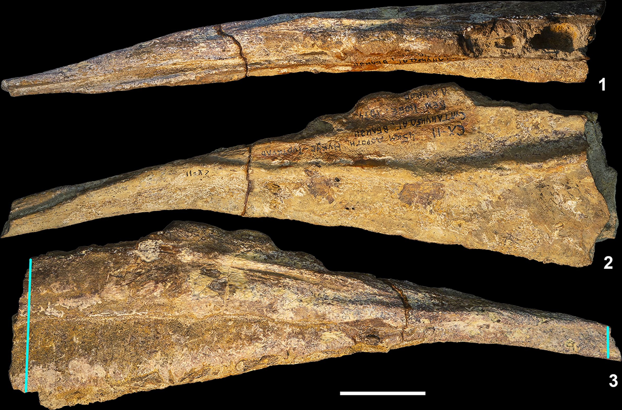
Ulughbegsaurus sp., ZIN PH 2368/16, right maxillary fragment, photographs in (1) dorsal, (2) lateral, and (3) medial views. Khodzhakul, Karakalpakistan, Uzbekistan; Khodzhakul Formation (Upper Cretaceous: Cenomanian). The blue vertical lines in (3) show the positions of the sections for histological analysis at the anterior and posterior ends of the bone, respectively. Scale bar = 3 cm.

Repositories and institutional abbreviations
Chernyshev’s Central Museum of Geological Exploration, Saint Petersburg, Russia (CCMGE); Dinosaur National Monument, Jensen, USA (DINO); Field Museum of Natural History, Chicago, USA (FMNH); State Geological Museum of the State Committee of the Republic of Uzbekistan on Geology and Mineral Resources, Tashkent, Uzbekistan (UzSGM); Paleoherpetological Collection, Zoological Institute, Russian Academy of Sciences, Saint Petersburg, Russia (ZIN PH).
Other abbreviation
URBAC, acronym for the Uzbekistan/Russian/British/American/Canadian joint paleontological expeditions to the Kyzylkum Desert, Uzbekistan (1997–2006).
Systematic paleontology
Dinosauria Owen, Reference Owen1842
Saurischia Seeley, Reference Seeley1887
Theropoda Marsh, Reference Marsh1881
Tetanurae Gauthier, Reference Gauthier1986
Allosauroidea Marsh, Reference Marsh1878
Carcharodontosauridae Stromer, Reference Stromer1931
Ulughbegsaurus Tanaka et al., Reference Tanaka, Anvarov, Zelenitsky, Ahmedshaev and Kobayashi2021
Type species
Ulughbegsaurus uzbekistanensis Tanaka et al., Reference Tanaka, Anvarov, Zelenitsky, Ahmedshaev and Kobayashi2021.
Ulughbegsaurus sp.
Occurrence
Khodzhakul (local site SKH-11), Karakalpakstan, Uzbekistan; lower part of Khodzhakul Formation, Upper Cretaceous (lower Cenomanian).
Description
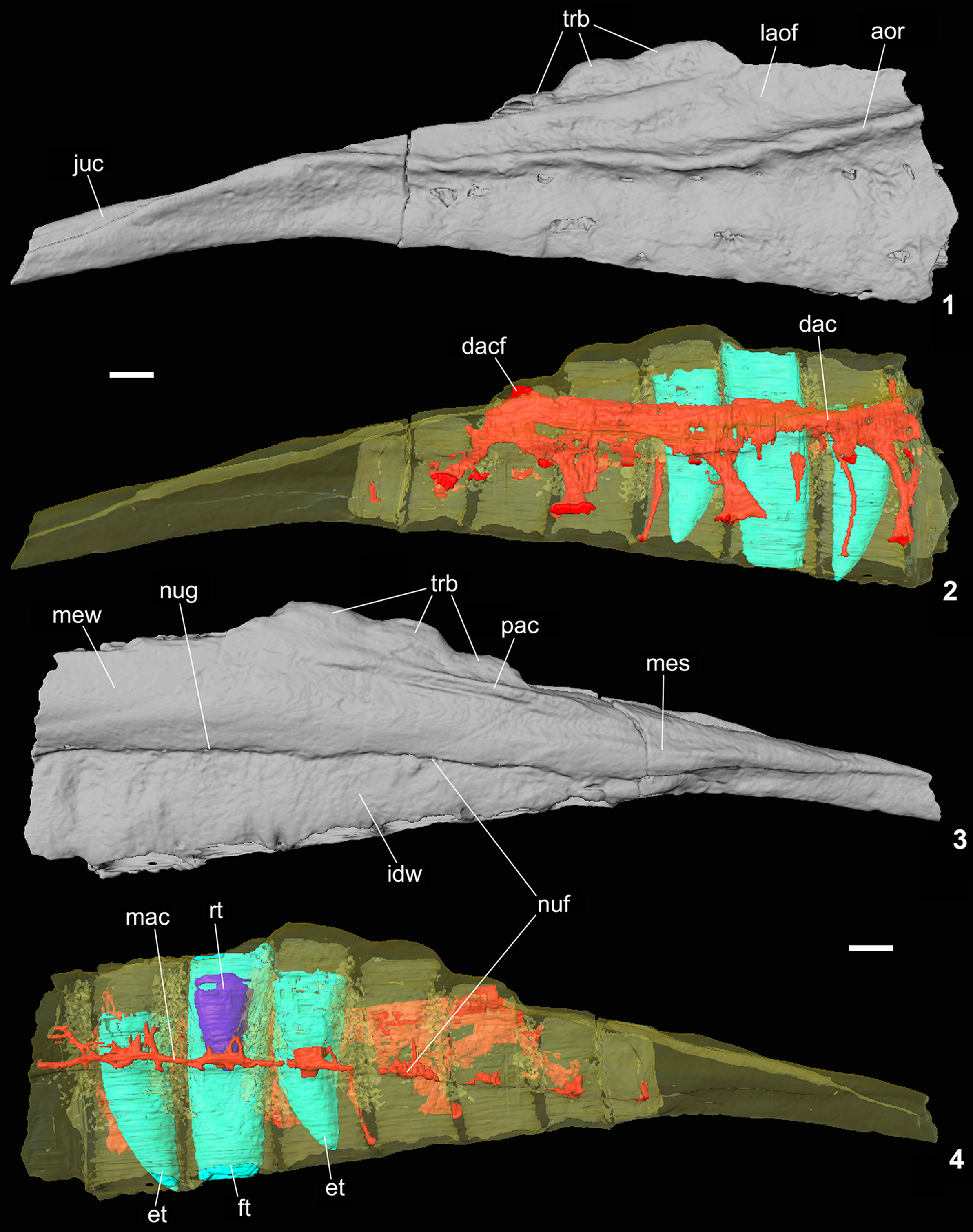
Specimen ZIN PH 2368/16 represents about one-half of a right maxilla and comprises the posterior part of the maxillary body (Figs. 2–4). The length of the fragment is 21.6 cm. The maxilla is anteroposteriorly elongated, with a dorsoventrally low jugal ramus. On the lateral side above the alveolar border, a high antorbital ridge borders the antorbital fossa ventrolaterally. The ridge is elevated above the adjacent dorsal and ventral portions of the lateral surface of the maxilla and extends slightly above the mid-height of that surface. The lateral surface of the antorbital fossa is slightly depressed relative to the lateral surface ventral to the lateral ridge. Ventral to the antorbital ridge, there are a few maxillary foramina. On the medial side, a pronounced ridge extending above the four posterior alveoli (excluding the posteriormost alveolus) contacted the palatine and borders the medial antorbital fossa ventrally. This ridge extends close to the dorsal margin of the maxilla. The medial wall of the bone is slightly convex anteriorly and forms a medial shelf posteriorly, which is separated from the ridge contacting the palatine. It is bordered ventrally by a prominent vascular groove. The interdental wall comprises almost completely fused interdental plates. Short vertical grooves at the alveolar border separate individual interdental plates. The interdental wall is less deep dorsoventrally than the alveolar margin on the lateral side, so that the alveoli are exposed in medial view (Fig. 3.3). The dorsal margin of the jugal ramus appears crenulated due to the bulges formed by protrusion of the tooth roots into the antorbital fenestra (Figs. 3, 4). These tooth-root bulges are preserved for three of the posterior teeth (excluding the ultimate and penultimate teeth), but those for more anterior teeth are broken. The alveolar margin of the maxilla is straight. There are eight preserved alveoli, which in occlusal view are rectangular, mediolaterally narrow, and slightly decreased in size posteriorly. The mesiodistal length of the anterior alveoli is more than twice their labiolingual width. The jugal ramus posterior to the last alveolus is deflected ventrolaterally. A deep groove for contact with the jugal extends along the dorsal side of this process. It is close to the lateral side and terminates anteriorly above the penultimate alveolus.
Ulughbegsaurus sp., ZIN PH 2368/16, right maxillary fragment, surface rendering based on X-ray computed tomography scans (1, 3) and segmented surface visualization of functional and erupting teeth (bluish-green), replacing tooth (purple), neurovascular system (red), and maxilla (translucent yellow) (2, 4) in lateral (1, 2) and medial (3, 4) views. Khodzhakul, Karakalpakistan, Uzbekistan; Khodzhakul Formation (Upper Cretaceous, Cenomanian). aor = antorbital ridge; dac = dorsal alveolar canal; dacf = entrance foramen for dorsal alveolar canal; et = erupting tooth; ft = functional tooth; idw = interdental wall; juc = jugal contact; laof = lateral antorbital fossa; mac = medial alveolar canal; mes = medial shelf; mew = medial wall; nuf = nutrient foramen; nug = nutrient groove; pac = palatine contact; rt = replacing tooth; trb = tooth-root bulges. Scale bars = 1 cm.

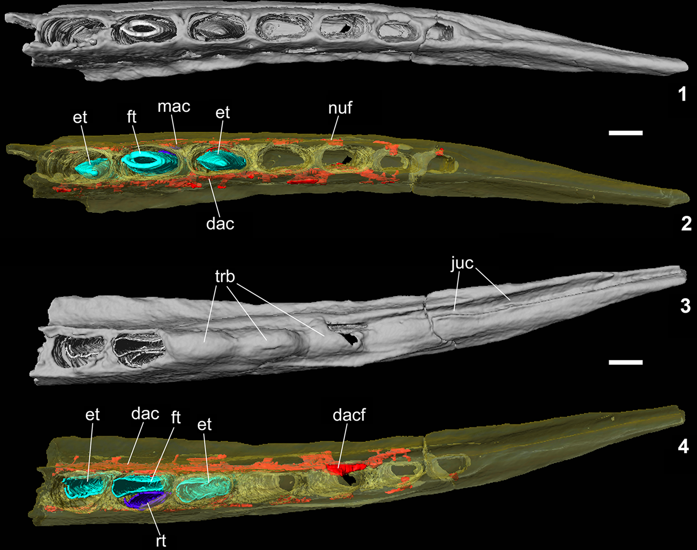
Ulughbegsaurus sp., ZIN PH 2368/16, right maxillary fragment, surface rendering based on X-ray computed tomography scans (1, 3) and segmented surface visualization of functional and erupting teeth (bluish-green), replacing tooth (purple), neurovascular system (red), and maxilla (translucent yellow) (2, 4), in ventral (1, 2) and dorsal (3, 4) views. Khodzhakul, Karakalpakistan, Uzbekistan; Khodzhakul Formation (Upper Cretaceous, Cenomanian). dac = dorsal alveolar canal; dacf = entrance foramen for dorsal alveolar canal; et = erupting tooth; ft = functional tooth; juc = jugal contact; mac = medial alveolar canal; nuf = nutrient foramen; rt = replacing tooth; trb = tooth-root bulges. Scale bars = 1 cm.

Three teeth are preserved in the second and third alveoli counting from the preserved anterior end (Figs. 3, 4). The first and the third teeth are unerupted, and their roots are incompletely formed. The second tooth was fully erupted and has a complete root; its crown was broken off above the alveolar margin. In this position, there is a replacing tooth directly medial to the functional tooth (Figs. 3.4, 4.4). This pattern of teeth eruption and replacement corresponds to the alternating tooth replacement typical for theropods (Edmund, Reference Edmund1960). Unfortunately, the resolution of the scans was not high enough to show details of the tooth crowns.
Internally, ZIN PH 2368/16 preserves the posterior portion of the complex neurovascular system. The dorsal alveolar (or maxillary) canal enters the maxilla posteriorly by a foramen at the anterior end of the groove for the reception of the jugal (Figs. 3.2, 4.4). This opening is located at the level of the third alveolus counting from the posterior end. The dorsal alveolar canal is straight and extends anteriorly for the remainder of the maxilla. It extends lateral to the alveoli, just above the antorbital ridge. Medially, the dorsal alveolar canal is adjacent to the lateral walls of the maxillary alveoli. Each alveolus anterior to the third one (counting from the posterior end) has a foramen opening into the lateral wall of the dorsal alveolar canal. The penultimate alveolus is connected via a posterolaterally directed branch of the dorsal alveolar canal; the posteriormost alveolus does not directly communicate with the canal. Several branches extend ventrolaterally from the dorsal alveolar canal and open in neurovascular foramina on the lateral surface of the maxilla. There are up to six longer branches, which exited through external foramina just dorsal to the alveolar margin (maxillary alveolar foramina of Hendrickx and Mateus, Reference Hendrickx and Mateus2014), and six shorter branches, which exit the maxilla just ventral to the antorbital ridge (maxillary circumfenestral foramina of Hendrickx and Mateus, Reference Hendrickx and Mateus2014). The dorsal alveolar canal probably contained the maxillary branch of the trigeminal nerve and the maxillary artery and vein (Barker et al., Reference Barker, Naish, Newham, Katsamenis and Dyke2017; Bouabdellah et al., Reference Bouabdellah, Lessner and Benoit2022), which supplied the alveoli medially and the external soft tissues of the snout laterally.
A second neurovascular canal extends medial to the alveoli. To the best of our knowledge, such a feature has not been previously described in theropods, and we refer to it here as the medial alveolar canal. It is located just dorsal to the vascular groove and extends parallel to it. This groove is generally present on the medial surface of theropod maxillae at the contact of the maxillary body with the interdental plates (Currie, Reference Currie1995; Hendrickx and Mateus, Reference Hendrickx and Mateus2014). Numerous neurovascular foramina pierce the wall of the groove. The groove and foramina are osteological correlates for the palatine artery and vein (Porter and Witmer, Reference Porter and Witmer2020). We interpret the medial alveolar canal as mainly containing tributaries of the palatine vessels but also potentially connected with the dental lamina within the maxilla (see Discussion). In ZIN 2368/16, each preserved alveolus is connected to this canal by a single foramen. The neurovascular branches supplied the four posterior alveoli and are not connected to each other within the maxilla; however, they are connected more anteriorly where they form the medial alveolar canal. Consequently, in ZIN PH 2368/16, each alveolus, starting from the penultimate preserved one, received branches of the trigeminal nerve and maxillary vessels from the lateral side and branches of the palatine vessels from the medial side.
Material
ZIN PH 2368/16, posterior fragment of a right maxilla.
Histology
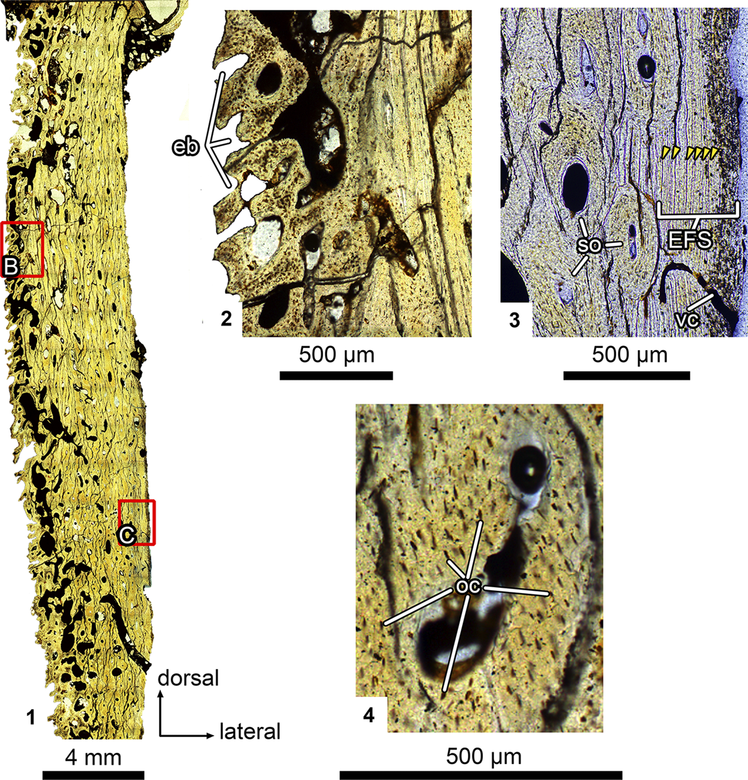
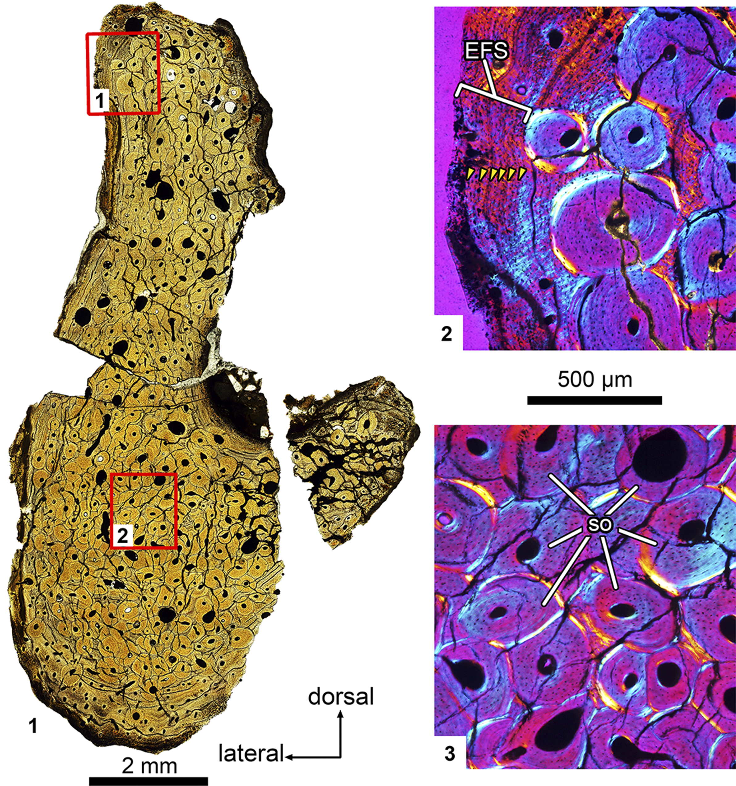
The maxilla ZIN PH 2368/16 was almost completely remodeled by secondary osteons (with Haversian bone formation). The only remaining primary bone is restricted to a thin band along the periphery of the bone (Figs. 5.1, 5.3, 6.1, 6.3). It comprises parallel-fibered bone with rare longitudinally oriented primary vascular canals and contains a sequence of multiple closely spaced lines of arrested growth (LAGs). These closely spaced LAGs constitute an external fundamental system (EFS) (Figs. 5.3, 6.2). Haversian bone was formed by two or three generations of densely packed, unevenly arranged secondary osteons (Fig. 6.1, 6.3).
Ulughbegsaurus sp., ZIN PH 2368/16, right maxillary fragment, histological sections under normal light. (1) Microanatomical overview. Note the layer of cancellous bone along the alveolar border. (2) Close-up of the cancellous bone along the alveolar border (rectangle B on (1)) showing the erosion bays and thick trabeculae composed of primary bone. (3) Close-up of the periphery of the bone (rectangle C on (1)). Note the presence of primary bone with closely spaced LAGs (yellow arrows) forming the external fundamental system. (4) Close-up of a secondary osteon showing the numerous osteocytic lacunae. eb = erosion bay; EFS = external fundamental system; oc = osteocyte lacuna; so = secondary osteons; vc = primary vascular canal.

Ulughbegsaurus sp., ZIN PH 2368/16, right maxillary fragment, histological sections viewed under normal light (1) and under polarized light with lambda waveplate (2, 3). (1) Microanatomical overview. (2) Close-up of the periphery of the bone (rectangle 1 on (1)). Note the presence of primary bone with closely spaced LAGs (yellow arrows) forming external fundamental system. (3) Close-up of the central part of the bone (rectangle 2 on (1)). Note the dense Haversian bone with two generations of secondary osteons. EFS = external fundamental system; so = secondary osteons.

The inner part of the bone at the alveolar border has undergone intensive remodeling, presumably due to active tooth development, and thick trabeculae and large erosion bays formed a layer of a cancellous bone along the alveolar border (Fig. 5.1, 5.2). This cancellous layer consists of primary bone without secondary lamellar bone lining. The numerous osteocytic lacunae vary in outline from rounded to ellipsoid (Fig. 5.4).
The presence of primary, poorly vascularized, and parallel-fibered bone containing the EFS at the periphery indicates a decrease in growth rate and attainment of skeletal maturity (Woodward et al., Reference Woodward, Horner and Farlow2011; Rainwater et al., Reference Rainwater, Woodward, Woodward and Wilkinson2022). The active secondary remodeling leading to formation of the widespread dense Haversian bone also suggests an adult growth stage (Horner et al., Reference Horner, de Ricqlès and Padian2000; Klein and Sander, Reference Klein and Sander2008; Scannella and Horner, Reference Scannella and Horner2010; Werning, Reference Werning2012; Padian et al., Reference Padian, Werning and Horner2016). As a result, we interpret the maxilla ZIN PH 2368/16 as representing a mature adult.
Remarks
Specimen ZIN PH 2368/16 can be referred to Ulughbegsaurus on the basis of shared derived characters such as the raised and “tuberculated” antorbital rim and the prominent tooth-root bulges. It is identified as Ulughbegsaurus sp. because it was recovered from a different, slightly older geological formation than the holotype of Ulughbegsaurus uzbekistanensis and represents a different portion of the maxilla.
Identification of ZIN PH 2368/16 from the Khodzhakul Formation as Ulughbegsaurus sp. extends the known temporal range of Carcharodontosauridae in West Asia to the early Late Cretaceous (early Cenomanian). The Khodzhakul Formation is now the third reported location worldwide where carcharodontosaurids and tyrannosauroids co-occur. The two other occurrences are in the Cenomanian Mussentuchit Member of the Cedar Mountain Formation of Utah, USA (Zanno and Makovicky, Reference Zanno and Makovicky2013; Zanno et al., Reference Zanno, Tucker, Canoville, Avrahami, Gates and Makovicky2019) and the Turonian Bissekty Formation of Uzbekistan (Averianov and Sues, Reference Averianov and Sues2012; Brusatte et al., Reference Brusatte, Averianov, Sues, Muir and Butler2016; Tanaka et al., Reference Tanaka, Anvarov, Zelenitsky, Ahmedshaev and Kobayashi2021). Furthermore, this identification further supports the hypothesis by Tanaka et al. (Reference Tanaka, Anvarov, Zelenitsky, Ahmedshaev and Kobayashi2021) that carcharodontosaurids occupied the apex predator role during the early Late Cretaceous of Uzbekistan.
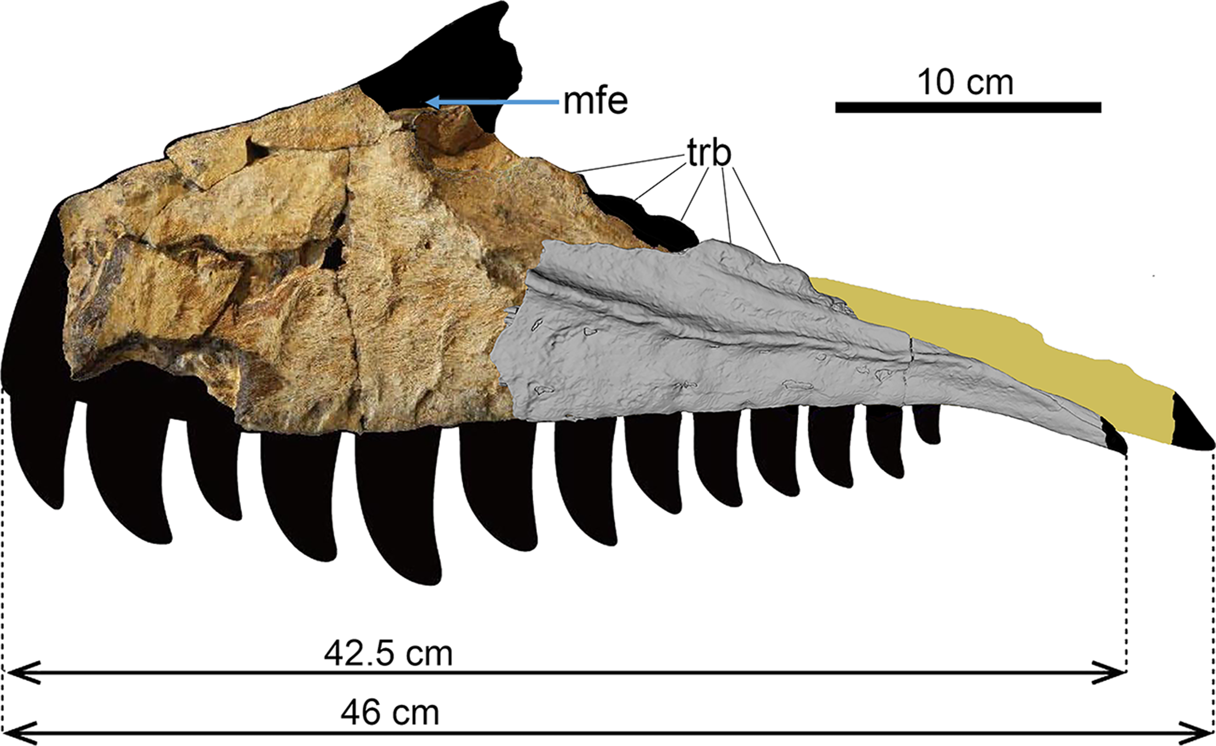
Tanaka et al. (Reference Tanaka, Anvarov, Zelenitsky, Ahmedshaev and Kobayashi2021) identified a maxillary fenestra and a promaxillary fossa on UzSGM 11-01-02, the holotype of Ulughbegsaurus uzbekistanensis. We offer alternative interpretations of these structures. What these authors considered the interfenestral strut, the bony wall separating the maxillary fenestra from the antorbital fenestra, is a tooth-root bulge extending into the antorbital fenestra. Such tooth-root bulges are also well preserved in ZIN PH 2368/16 (Figs. 3, 4). The “promaxillary fossa” of Tanaka et al. (Reference Tanaka, Anvarov, Zelenitsky, Ahmedshaev and Kobayashi2021) is not associated with any foramen that can be interpreted as a promaxillary fenestra. Apparently, the maxillary fenestra, which is not preserved in UzSGM 11-01-02, was small, confined to the anterior margin of the antorbital fossa, and confluent with the promaxillary fenestra, as in other carcharodontosaurids (Brusatte and Sereno, Reference Brusatte and Sereno2007; Hendrickx and Mateus, Reference Hendrickx and Mateus2014). This distinctive morphology is consistent with attribution of Ulughbegsaurus to Carcharodontosauridae. A possible position for the maxillary fenestra in UzSGM 11-01-02 is shown in our new reconstruction (Fig. 7).
Attempted reconstruction of the maxilla of Ulughbegsaurus in lateral view, combining the left maxillary fragment UzSGM 11-01-02 (holotype of Ulughbegsaurus uzbekistanensis modified from Tanaka et al., Reference Tanaka, Anvarov, Zelenitsky, Ahmedshaev and Kobayashi2021, fig. S7) and the right maxillary fragment (reversed) ZIN PH 2368/16 of Ulughbegsaurus sp. (surface rendering based on X-ray computed tomography scans in gray). Note that this composite was created solely to provide an idea of the overall proportions and shape of the maxilla. The original reconstruction of the jugal ramus by Tanaka et al. (Reference Tanaka, Anvarov, Zelenitsky, Ahmedshaev and Kobayashi2021) is highlighted in yellow. The ascending ramus is reconstructed on the basis of that of Carcharodontosaurus saharicus (Brusatte and Sereno, Reference Brusatte and Sereno2007, fig. 2C). mfe = maxillary fenestra; trb = tooth-root bulges.

Tanaka et al. (Reference Tanaka, Anvarov, Zelenitsky, Ahmedshaev and Kobayashi2021, fig. S7) reconstructed the length of the maxilla of U. uzbekistanensis as 46 cm, combining the holotype maxilla UzSGM 11-01-02 and the posterior jugal ramus fragment CCMGE 600/12457 from Dzharakuduk. However, there is no overlap between these two specimens, casting doubt on this estimate (Sues et al., Reference Sues, Averianov and Britt2023). However, there is now considerable overlap between the maxillary fragments UzSGM 11-01-02 and ZIN PH 2368/16, allowing for a more reliable reconstruction of the maxilla of Ulughbegsaurus (Fig. 7). Assuming closely similar proportions, the estimated length of maxilla in U. uzbekistanensis would be 42.5 cm. Using this new estimate of the maxillary length and the equation provided by Tanaka et al. (Reference Tanaka, Anvarov, Zelenitsky, Ahmedshaev and Kobayashi2021), we obtained a slightly shorter total body length, 7.4 m (7.5–8.0 m in Tanaka et al., Reference Tanaka, Anvarov, Zelenitsky, Ahmedshaev and Kobayashi2021). This new estimate does not challenge the assumption that the carcharodontosaurid Ulughbegsaurus was the largest dinosaurian predator in the early Late Cretaceous terrestrial ecosystems of West Asia.
Discussion
Antorbital fossa
The extent of the antorbital fossa on the lateral side of the maxilla varies among carcharodontosaurids. In Ulughbegsaurus, the antorbital fossa extends for about half the height of the jugal ramus (Figs. 3.1, 7). A similar condition is present in Acrocanthosaurus atokensis Stovall and Langston, Reference Stovall and Langston1950, Concavenator corcovatus Ortega et al., Reference Ortega, Escaso and Sanz2010, Mapusaurus roseae Coria and Currie, Reference Coria and Currie2006, and Meraxes gigas Canale et al., Reference Canale, Apesteguía, Gallina, Mitchell, Smith, Cullen, Shinya, Haluza, Gianechini and Makovicky2022 (Currie and Carpenter, Reference Currie and Carpenter2000; Eddy and Clarke, Reference Eddy and Clarke2011; Cuesta et al., Reference Cuesta, Vidal, Ortega and Sanz2018). In Eocarcharia, the antorbital fossa occupies a relatively larger region on the anterior body of the maxilla, but it also takes up about half of the dorsoventral height on the jugal ramus (Sereno and Brusatte, Reference Sereno and Brusatte2008). The antorbital fossa is greatly reduced in extent to nearly absent in Shaochilong maortuensis (Hu, Reference Hu1964), Carcharodontosaurus spp., and Gigantosaurus carolinii Coria and Salgado, Reference Coria and Salgado1995 (Sereno et al., Reference Sereno, Dutheil, Iarochene, Larsson, Lyon, Magwene, Sidor, Varricchio and Wilson1996; Brusatte and Sereno, Reference Brusatte and Sereno2007; Brusatte et al., Reference Brusatte, Benson, Chure, Xu, Sullivan and Hone2009, Reference Brusatte, Chure, Benson and Xu2010). The condition in Ulughbegsaurus, with the antorbital fossa occupying about half of the jugal ramus height, is likely plesiomorphic for Carcharodontosauridae because it is also present in the neovenatorid carcharodontosaurian Neovenator salerii Hutt et al., Reference Hutt, Martill and Barker1996 (Brusatte et al., Reference Brusatte, Benson and Hutt2008; Barker et al., Reference Barker, Naish, Newham, Katsamenis and Dyke2017).
Antorbital rim
The ridged antorbital rim bordering the antorbital fossa anteriorly and ventrally is the most conspicuous diagnostic feature of Ulughbegsaurus. In both UzSGM 11-01-02 and ZIN PH 2368/16, the ridge is elevated above the lateral antorbital fossa and the ventral portion of the lateral surface of the maxilla. In the majority of theropods, this junction is either smooth or step-like, with a “lip” (ridge) formed by the flat lateral surface and the depressed surface of the antorbital fossa (e.g., Powers et al., Reference Powers, Fabbri, Doschak, Bhullar, Evans, Norell and Currie2022). A similarly elevated antorbital ridge is present in Carcharodontosaurus saharicus (Stromer, Reference Stromer1931, pl. 1, fig. 6b; Brusatte and Sereno, Reference Brusatte and Sereno2007, fig. 2C) and certain other theropods (Rauhut, Reference Rauhut2003, character 15). This resemblance to C. saharicus further supports referral of Ulughbegsaurus to Carcharodontosauridae. Among other carcharodontosaurids, the antorbital ridge is strongly developed in Acrocanthosaurus, Concavenator, and Eocarcharia dinops Sereno and Brusatte, Reference Sereno and Brusatte2008 (Currie and Carpenter, Reference Currie and Carpenter2000; Eddy and Clarke, Reference Eddy and Clarke2011; Cuesta et al., Reference Cuesta, Vidal, Ortega and Sanz2018). Shaochilong and Carcharodontosaurus iguidensis Brusatte and Sereno, Reference Brusatte and Sereno2007 both have a reduced antorbital fossa and lack an antorbital rim (Brusatte et al., Reference Brusatte, Benson, Chure, Xu, Sullivan and Hone2009, Reference Brusatte, Chure, Benson and Xu2010).
The surface of the antorbital ridge in ZIN PH 2368/16 is uneven, with bumps that likely correspond to the tubercles along the antorbital ridge described for the holotype of Ulughbegsaurus uzbekistanensis (Tanaka et al., Reference Tanaka, Anvarov, Zelenitsky, Ahmedshaev and Kobayashi2021).
Interdental plates
The fusion of the interdental plates into an interdental wall is present in both the holotype of Ulughbegsaurus uzbekistanensis and ZIN PH 2368/16. An interdental wall composed of fused interdental plates is present in a variety of theropods, including abelisaurids, the megalosauroid Torvosaurus gurneyi Hendrickx and Mateus, Reference Hendrickx and Mateus2014, dromaeosaurids, and troodontids (Currie, Reference Currie1995; Coria and Currie, Reference Coria and Currie2006; Brusatte et al., Reference Brusatte, Benson and Xu2012; Sues et al., Reference Sues, Averianov and Britt2023). However, Brusatte and Sereno (Reference Brusatte and Sereno2008) considered fully co-ossified posterior interdental plates a synapomorphy for Carcharodontosauridae.
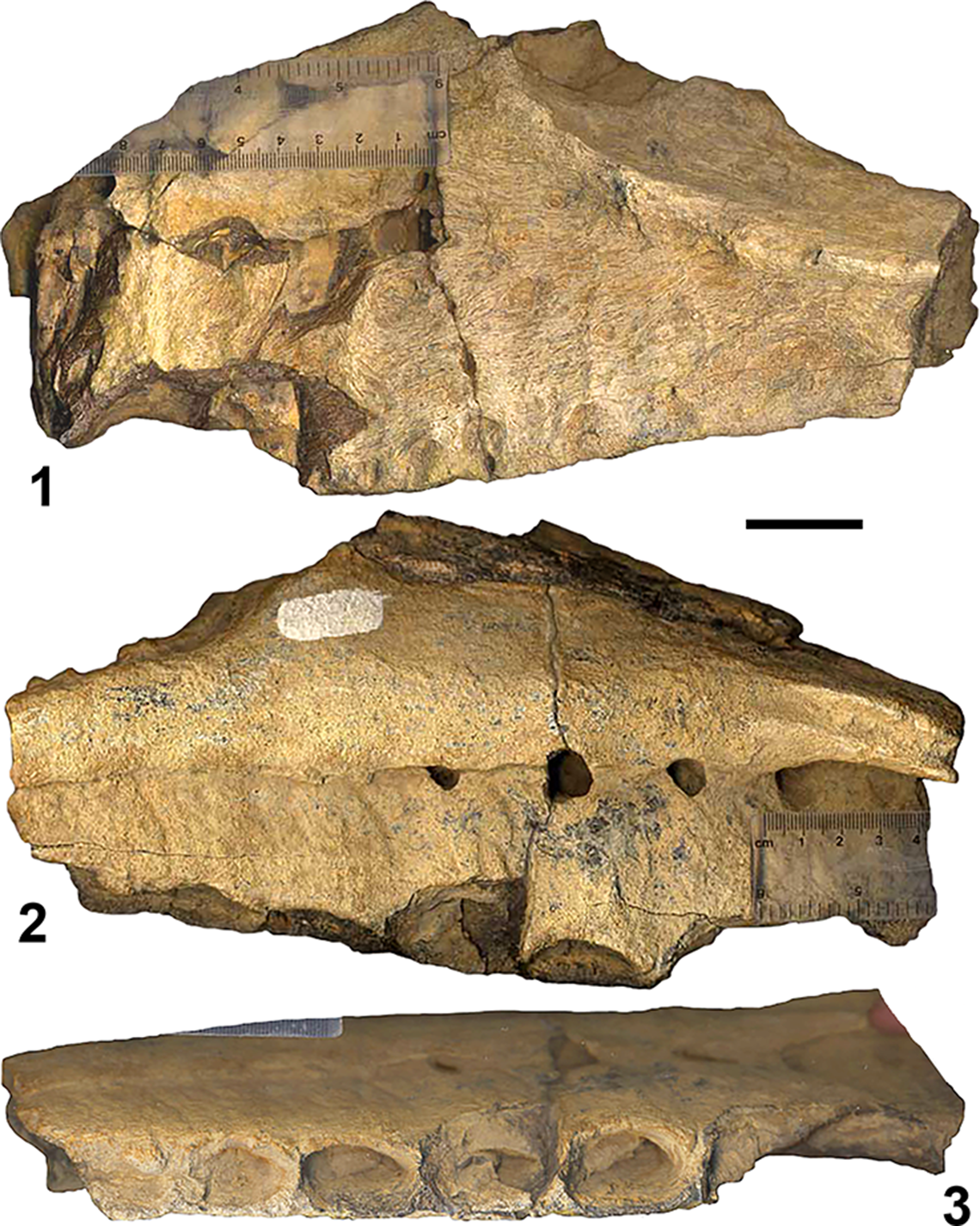
Tanaka et al. (Reference Tanaka, Anvarov, Zelenitsky, Ahmedshaev and Kobayashi2021) described the surface of the internal plates in UzSGM 11-01-02 as slightly rugose. However, as is evident on photographs of this specimen provided by the late Yu.I. Chikin to the first author in 2009 (Fig. 8.2), the interdental plates are covered with shallow, wide, sometimes anastomizing, and vertical grooves. A similar pattern of grooves, although less pronounced, is also present in ZIN PH 2368/16 (Fig. 3.3). These grooves extend ventral to the nutrient groove but not dorsal to it. They are likely impressions of the tributaries of the palatine vessels that branched off ventrally to supply soft tissues along the medial surface of the maxilla. Deep, dorsoventrally oriented grooves located dorsally on the maxillary interdental plates were considered an autapomorphy of Shaochilong (Brusatte et al., Reference Brusatte, Benson, Chure, Xu, Sullivan and Hone2009, Reference Brusatte, Chure, Benson and Xu2010). These grooves closely resemble those of Ulughbegsaurus.
Ulughbegsaurus uzbekistanensis, UzSGM 11-01-02, holotype, left maxillary fragment, photographs in (1) lateral, (2) medial, and (3) ventral views. Dzharakuduk, Uzbekistan; Bissekty Formation (Upper Cretaceous, Turonian). Photos courtesy of the late Yu. A. Chikin. Scale bar = 3 cm.

Interdental wall
The interdental wall is very high dorsoventrally in Ulughbegsaurus (Tanaka et al., Reference Tanaka, Anvarov, Zelenitsky, Ahmedshaev and Kobayashi2021, fig. 1b; Fig. 8.2). In ZIN PH 2368/16, representing the posterior region of the maxilla, it undoubtedly further increased in height more anteriorly. High anterior interdental plates, with a dorsoventral height more than twice the anteroposterior width, are a synapomorphy for Carcharodontosauridae (Brusatte and Sereno, Reference Brusatte and Sereno2008; Brusatte et al., Reference Brusatte, Benson, Chure, Xu, Sullivan and Hone2009, Reference Brusatte, Chure, Benson and Xu2010, Reference Brusatte, Benson and Xu2012). Dorsoventrally lower interdental plates are present in the non-carcharodontosaurid carcharodontosaurian Neovenator (Brusatte et al., Reference Brusatte, Benson and Hutt2008).
In Ulughbegsaurus (Fig. 3.3), Shaochilong (Brusatte et al., Reference Brusatte, Benson, Chure, Xu, Sullivan and Hone2009, Reference Brusatte, Chure, Benson and Xu2010), Eocarcharia (Sereno and Brusatte, Reference Sereno and Brusatte2008), Carcharodontosaurus saharicus (Brusatte and Sereno, Reference Brusatte and Sereno2007), and Mapusaurus (Coria and Currie, Reference Coria and Currie2006), the interdental wall extends not as far ventrally as the lateral wall of the maxilla, and thus the alveoli are exposed in medial view. This condition is also present in the megalosauroid Torvosaurus and the carcharodontosaurian Neovenator (Brusatte et al., Reference Brusatte, Benson and Hutt2008; Hendrickx and Mateus, Reference Hendrickx and Mateus2014). In Carcharodontosaurus iguidensis, the medial exposure of the alveoli is greater than in other carcharodontosaurids (Brusatte and Sereno, Reference Brusatte and Sereno2007).
Jugal process
The long jugal process of the maxilla of Ulughbegsaurus is deflected ventrolaterally just posterior to the tooth row (Figs. 3, 7). A similar condition is present in the carcharodontosaurids Acrocanthosaurus, Eocarcharia, Concavenator, and Shaochilong as well as in a few early-diverging tetanurans such as Afrovenator (Currie and Carpenter, Reference Currie and Carpenter2000; Sereno and Brusatte, Reference Sereno and Brusatte2008; Brusatte et al., Reference Brusatte, Chure, Benson and Xu2010; Eddy and Clarke, Reference Eddy and Clarke2011; Cuesta et al., Reference Cuesta, Vidal, Ortega and Sanz2018). A long, ventrally deflected jugal process is absent in other allosauroids, including the carcharodontosaurian Neovenator, and in most early tetanurans (Brusatte et al., Reference Brusatte, Benson and Hutt2008, Reference Brusatte, Chure, Benson and Xu2010; Eddy and Clarke, Reference Eddy and Clarke2011).
Tooth-root bulges
An unusual feature, present in both ZIN PH 2368/16 and UzSGM 11-01-02, is the tooth-root bulges that extend above the dorsal margin of the maxillary body (= eminentia radicis dentis of Witmer (Reference Witmer1997, fig. 29C; Figs. 3, 4). In UzSGM 11-01-02, two bulges are preserved and were incorrectly identified as the strut separating the maxillary and antorbital fenestrae (Tanaka et al., Reference Tanaka, Anvarov, Zelenitsky, Ahmedshaev and Kobayashi2021; fig. 1a, e). The tooth-root bulges can be clearly recognized on the maxilla of the lost holotype of Carcharodontosaurus saharicus (Stromer, Reference Stromer1931, pl. 1, fig. 6a), but they are absent on the neotype maxilla of that species (Brusatte and Sereno, Reference Brusatte and Sereno2007, fig. 2). Among other theropods, tooth-root bulges are known on the maxillae of the ceratosaurid Ceratosaurus and the piatnitzkysaurid Marshosaurus bicentesimus Madsen, Reference Madsen1976 (Hendrickx and Mateus, Reference Hendrickx and Mateus2014, p. 5, fig. 3B1). As previously noted, Nesov (Reference Nesov1995) compared the tooth-root bulges of ZIN PH 2368/16 with those on the dentary of Ceratosaurus. Maxillary tooth-root bulges have not been reported for any dromaeosaurid (Powers et al., Reference Powers, Fabbri, Doschak, Bhullar, Evans, Norell and Currie2022).
Surface texture
As noted by Sues et al. (Reference Sues, Averianov and Britt2023), the rugose lateral surface in the holotype maxilla of U. uzbekistanensis is a preservational artifact due to erosion. In ZIN PH 2368/16, the lateral surface is intact and smooth. A series of oval depressions on the lateral surface along the alveolar border in UzSGM 11-01-02 is also likely an artifact of the badly eroded surface. There are no such depressions in ZIN PH 2368/16. Among known carcharodontosaurids, the lateral surface of the maxilla is smooth in Acrocanthosaurus, Eocarcharia, Concavenator, Kelmayisaurus petrolicus Dong, Reference Dong1973, and Shaochilong (Currie and Carpenter, Reference Currie and Carpenter2000; Sereno and Brusatte, Reference Sereno and Brusatte2008; Brusatte et al., Reference Brusatte, Benson, Chure, Xu, Sullivan and Hone2009, Reference Brusatte, Chure, Benson and Xu2010, Reference Brusatte, Benson and Xu2012; Benson et al., Reference Benson, Carrano and Brusatte2010; Cuesta et al., Reference Cuesta, Vidal, Ortega and Sanz2018). By contrast, the pronounced grooved sculpturing occupies nearly the entire lateral surface of the maxilla in Carcharodontosaurus (Sereno et al., Reference Sereno, Dutheil, Iarochene, Larsson, Lyon, Magwene, Sidor, Varricchio and Wilson1996; Brusatte and Sereno, Reference Brusatte and Sereno2007). The maxillary surface texture is composed of irregular rugosities that do not form distinct ridges or grooves as present in the carcharodontosaurids Mapusaurus and Giganotosaurus and in the carcharodontosaurian Neovenator (Brusatte et al., Reference Brusatte, Benson and Hutt2008, Reference Brusatte, Chure, Benson and Xu2010).
Neurovascular features and position of dental lamina
Specimen ZIN PH 2368/16 preserves the posterior portion of the complex neurovascular system documented for the maxillae of various other non-avian theropods (Barker et al., Reference Barker, Naish, Newham, Katsamenis and Dyke2017; Cau, Reference Cau2020; Porter and Witmer, Reference Porter and Witmer2020; Bouabdellah et al., Reference Bouabdellah, Lessner and Benoit2022). In ZIN PH 2368/16, the dorsal alveolar (maxillary) canal lies lateral to the alveoli and is connected to the external surface by a series of laterally directed branches, which is consistent with the anatomy in other theropods such as Majungasaurus, Neovenator, Spinosaurus and Tyrannosaurus (Barker et al., Reference Barker, Naish, Newham, Katsamenis and Dyke2017; Cau, Reference Cau2020; Porter and Witmer, Reference Porter and Witmer2020; Bouabdellah et al., Reference Bouabdellah, Lessner and Benoit2022). The entrance foramen of the dorsal alveolar canal is located at the level of the antepenultimate alveolus in ZIN PH 2368/16. A similarly located neurovascular foramen was described in Torvosaurus gurneyi (Hendrickx and Mateus, Reference Hendrickx and Mateus2014, fig. 6B).
An interesting novel feature of ZIN PH 2368/16 is the presence of the medial alveolar canal, which supplies the alveoli medially. This feature has not been previously reported in theropods such as Neovenator and Tyrannosaurus (Barker et al., Reference Barker, Naish, Newham, Katsamenis and Dyke2017; Bouabdellah et al., Reference Bouabdellah, Lessner and Benoit2022). Barker et al. (Reference Barker, Naish, Newham, Katsamenis and Dyke2017) emphasized the absence of neurovascular canals medial to the alveoli in Neovenator. However, alveoli of Neovenator (Barker et al., Reference Barker, Naish, Newham, Katsamenis and Dyke2017, fig. 3C) and some other sampled theropods (Allosaurus, Majungasaurus, Tyrannosaurus) receive short canals from the nutrient groove on the medial surface of the maxilla. The groove and canals are osteological correlates for the palatine artery and vein (Porter and Witmer, Reference Porter and Witmer2020). We interpret the medial alveolar canal of ZIN PH 2368/16 as carrying tributaries of the palatine vessels and perhaps homologous to the nutrient groove sensu Hendrickx and Mateus (Reference Hendrickx and Mateus2014). In Allosaurus (DINO 2560) and Tyrannosaurus (FMNH PR 2081), a separate medial alveolar canal is absent, and there are only short neurovascular canals between the nutrient groove and each alveolus (I.T.K., personal observation of CT data available at Morphosource.org, 2024). On several maxillae of Majungasaurus (FMNH PR 2100, 2278), the nutrient groove is deeply incised into the medial surface of the bone and almost forms a separate canal in some specimens (I.T.K., personal observation of CT data available at Morphosource.org, 2024). Short canals branch off from it within the maxilla of Majungasaurus and approach each alveolus, resembling the condition in ZIN PH 2368/16. Thus, the presence of vascular canals supplying the maxillary alveoli from the medial surface of the element appears to be a more broadly distributed feature among theropods.
The nutrient groove and canals between it and each alveolus were considered osteological correlates for the palatine artery and vein (Porter and Witmer, Reference Porter and Witmer2020). However, it was sometimes previously referred to as the groove for the dental lamina in theropods, and the canals entering the maxilla were thought of as for entrance of the developing tooth buds (e.g., Brusatte and Sereno, Reference Brusatte and Sereno2007). Moreover, an external position of the dental lamina, medial to the tooth-bearing bones, has been inferred for ornithischians (Chen et al., Reference Chen, LeBlanc, Jin, Huang and Reisz2018). We do not support this hypothesis on the basis of comparison with crocodylians and mammals that also have the thecodont tooth implantation. Adults of extant crocodylians possess the dental lamina—a band of an epithelial tissue considered to be the source of odontogenic stem cells for tooth renewal (Wu et al., Reference Wu, Wu, Jiang, Elsey and Temple2013). The dental lamina of crocodylians is adjacent to the lingual surface of the replacement teeth deep within each alveolus and is disconnected from the oral epithelium. It is expanded at each alveolus and is constrained and string-like between alveoli, continuing in a canal within tooth-bearing bones (Wu et al., Reference Wu, Wu, Jiang, Elsey and Temple2013). Remnants of the dental lamina of mammals are also located within the boundaries of alveoli and do not protrude externally (Wu et al., Reference Wu, Wu, Jiang, Elsey and Temple2013, fig. 8). Consequently, we consider the position of the dental lamina within the nutrient groove, at the medial surface of the maxilla, unlikely in theropods. The groove is better interpreted as the osteological correlate for the palatine vessels, following Porter and Witmer (Reference Porter and Witmer2020).
Alternatively, the structure of the medial alveolar canal of ZIN PH 2368/16 is more consistent with the position of the dental lamina as the canal extends within the bone and is expanded adjacent to the medial walls of alveoli. The medial alveolar canal is connected to the nutrient groove, and it likely carried tributaries of the palatine vessels to the developing teeth. But it may also correspond to the position of the dental lamina within the maxilla. Yet the presence of a separate medial alveolar canal parallel and connected to the nutrient groove appears unique to ZIN PH 2368/16 at present. We require more comparative data on the arrangement of the dental lamina in extant reptiles and internal features of tooth-bearing elements in dinosaurs to test this hypothesis.
Conclusion
The carcharodontosaurid theropod dinosaur Ulughbegsaurus uzbekistanensis was initially described on the basis of an incomplete large maxilla from the lower Upper Cretaceous (Turonian) Bissekty Formation of Uzbekistan (Tanaka et al., Reference Tanaka, Anvarov, Zelenitsky, Ahmedshaev and Kobayashi2021). Originally, Ulughbegsaurus was interpreted as a neovenatorid carcharodontosaurian on the basis of the rugose lateral surface of the maxilla, the position of the promaxillary fossa comparable to the condition in allosauroids, fused, slightly rugose interdental plates, and alveoli that are labiolingually narrower than mesiodistally long (Tanaka et al., Reference Tanaka, Anvarov, Zelenitsky, Ahmedshaev and Kobayashi2021). Sues et al. (Reference Sues, Averianov and Britt2023) questioned these characters. The rugose lateral surface of the maxilla was considered a taphonomical artifact due to erosion. The position of the promaxillary fossa is highly variable in theropods. The fused interdental plates are found in a variety of theropods, including dromaeosaurids. The labiolingually narrower and mesiodistally long dental alveoli are found in other theropods, including dromaeosaurids. According to Sues et al. (Reference Sues, Averianov and Britt2023), three of the purported carcharodontosaurian synapomorphies of Ulughbegsaurus are also shared by dromaeosaurids. Sues et al. (Reference Sues, Averianov and Britt2023) also noted that the sub-vertical contact between the premaxilla and maxilla and the smooth junction between the ventral margin of the antorbital fossa and the lateral surface of the maxilla ventral to this margin are present in both Ulughbegsaurus and some dromaeosaurids such as Utahraptor. In this report, we found that the structure of U. uzbekistanensis holotype maxilla, interpreted by Tanaka et al. (Reference Tanaka, Anvarov, Zelenitsky, Ahmedshaev and Kobayashi2021) as an interfenestral strut, the bony wall separating the maxillary fenestra from the antorbital fenestra, is a tooth-root bulge extending into the antorbital fenestra. These bulges are well preserved on the maxilla of Ulughbegsaurus sp. from the Khodzhakul Formation. Thus, the promaxillary fenestra is not preserved on the holotype of U. uzbekistanensis. The maxillary fenestra was likely small, confined to the anterior margin of the antorbital fossa, and confluent with the promaxillary fenestra, as in other carcharodontosaurids. The most important diagnostic character of both maxillae of Ulughbegsaurus is the ridged antorbital rim bordering the antorbital fossa anteriorly and ventrally. Such a ridge is present also in Carcharodontosaurus, which strongly supports attribution of Ulughbegsaurus to Carcharodontosauridae.
Acknowledgments
We thank S. Brusatte and R.A. Coria for reviewing the paper and for useful comments. We are indebted to the staff of the Resource Center “X-ray Diffraction Methods of Research” (Saint Petersburg State University, Saint Petersburg) for help with the use of the Leica 2500P polarization microscope, to K.M. Lyubarov (OOO “LIMS”, Saint Petersburg) for preparation of thin sections of ZIN PH 2368/16, to L.А. Tarasova and V.V. Kolchanov (Saint Petersburg State University, Saint Petersburg) for photographing the thin sections and preparing Figures 5 and 6, and R.A. Bapinaev for taking the photographs used in Figure 2. We thank the late Y.I. Chikin for providing us with photographs of UzSGM 11-01-02, which are reproduced here in Figure 8. The work of A.O.A. is supported by the Zoological Institute, Russian Academy of Sciences (project 122031100282-2).
Competing interests
The authors declare none.