Anomalous origin of the left coronary artery from the pulmonary artery is a rare CHD with very high morbidity and mortality during infancy, and 85% of the patients die if left surgically untreated.Reference KO, Chu, Young, Cheung and Lun 1 However, there is a small group of patients who will have late presentation beyond infancy, and even in adulthood, and may present with signs and symptoms of heart failure, exertional dyspnoea, angina, mitral regurgitation, arrhythmia, and sudden cardiac death.Reference Wesselhoeft, Fawcett and Johnson 2 Nowadays, establishment of a dual coronary system with reimplantation of the left coronary artery into the aorta is the preferred method for treatment of this anomaly whenever diagnosis is confirmed.Reference Lange, Vogt and Horer 3
After corrective surgery, usually the outcome is good and left ventricular function improvement is marked within the first postoperative years.Reference Lange, Vogt and Horer 3 , Reference Shavilkar, Borgers, Daenen, Gewillig and Flameng 4 Nevertheless, left ventricular dysfunction with residual mitral valve regurgitation may necessitate mitral valve repair or replacement.Reference Azakie, Russell and Mccrindle 5 , Reference Kazmierczak, Ostrowska, Dryzek, Moll and Moll 6
Improvement of the left ventricular function is most often overestimated by conventional echocardiographic studies. This method cannot detect the residual myocardial damage and can only assess the global function of the left ventricle with the estimation of ejection fraction.Reference Sileikiene, Vaskelyte and Mizariene 7 Although regional myocardial damage can be directly detected by late gadolinium enhancement and cardiac MRI as a standard method, this method is time consuming, needs anaesthesia in children, and is expensive.Reference Fratz, Hauser and Bengel 8 , Reference Shan, Constantine, Sivananthan and Scott 9 Strain and strain rate, which are measured by speckle tracking echocardiography and tissue Doppler imaging, are non-invasive methods for the assessment of myocardial function. These methods can also differentiate between active and passive movements of the myocardial segments, which are not visually assessable.Reference Dandel, Lehmkuhl and Knosalla 10
Considering the sequelae of anomalous origin of the left coronary artery from the pulmonary artery, and despite the acceptable left ventricular function measured by conventional echocardiography after surgical correction, the possibility of abnormal segmental and global left ventricular myocardial deformation is still high. Strain and strain rate measurements are important tools for the evaluation of regional and global ventricular function in paediatric patients with CHD defects and cardiomyopathies.Reference LHB 11
The present study aims to assess regional and global left ventricular function by tissue Doppler imaging and speckle tracking echocardiography in surgically corrected patients with late presentation of anomalous origin of the left coronary artery from the pulmonary artery.
Materials and methods
This study was conducted on 12 patients diagnosed with anomalous origin of the left coronary artery from the pulmonary artery, who had been operated upon between 2001 and 2013 in the medical centres of Shiraz University of Medical Sciences, Shiraz, Iran.
Of the 13 patients diagnosed with anomalous origin of the left coronary artery from the pulmonary artery, who were under our follow-up, 12 underwent reimplantation of the left main coronary artery into the aorta and repair of the main pulmonary artery by pericardial patch.
One of the patients whose family refused surgical correction is now 22 years old and was excluded from the study. Measurements of left ventricular function of this patient are demonstrated in Table 1.
Measurement of the left ventricular function in patient who refused surgical correction.

ASS=anteroseptal strain; AS=anterior strain; GS=global strain; EF=ejection fraction; LS=lateral strain; LSR=lateral strain rate; PS=posterior strain; SR=septal strain; SS=septal strain
The mean age of the patients at the time of surgical correction was 12.6 years (6 months to 43 years), and three patients were below 2 years of age.
The study population included nine female and three male patients with postoperative follow-up of 1–12 years. The medical charts of all the patients were reviewed and the preoperative data, including electrocardiography and conventional trans-thoracic echocardiography, were collected.
A total of age- and sex-matched healthy individuals were chosen as the control group and underwent similar echocardiographic studies. They were referred for checkup and did not have cardiac problem and accepted to participate in the study.
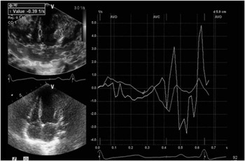
The study protocol was approved by the Ethics Committee of Shiraz University of Medical Sciences and written informed consents were obtained from the patients or their parents. Tissue Doppler imaging was carried out from basal to mid-septum and the lateral wall to measure the strain rate of the septal and lateral walls of the left ventricle (Fig 1).
Tissue velocity-based strain rate from the basal to mid-septum and lateral walls of one of our patients with acceptable ejection fraction mentioned in discussion.

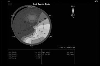
In addition, the segmental and global longitudinal two-dimensional strains of the left ventricle were obtained by means of speckle tracking echocardiography method from apical four-chamber, apical two-chamber, and parasternal long-axis views, and were illustrated on bull’s eye map (Fig 2).
Calculation of segmental and global strain by speckle tracking method demonstrated by Bull’s eye map from one of our patients mentioned in discussion.

Then, we made a comparison between the patient group with acceptable ejection fraction and the healthy control group with respect to the left ventricular systolic function, assessed by tissue Doppler imaging and speckle tracking echocardiography. Acceptable ejection fraction was defined as ⩾55% in M-mode from parasternal long-axis view of the left ventricle just inferior to the mitral valve leaflets.
All the echocardiographic studies were conducted in the supine and left lateral decubitus positions using a Vivid E9 ultrasound machine (Vivid E9; GE Health Care, Milwaukee, Wisconsin, United States of America).
Two-dimensional Cin-loop recordings in 50–70 frame rates for the echocardiographic views of all the patients were stored for offline analysis.
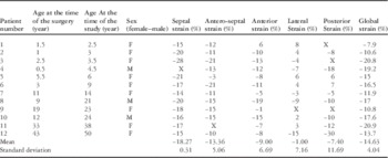
Furthermore, custom acoustic-tracking software (Echo Pac Advanced Analysis Technologies; GE Health care) was used to trace the endocardial cavity interface manually. Of the 60 segments, 6 with poor image quality were excluded from the study as shown in Table 2. The echocardiographic measurements were assessed by two observers at the same time and the best image of the three was chosen.
Age, sex, and myocardial strains of all operated patients using speckle tracking method sorted by age at the time of the study.

Statistical analysis
SPSS for windows version 19 (SPSS Inc., Chicago) was used for statistically analysing the data. Continuous variables were described through the values, means, and standard deviations.
The differences between variables in the study groups were compared by using Mann–Whitney U-test to compare the data within groups. Besides, p-value⩽0.05 was considered as statistically significant.
Results
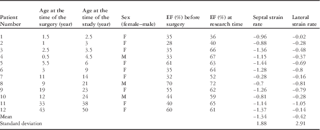
The clinical characteristics and echocardiographic data of the all patients are presented in Tables 2 and 3.
Age, sex, year after surgery, ejection fraction, and myocardial strain rate in all operated patients using conventional echocardiography and tissue Doppler imaging sorted by age at the time of the study.

EF=ejection fraction
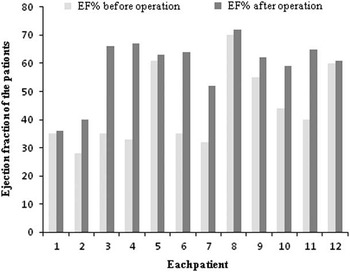
In this study, three patients did not achieve desirable improvement of ventricular function after the surgery. Therefore, nine patients who had acceptable ejection fraction postoperatively in comparison with preoperative values (p=0.035) were selected for the study (Fig 3).
Ejection fraction of each patient before and after the operation sorted by age at the time of the study.

The results of tissue Doppler imaging in this study revealed no significant difference between the patients and controls with regard to the septal strain rate (p=0.65). However, a significant difference was found between the two groups concerning the lateral strain rate (p=0.001; Table 4).
Comparison of mean septal and lateral wallstrain rate of the left ventricle between the patients who had acceptable ejection fraction and control group by tissue Doppler imaging.

MW-U=Mann–Whitney U-test
In addition, a significant difference was observed between the patients and the control group in terms of the strain of the anterior, lateral, and posterior segments of the left ventricle by speckle tracking echocardiography. On the other hand, suboptimal differences were observed between the two groups concerning the septal and anteroseptal segments of the ventricle. Meanwhile, the global strain values were significantly different among the patients and the control group (p=0.016; Table 5).
Comparison of mean segmental and global strain of the left ventricle in the patients who had acceptable ejection fraction and control group by speckle tracking echocardiography.

MW-U=Mann–Whitney U-test
Besides, according to Table 3, we could not define any correlation between age at the time of surgery and changes in echocardiographic data, such as ejection fraction, strain, and strain rate.
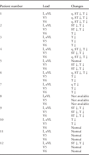
Table 6 reveals the important electrocardiographic data of each patient at the time of the surgery.
Important electrocardiography data of the operated patients at the time of the operation.

Discussion
After surgical correction of anomalous origin of the left coronary artery from the pulmonary artery, most patients have good global left ventricular function assessed by conventional echocardiography. However, a small number of patients with late presentation may not show enough improvement of ventricular function owing to the severity of myocardial ischaemia before the operation and also progression of segmental left ventricular dysfunction, which may necessitate mitral valve annuloplasty or mitral valve replacement after the initial surgical correction.Reference Lange, Vogt and Horer 3 , Reference Shavilkar, Borgers, Daenen, Gewillig and Flameng 4 , Reference Kazmierczak, Ostrowska, Dryzek, Moll and Moll 6
One of the interesting issues among the delayed surgically corrected patients is whether they have segmental left ventricular dysfunction or not, especially in the patients with acceptable ejection fraction in conventional echocardiography.
Our study findings revealed that the strain rate values of the septal and lateral walls of the left ventricle by tissue Doppler imaging had proper and predictable results for detection of abnormal left ventricular systolic function in the presence of acceptable ejection fraction (Table 3). Yet, the strain rate by tissue Doppler imaging for the left ventricular function is angle dependent and the values may not be reproducible.Reference Vrain, Bilhorn, Kurup and Linda 12
Two-dimensional speckle tracking echocardiography is a novel method for the determination of the left ventricular myocardial deformation and also a reliable, angle-independent tool for the evaluation of regional and global ventricular function.Reference Lorch, Ludomirsky and Singh 13
According to the results presented in Table 4, there were no significant differences between the patient and the control group with regard to the strain values of the septal and anteroseptal segments of the left ventricle. However, significant differences were observed between the two groups concerning the strain values of the anterior, lateral, and posterior segments of the left ventricle.
In our study, the results obtained by speckle tracking echocardiography were in accordance with the left coronary artery territories, that is, the anterior and lateral walls of the left ventricle, which are involved in the patients with anomalous origin of the left coronary artery from the pulmonary artery disorder.Reference Hauser 14
In their report on a patient with this anomaly by two-dimensional strain, Iriart et alReference Iriart, Jalal, Derval, Latrabe and Thambo 15 preoperatively demonstrated that myocardial ischaemia was the aetiology of anterior wall dysfunction of the left ventricle. In another study, speckle tracking echocardiography correctly identified the segmental left ventricular dysfunction induced by scarring in an animal model of myocardial infarction.Reference Popovic, Benejam and Bian 16
In patients with this anomaly, segmental and global myocardial dysfunction can be identified by cardiac magnetic resonance. In a report by Aurelio et al, left ventricular function of the six repaired patients was assessed by cardiac magnetic resonance to detect myocardial ischaemia and fibrosis. Although only one of the patients had hypokinesia of the anterior segment of the left ventricle by conventional echocardiography, all of them had basal and antero-lateral subendocardial fibrosis.Reference Secinaro, Ntsinjana and Tann 17
It is worthy to mention that one of our patients, who was operated upon at 2 years of age, presented with a syncopal attack 5 years later because of the ventricular tachycardia and fibrillation that was terminated by cardiac defibrillator in the hospital. She was in NYHA functional class I before this attack.
Although she had acceptable ejection fraction by conventional echocardiogphray, tissue Doppler imaging of this patient revealed septal and lateral strain rates of −0.97 and −0.27, respectively (Fig 1). Moreover, the strain of the anterior, lateral, and posterior segments of her left ventricle by speckle tracking were −11, 4, and 7, respectively, which are compatible with severe segmental left ventricular dysfunction (Fig 2).
In conclusion, although our study was limited relatively by small number of the patients with late presentation, it revealed abnormal deformation of the anterior and lateral walls of the left ventricle with regional and global myocardial dysfunction. Such abnormalities may affect the outcome of patients with this anomaly and prone them to progressive ventricular dysfunction, heart failure, arrhythmia, or even sudden death. Therefore, we may recommend speckle tracking echocardiography as a tool for helpful additional information in the care of these patients. Speckle tracking method is more reliable than tissue Doppler imaging because of its segmental approach and angle independency.
Acknowledgements
Hereby, the authors would like to thank Professor Mahmood Zamirian, MD and Maryam Soroush, PHD for their assistance in this study. They are grateful to Miss Afsaneh Keivanshekouh at the Research Improvement Center of Shiraz University of Medical Sciences for improving the use of English in the manuscript. The present article was extracted from dissertation number 6219 in Shiraz University of Medical Sciences, Shiraz, Iran.
Ethical Standards
The study was approved by the Ethics Committee of the University, and written informed consent was obtained from the patients’ parents.
Conflicts of Interest
None.