Introduction
The granary weevil is a serious pest of stored grains, and it is dispersed worldwide. Pest infestations of food facilities lead to significant losses due to pest grain consumption and mold development, including toxigenic species (Sauer et al., Reference Sauer, Storey and Walker1984; Magan et al., Reference Magan, Hope, Cairns and Aldred2003). Host selection and mating in Sitophilus granarius is based on chemosensation, which guides the behavior of all insects possessing chemosensory systems. S. granarius males produce aggregation pheromones, which are attractive to both sexes (Faustini et al., Reference Faustini, Giese, Phillips and Burkholder1982).
The use of semiochemicals has stimulated interest because they may improve integrated pest management strategies, including those for stored-product insect pests (Phillips, Reference Phillips1997; Cox, Reference Cox2004). However, despite its economic importance, the physiology of the antennal sensory apparatus of S. granaries remains largely unexplored. Insect antennae, which bear sensory sensilla, play important roles in the detection of various stimuli involved in finding suitable habitats, food sources and mates. Numerous studies have characterized the antennal sensilla of various insects, as well as their implication in odor detection (Merivee et al., Reference Merivee, Rahi, Bresciani, Ravn and Luik1998; Renthal et al., Reference Renthal, Velasquez, Olmos, Hampton and Wergin2003; Sukontason et al., Reference Sukontason, Methanitikorn, Chaiwong, Kurahashi, Vogtsberger and Sukontason2007; Onagbola & Fadamiro, Reference Onagbola and Fadamiro2008; Sun et al., Reference Sun, Wang and Zhang2011).
Insects can identify and respond selectively to cues from a heterogeneous environment; they can differentiate between hosts, both plant and animal, and distinguish among many microclimatic factors, such as variations in humidity, temperature and air flow (Gullan & Cranston, Reference Gullan and Cranston2009). Antennae of insects have sensory organs with different functions that play crucial roles during their entire lifespan (Schneider, Reference Schneider1964). Sensory organs protrude from the cuticle, or sometimes lie within or beneath it, and specialized cells detect stimuli that can be categorized as mechanical, thermal, chemical and visual. The nervous system instigates and controls appropriate behavior, such as posture, movement, feeding and behavior associated with mating and oviposition (Gullan & Cranston, Reference Gullan and Cranston2009).
Several studies have been carried out in various beetle species on their antennal sensilla and ultrastructure. Nevertheless, no work has been performed so far on the antenna sensilla of S. granarius. In our current study, scanning electron microscopy (SEM) and transmission electron microscopy (TEM) were used to examine antennal morphology, and the typology and locations of antennal sensilla on both sexes, as well as the fine structures of the cuticular wall (CW) and dendrites of each sensilla type in S. granarius. A better understanding of the antennal morphology can provide significant information that will advance studies on S. granarius’ behavior, predation, ability to locate hosts and electrophysiological responses.
Materials and methods
Insects
Adult male and female trial beetles were obtained from a laboratory culture maintained on wheat grains at 28°C, 75% relative humidity with 8:16 light:dark photoperiod at the College of Plant Sciences and Technology at Huazhong Agricultural University, Hubei Province, People's Republic of China. Alcohol was used to wash the insects before using a binocular microscope for sex identification, following the method described by Bousquet (Reference Bousquet1990).
SEM
Before using SEM, the antennae were cleaned in an ultrasonic bath (250 W) for 5 s. The antennae were dehydrated through a graded ethanol series of 30, 45, 60, 75, 90 and 95% (each for 15 min), and then twice fully dehydrated in 100% ethanol solution (each for 15 min). Following drying to the critical point, the antennal preparations were mounted on a holder using double-sided adhesive tape and sputter-coated with gold/palladium (40:60). In total, 16 antennae from males and females (eight for each gender) were examined using a Hitachi jsm-6390l (Tokyo, Japan) SEM at 20 kV. The morphological terms used in the ultrastructural observations have been used previously (Altner & Loftus, Reference Altner and Loftus1985; Zacharuk, Reference Zacharuk, Kerkut and Gilbert1985).
The antennal sensilla length, diameter and sensilla number per segment were measured directly from the printed SEM images. The measurements (μm) obtained from photomicrographs of individual sensilla of the same type (Sukontason et al., Reference Sukontason, Methanitikorn, Chaiwong, Kurahashi, Vogtsberger and Sukontason2007) were used to calculate the means. The data obtained were analyzed to determine the standard error (SE). Photoshop 7.0 software was used to improve the quality of the images.
TEM
Antennae of S. granarius for TEM were excited and fixed for 24 h with paraformaldehyde (4%) and glutaraldehyde (2%) in 0.1 mol l−1 phosphate buffer solution pH 7.4, followed by dehydration in an ethanol series. The antennae of 50 males and 50 females of both untreated and treated groups were placed separately in a solution of 1% glutaldehyde. After fixation, specimen were washed in cacodylate buffer at pH 6.8 three times (each for 20 min), and then dehydrated in 30, 50, 70, 90 and 100% (three times each for 15 min) ethanol. Afterwards, they were dried to the critical point and finally coated with gold using sputter-coating.
Results
Sensilla basiconica (SB)
Type I SB (SBI)
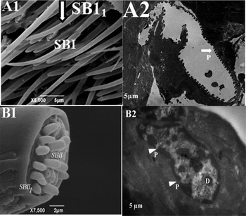
SBI, found on the male antennae of S. granarius, can be distinguished by their finger-like appearance, and very long and proximally wide inward curved shape (fig. 1, A1). They have one subtype whose sensilla have a distally wide inward curved shape. The sensilla of this subtype are found only on the tip of the most terminal segment, and were too numerous to count. TEM of their cross-sections showed that SBI was characterized by a pore structure surrounding the CW covering the cuticular surface, and no dendrites were seen within the sensilla lumen (fig. 1, SBI, A2). TEM photomicrographs showed that, on the antennae of S. granarius, types of SB are characterized by strongly corrugated pores around the cuticle, suggesting an olfactory function.
SEM and TEM photomicrographs of SB on the antennae of S. granarius. SEM of antennae of S. granarious is showing two types of SB with two subtypes: (SBI. A1) which are finger-like, very long and proximally widely inward curved and (SBI1.A1) is subtype of SB in Male and SB type II which are thick, conical with rounded tip, found exclusively on the tip of rostrum in Female. CW, cuticle wall; P, Pores structure; D, dendrites. SBI, Sensilla basiconica type 1, SBI1, subtype of Sensilla basiconica and SBII, Sensilla basiconica type 1I.

Type II SB (SBII)
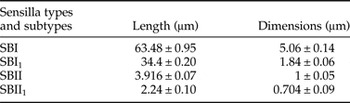
SBII, found on female antennae of S. granarius, are thick and conical with rounded tips. Unlike SBI, SBII are found exclusively on the tip of the rostrum (fig. 1, B1). Their number varies between 6 ± 0.331 (SBII) and 6.4 ± 0.4 (SBII1). They have one subtype (SBII1), in which the sensilla are more peg-shaped (fig 1, B2). Furthermore, SBI (63.48 ± 0.95 µm) and SBII (34.4 ± 0.20 µm) were the longest SB types found in S. granarius (table 1).
SB average length and width (n = 5).

Sensilla coeloconica (SC)
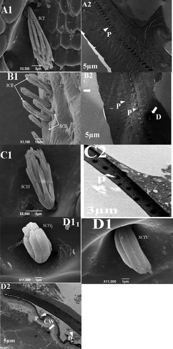
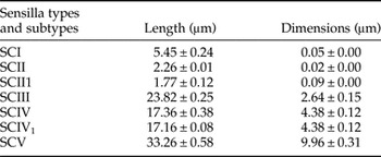
The SC types found in S. granarius are characterized by peg-shaped projections located within deeply sunken depressions with recessed bases. This sensilla commonly occurred on the rostrum of males (fig. 2, A1 and B1) and females (fig. 2, C1and D1). The sensilla are variable in length and basal diameter (fig. 2). Their average length and width range from 1.77 ± 0.12 to 33.26 ± 0.58 µm and 0.02 ± 0.00 to 9.96 ± 0.31 µm, respectively (table 2).
SEM and TEM photomicrographs of SC on the antennae of S. granarius. SEM of the antennae of S. granarious shows four types of SB with two subtypes: (A1, B1 and C1) SC type are characterized by peg-shaped projections located within deeply sunken depressions with recessed bases, while (D1) SCIV types are raised from very large and deep pits. The TEM micrograph of longitudinal sections of SC shows dendrites branches (B2 and D2) and cuticular pores arrow head. CW, cuticle wall; P, Pores structure; D, dendrites. SCI, Sensilla Coeloconica type I; SCII, Sensilla Coeloconica type II; SCIII, Sensilla Coeloconica type III; SCIV, Sensilla Coeloconica type IV.

SC length and width of S. granarious (n = 5).

Type I SC (SCI)
This sensilla type appears shrub-like and proximally multi-branched. These are differently ramified (fig. 2, A1) micrographs of SC longitudinal sections showing cuticular pores with row heads, smooth-surfaced pegs bearing an apical pore suggestive of a gustatory function (fig. 2, A2).
Type II SC (SCII)
This sensilla type also has a shrub-like shape, and it is proximally multi-branched. These are differently ramified (fig. 2, B1). The longitudinal section in the figure shows four dendrites (d) within the sensillum lumen and cuticular pores with arrow heads distributed regularly around the cuticular surface (fig. 2, B2).
Type III SC (SCIII)
This sensilla type is shrub-like and proximally multi-branched. They are differently ramified (fig. 2) TEM micrographs of longitudinal sections of SCIII showed a smooth surface, with pointed pore structures irregularly distributed around the cuticular surface (fig. 2, C2).
Type IV SC (SCIV)
SCIV are raised from very large and deep pits, and are closely opposed with band-like longitudinal furrows. They are also a little outwardly bent (fig. 2, D1). One subtype has been found in which sensilla are relatively straight (fig. 2, D11). Furthermore, SCV (33.26 ± 0.58 µm), SCIII (23.82 ± 0.25 µm) and SCIV (17.36 ± 0.38 µm) are the longest SC types found in S. granarius (table 3). TEM micrographs of longitudinal sections showed thick-walled cuticles with pore structure and dendrites divided into many branches (arrows) (fig. 2, D2).
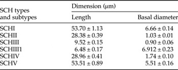
S. granarious SCH average length and width (n = 5).

Sensilla chaetica (SCH)
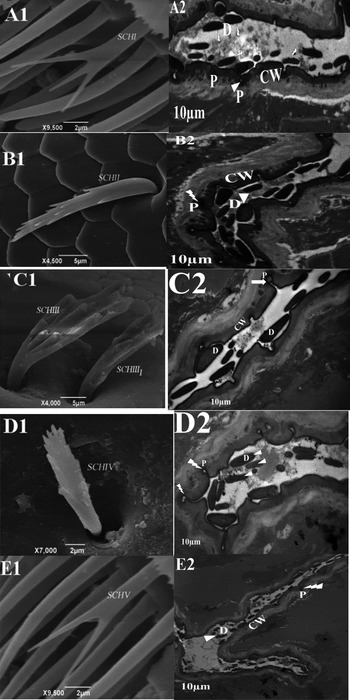
The SCH types found on S. granarius antennae are peculiar because their external structures and their shape (fig. 3). The average length and width ranged from 6.48 ± 0.17 to 53.70 ± 1.13 and 0.90 ± 0.06 to 6.66 ± 0.14 µm, respectively (table 3).
SEM and TEM photomicrographs of SCH on the antennae of S. granarius. SEM of antennae of S. granarious showed five types of SCH: (A1, B1, C1, D1 and E1) SCH type SCHI.A1 and SCHV.E1 are similar, both having their setae rounded and straight while SCHII.B1 are proximally bent toward the cuticle surface and project from deep pits. SCHIII.C1 are little outwardly curved and cut unevenly, while SCHIV.D1 are like toothed-tree leaves. TEM micrographs of longitudinal sections of SCH showed many dendrite branches with cuticular pores arrow head (A2, B2, C2, D2 and E2). CW, cuticle wall; P, Pores structure; D, dendrites. SCHI, Sensilla Chaetica type I; SCHII, Sensilla Chaetica type II; SCHIII, Sensilla Chaetica type III; SCHIV, Sensilla Chaetica type IV; SCHV, Sensilla Chaetica type V.

SCH, types I and V
These are sparsely distributed on the male flagellum (fig. 3, A1 and E1) and not observed on female antennae. These sensilla are morphologically similar. These have their setae rounded and straight before being ramified into two branches or three branches, which are uneven in length (fig. 3, A1). TEM micrographs of longitudinal sections of type I SCH revealed thick porous CWs surrounded by an inner lumen innervated by many small cylindrical dendritic branches (fig. 3, A2).
SCH, types II–IV
These three sensilla types are morphologically similar and found on female antennae of S. granarius (fig. 3, B1, C1 and D1). Type II SCH were distinguished by their fishbone-like, flattened or concave shape that is expanded, distally or proximally bent toward the cuticle surface and projected from deep pits (fig. 2, B1). Types II SCH have almost no projection angle. They arise from small and deep pits and lay on the cuticular surface. TEM micrographs of longitudinal sections of type II SCH showed thick CWs and multiple dendritic branches within the cuticle (fig. 3, B2).
Type III SCH
Type III SCH and its sole subtype are shaped like the inner part of the human palm. They have a small outward curve and are cut unevenly. The palms have cuticular ridges, which are separated by furrows (fig. 3, C1). TEM micrographs of longitudinal sections of type III SCH have thick CWs with pore structures and eight dendritic branches apparent within the cylindrically shaped cuticle (fig. 3, C2).
Type IV SCH
Types IV SCH are shaped like toothed-tree leaves (fig. 3, DI). S. granarius antennae possess only type II SCH with an average number per segment ranging from 5.4 ± 0.24 to 23.2 ± 0.86 (table 2), while the other SCH types are distributed around the eyes, head and the rostrum. Furthermore, TEM of type I SCH (53.70 ± 1.13 µm) revealed the presence of slightly longitudinal grooves on the sensillary surface, showing the presence of thick CWs with many pore structures around the cuticle's surface dendrites (fig. 3, D2).
Type V SCH
These sensilla were found to be morphologically similar to type I SCH. They have rounded setae that straighten before being ramified into two branches or three branches, which are uneven in length (fig. 3, E1). Type V SCH (53.51 ± 0.89 µm) is the longest type found in S. granarius (table 3).
The TEM micrographs of longitudinal sections of type V SCH showed relatively thick external process walls and small-sized dendritic branches within the sensilla. The pores were distributed around the cuticle, and spaced apart from each other (fig. 3, E2).
Discussion
This study used SEM and TEM of the antennae of S. granarius to reveal a complex arrangement of multiple sensillum types. This data can lead to opportunities for electrophysiological investigations into the capability of S. granarius to detect semiochemicals.
To our knowledge, this is the first study describing antennal sensillum types and distribution in S. granarius. Based on SEM observations, three types of antennal sensilla have been described. In the present study, SB were found to be the most widespread and numerous sensilla on the antennae of both sexes of S. granarius. SBI, with two subtypes, was distinguished by their finger-like appearance. They were very long, proximally wide and inwardly curved, and they were found in males (fig. 1, A1). However, the SBII found on the female antennae of S. granarius was thick and conical with rounded tips (fig. 1, B1). The cross-sections of SBI on the antennae of S. granarius showed porous sensilla (fig. 1, A2), and those of SBII (fig. 1, B2) revealed the presence on wall pores, suggesting that they play a role in the detection of various chemical compounds during courtship and host recognition (Zacharuk, Reference Zacharuk, Kerkut and Gilbert1985). S. granarius uses the SB to detect sex pheromones, possibly in conjunction with host plant odors, and higher numbers of sensilla may indicate higher levels of sensitivity (Chapman, Reference Chapman1982).
The SC types found in S. granarius were characterized by peg-shaped projections located within deeply sunken depressions with recessed bases. These sensilla commonly occurred on the rostrum of males (SCI and SCII are seen in fig. 2, A1 and B1, respectively) and females (SCIII and SCIV are seen in fig. 2, C1 and D1, respectively). These are variable in length and basal diameter (fig. 2). SC possess a deep pit, wall or depression inside and they raise from the base of the sensilla (Hunger & Steinbrecht, Reference Hunger and Steinbrecht1998; Onagbola et al., Reference Onagbola, Meyer, Boina and Stelinski2008; Roberto et al., Reference Roberto, Marco, Paola and Nunzio2009; Yang et al., Reference Yang, Yan and Liu2009; Vonnie, Reference Vonnie, Méndez-Vilas and Díaz2010; Maria et al., Reference Maria, Simone and Anna2011; Andrea et al., Reference Andrea, Emanuela, Marco and Roberto2012). We classified the S. granarius sensilla (fig. 2). Some of the SCH types found in S. granarius were similar to those found in Dendroctonus valens (Coleoptera: Curculionidae, Seolytinae) (Chen et al., Reference Chen, Zhang, Wang and Kong2010; Andrea et al., Reference Andrea, Emanuela, Marco and Roberto2012). Furthermore, types III and IV SCH, which were distributed in the same area (around the eyes), could not be sufficiently distinguished from one another to be counted. This was also true for SCII and SCIII, which were found to be abundantly distributed around the eyes and on the rostrum of male and female S. granarius, respectively (fig. 2, B1 and C1). Thus, the magnification of 10 µm or more, which allows for the viewing of a large number of antennal segment surfaces did not show enough detail to distinguish clearly the differences between some types and subtypes. That is why, in some cases, we have only counted the sensilla types without specifying the subtypes. TEM micrographs of SC longitudinal sections showed flat-tipped and smooth-surfaced pegs bearing apical pores (fig. 2, A2, B2, C2 and D2), suggesting their ability to detect various chemical compounds of gustatory function. There is a difference in the numbers of the sensilla in pore structures between the sexes, with female antennae having fewer than male antennae. This difference is thought to be related to sex-specific differences in behavior, such as those occurring during courtship and host recognition (Zacharuk, Reference Zacharuk, Kerkut and Gilbert1985). The antennae of adult insects typically have various types of sensilla that take part in essential roles in a number of behaviors (Hu et al., Reference Hu, Zhang and Wang2009).
The external morphology, types and distribution of sensilla on the antennae of female and male S. granarius were observed using SEM. The five types of SC, with one subtype, found on S. granarius antennae were peculiar due to their external structure, as well as their shape in both males (fig. 3, A1 and E1) and females (fig. 3, B1, C1 and D1). The SC longitudinal sections had thick CWs and pores were also observed on the surface (fig. 3, A2, B2, C2, D2 and E2). Our data also showed that the number of SC did not differ between females and males. This description agreed with the report from Borg & Norris (Reference Borg and Norris1971) that the dendrites did not penetrate the setae but remained at the base because of their function. SC longitudinal sections showed dendritic branches and cuticular pore arrow heads that may be used in the perception of humidity, temperature, heat and CO2 due to the distribution around the eyes, head and the rostrum. They may function as contact-chemoreceptors as has been suggested by Keil & Steinbrecht (Reference Keil and Steinbrecht1984).
Conclusion
In conclusion, the present study has identified and characterized the distribution of different sensilla types on S. granarius using SEM and TEM. The results obtained from our study supply direct morphological evidence that insect antennae possess structures that play pivotal roles in locating mates and host plants. Future studies on the functional morphology of the antennal sensilla using TEM coupled with electrophysiological recordings will likely confirm the functions of the different sensilla identified during this study. A more extensive study will provide a full understanding of the role of antennae in the particular behaviors of S. granarius.