Introduction
The aetiology of Bell's palsy is unknown, but viral infection, ischaemia and autoimmune disease have been proposed as possible mechanisms.Reference Finsterer1 The spread of inflammation and oedema around the nerve is known to play a major role in the pathophysiology of Bell's palsy, as it leads to blockage of axonal flow due to compression within the bony canal. Thus, visualisation of neural oedema and inflammation within the intratemporal course demonstrates the site of the lesion.Reference Leslie and Zinreich2, Reference Saito, Takeda and Kishimoto3
Magnetic resonance imaging (MRI) has greater accuracy for detecting nervous system lesions compared with other imaging modalities, and is aided by use of the contrast medium gadolinium, a paramagnetic imaging agent that enhances T1-weighted signal intensity. This contrast material is primarily distributed within the extracellular fluid; thus, its presence is most notable when extracellular fluid is increased, such as in areas of neoplasia, inflammation or oedema. In the case of facial nerve palsy, increased contrast enhancement results from breakdown of the barrier between the blood and the peripheral nerve, and/or from venous congestion due to increased intraneural pressure.Reference Engström, Abdsaleh, Ahlstöm, Johansson, Stalberg and Jonsson4, Reference Sartoretti-Schefer, Wichmann and Valavanis5
However, controversy exists regarding the prognostic value of MRI in cases of idiopathic facial palsy. Some have stated that MRI has no prognostic value, while others have reported that the presence of facial nerve enhancement on MRI is associated with a poor prognosis.Reference Kumar, Mafee and Mason6–Reference Mineta, Saitoh, Yoshikawa, Yamada, Aburano and Matoba10
Moreover, no consensus exists on the quantitative measurement of MRI signal intensity. Some studies have used subjective evaluation but this is thought to be unreliable. Others have reported results for quantitative analysis using region-of-interest measurements, which may enable more objective evaluation of the clinical significance of facial nerve enhancement.Reference Sartoretti-Schefer, Brandle, Wichmann and Valavanis11
The present study aimed to assess the clinical significance and prognostic value of quantitative analysis of MRI data in patients with acute facial nerve palsy.
Materials and methods
Forty-four patients with peripheral facial nerve palsy underwent gadolinium-enhanced MRI between January 2007 and December 2009. The group comprised 27 male and 17 female patients, aged 15–81 years (mean age, 46.1 years). Patients with a history of facial paralysis, temporal bone or brain surgery, head injury, or bilateral nerve palsy were excluded.
Temporal MRI scans were obtained using a 1.5 T MRI scanner with a head coil (Signa Excite; GE Healthcare, Milwaukee, Wisconsin, USA). For evaluation of the brain, the following T1 enhancement conditions were used: (TR 400/TE 13), 2 mm thickness and 0.5 mm gap.
The degree of facial nerve function was measured using the House–Brackmann grading system at the patient's first and last visits to the out-patient clinic, the latter being at least one year after the onset of facial palsy.
The mean delay between the onset of facial palsy and the first visit to the clinic was 2.55 days. The mean delay between the onset of facial palsy and the performance of MRI scanning was 6.02 days.
All patients were treated with 1 mg/kg prednisone for 5 days. Thereafter, the dose was reduced by 10 mg every 2 days.
The mean duration of follow up was 13.5 months.
Quantitative analysis assessed the following facial nerve segments: distal internal auditory canal, geniculate ganglion, tympanic and mastoid. The degree of enhancement was calculated for each segmental region of interest, on both sides. The signal intensity of each segment was calculated using the following equation: signal intensity increase = [(post-contrast signal intensity) – (pre-contrast signal intensity)/(pre-contrast signal intensity)] × 100.
The signal intensity of each segment was also analysed subjectively, based on visual inspection, and a visual grade assigned, with 4 representing the highest signal intensity and 1 the lowest (Figure 1).
Axial magnetic resonance imaging scans of the temporal bone of a patient with acute facial palsy (b) before and (a) after enhancement with gadolinium contrast, showing increased signal intensity of the geniculate ganglion (arrow), compared with same area pre-contrast (arrowhead).

The correlation between clinical facial nerve function and MRI facial nerve enhancement was statistically analysed, for both the region-of-interest and visual evaluation systems. The Spearman correlation test was used for statistical analysis, accessed via the Statistical Package for the Social Sciences for Windows version 15.0 software program (SPSS Inc, Chicago, Illinois, USA).
Results and analysis
At their initial visit, 17 of the 44 patients (39 per cent) had a House–Brackmann grade of IV, 12 (27 per cent) were grade II, 11 (25 per cent) were grade III and four (9 per cent) were grade V. At their final follow-up visit, 27 patients (61 per cent) had a House–Brackmann grade of I, 10 (23 per cent) were grade II, four (9 per cent) were grade III, two (5 per cent) were grade IV and one (2 per cent) was grade V.
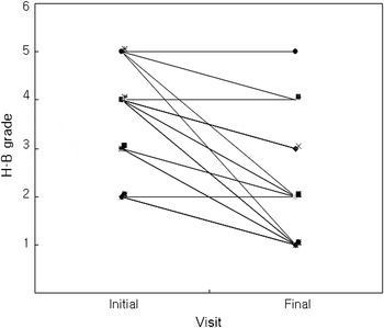
Correlative analysis of the patients’ House–Brackmann grades at their first and last hospital visits showed a significant correlation (p = 0.001) (Figure 2). In other words, the degree of facial palsy at the patients’ last hospital visit was significantly associated with that at their first visit.
Plot of hospital visit versus House–Brackmann (H-B) grade, showing a significant correlation between the two variables (p = 0.001).

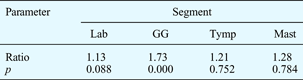
The degree of enhancement of the various facial nerve segments on the palsied side was compared with that on the normal side (Table I). Geniculate ganglion enhancement was significantly increased on the palsied side compared with the normal side (p = 0.000).
Enhancement of facial nerve segments: palsied vs normal side
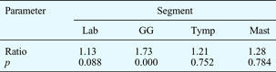
Lab = labyrinthine; GG = geniculate ganglion; Tymp = tympanic; Mast = mastoid; Ratio = signal intensity increase on palsied side vs normal side
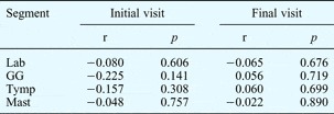
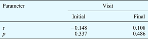
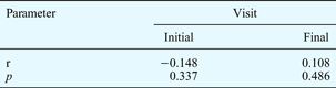
The correlation between the degree of enhancement of each facial nerve segment and the House–Brackmann grade was assessed, for both the early and late stages. There was no significant correlation for any segment, at either stage (p > 0.05) (Table II). Similarly, there was no correlation between House–Brackmann grading and subjective visual grading of facial nerve enhancement (Table III).
Correlation between objective facial nerve enhancement* and facial palsy severity†, by visit and segment

* Assessed using calculation of signal intensity increase for each region of interest.
† Assessed using House–Brackmann grades. r = Spearman's rho; Lab = labyrinthine; GG = geniculate ganglion; Tymp = tympanic; Mast = mastoid
Discussion
Bell's palsy is peripheral facial nerve palsy unaccompanied by any identifiable disease or external injury. It is the most frequent type of peripheral facial nerve palsy, and is thought to be mainly due to viral infection.
Patients with this condition have no identifiable abnormality of the facial nerve canal; therefore, no abnormal findings are seen on temporal bone computed tomography. However, such patients have been investigated with temporal bone MRI in order to identify nerve inflammation and, rarely, tumour around the facial nerve.Reference Kinoshita, Ishii, Okitsu, Okudera and Ogawa12
Although gadolinium cannot pass into the cranial nerves under normal conditions, it can penetrate through blood vessels in the presence of inflammation or oedema, resulting in nerve enhancement. Therefore, the degree of gadolinium enhancement indicates the increase in blood vessel permeability, which reflects the severity of facial nerve inflammation and oedema. Based on the hypothesis that the severity of facial nerve inflammation and oedema is correlated positively with the severity of facial nerve palsy, one could theorise that an increase in facial nerve contrast enhancement will indicate increased severity of facial nerve palsy.Reference Millen, Danniels and Meyer13–Reference Nakao, Sakihama and Kumagami15 In addition, it is known that facial nerve palsy which is more severe in the early stage has a worse prognosis; therefore, if early stage facial nerve palsy can be assessed objectively, its prognosis may be predictable.
Some studies have demonstrated gadolinium enhancement of facial nerve segments on temporal bone MRI performed under normal conditions, especially the geniculate ganglion, tympanic and mastoid segments. Therefore, our study compared the degree of enhancement of four distinct facial nerve segments on the lesional side versus the normal side. We observed a significantly greater degree of facial nerve geniculate ganglion enhancement on the lesional side. This suggests that the geniculate ganglion is the most vulnerable portion of the facial nerve under the pathological conditions which produce Bell's palsy.Reference Hong, Yi, Cha, Park, Kim and Lee16 Our results are similar to those of Kinoshita et al., who found that the geniculate ganglion and the tympanic–mastoid segment showed 21 per cent enhancement in normal controls but 91 per cent enhancement in Bell's palsy patients.Reference Kinoshita, Ishii, Okitsu, Okudera and Ogawa12
To enable a more precise assessment of the severity of facial nerve inflammation, our study calculated the signal intensity increase in each facial nerve segment, using the objective region-of-interest measurement method. However, we found no significant correlation between the degree of facial nerve enhancement, determined using this method, and the clinical severity of facial nerve palsy. We also assessed the overall degree of facial nerve enhancement using a subjective visual grading system involving four grades. However, similarly, there was no significant correlation between the degree of visually determined facial nerve enhancement and the clinical severity of facial nerve palsy. This indicates that the severity of facial nerve inflammation and oedema, which promotes blood vessel permeability and gadolinium influx, does not adequately reflect the clinical severity of facial nerve palsy at that time.
• Temporal bone magnetic resonance imaging is not essential in acute facial nerve palsy
• It cannot accurately indicate initial facial nerve inflammation or future prognosis
• It is recommended if surgical intervention is considered
• It is also recommended for unusual cases, to exclude facial nerve tumour
Furthermore, predicting the prognosis of facial nerve palsy from the degree of facial nerve enhancement at the initial stage is also difficult. Although some studies have reported that the degree of enhancement is proportional to the clinical severity of facial nerve palsy in the early stage, others (including ourselves) have found no such association. Thus, the clinical effectiveness of temporal bone MRI for patients with facial nerve palsy is still open to question.
Another important justification for the necessity of temporal bone MRI in such cases is the identification of facial nerve tumour. However, the probability of occurrence of such a tumour is very low (less than 5 per cent); moreover, 50 per cent of facial nerve tumour cases do not show facial nerve palsy.Reference O'Donoghue, Brackmann, House and Jackler17, Reference McMonagle, Al-Sanosi, Croxson and Fagan18 In our study, no cases of facial nerve tumour were discovered.
Thus, the current information on the clinical effectiveness of temporal bone MRI in cases of facial nerve palsy raises many questions.
One limitation of our study was its shortage of data; thus, studies with a larger number of cases are recommended.
Conclusion
This study found no significant correlation between the clinical severity of facial nerve palsy and the degree of facial nerve contrast enhancement on temporal bone MRI.
Temporal bone MRI is recommended in the limited number of cases of complete facial palsy in which surgical intervention is considered. Temporal bone MRI is also recommended for unusual cases of facial palsy in which symptoms are prolonged and/or aggravated, in order to exclude a facial nerve tumour.