Introduction
Imaging studies have demonstrated neuroanatomical abnormalities in bipolar disorder (BD) patients (Hibar et al., Reference Hibar, Westlye, Doan, Jahanshad, Cheung, Ching and Mwangi2018). However, interpretation of these abnormalities is challenging as they may constitute either biological risk factors conveying vulnerability for BD, or consequences of the illness or its treatment. Neuroimaging studies of first-degree relatives of patients with BD who have not developed BD themselves provide an opportunity to differentiate these factors. Most studies examining high-risk cohorts have reported structural pathology using measures of brain volume. Cohorts of unaffected high-risk subjects who have not passed the peak age of onset for developing BD (⩽30 years) (Bellivier et al., Reference Bellivier, Etain, Malafosse, Henry, Kahn, Elgrabli-Wajsbrot and Scott2014; Goodwin & Jamison, Reference Goodwin and Jamison2007; Kessler et al., Reference Kessler, Amminger, Aguilar-Gaxiola, Alonso, Lee and Ustun2007) are particularly valuable to study as they are more likely to include a higher proportion of individuals who will later convert to BD. Most studies examining high-risk cohorts have reported structural pathology using measures of brain volume. While the majority of such studies have reported differences in gray matter (GM) in frontotemporal and sub-cortical regions, some findings have been inconsistent (Nery, Monkul, & Lafer, Reference Nery, Monkul and Lafer2013; Özerdem, Ceylan, & Can, Reference Özerdem, Ceylan and Can2016). The few studies that have examined cortical thickness variously report larger (van Erp et al., Reference van Erp, Thompson, Kieseppä, Bearden, Marino, Hoftman and Kaprio2012) or smaller hippocampal thickness, and thinner orbitofrontal cortex (Hulshoff Pol et al., Reference Hulshoff Pol, van Baal, Schnack, Brans, van der Schot, Brouwer and Kahn2012) and inferior frontal gyrus (IFG) (Roberts et al., Reference Roberts, Lenroot, Frankland, Yeung, Gale, Wright and Mitchell2016). A larger IFG has been repeatedly observed in high-risk subjects (Drobinin et al., Reference Drobinin, Slaney, Garnham, Propper, Uher, Alda and Hajek2019; Hajek, Alda, Hajek, & Ivanoff, Reference Hajek, Alda, Hajek and Ivanoff2013; Macoveanu et al., Reference Macoveanu, Baaré, Madsen, Kessing, Siebner and Vinberg2018; Roberts et al., Reference Roberts, Lord, Frankland, Wright, Lau, Levy and Breakspear2017; Saricicek et al., Reference Saricicek, Yalin, Hidiroglu, Cavusoglu, Tas, Ceylan and Ozerdem2015) and those in early stages of BD (Adler, Levine, DelBello, & Strakowski, Reference Adler, Levine, DelBello and Strakowski2005). Functional (Roberts et al., Reference Roberts, Lord, Frankland, Wright, Lau, Levy and Breakspear2017) and structural (Roberts et al., Reference Roberts, Perry, Lord, Frankland, Leung, Holmes-Preston and Breakspear2018) dysconnectivity of the IFG has also been observed in high-risk subjects. Additionally, previous studies have documented IFG changes over time in participants at high-risk for BD (Papmeyer et al., Reference Papmeyer, Giles, Sussmann, Kielty, Stewart, Lawrie and McIntosh2015) and a negative correlation between the duration of illness and GM volume in this region (Hajek, Cullis, et al., Reference Hajek, Cullis, Novak, Kopecek, Blagdon, Propper and Alda2013; Matsuo et al., Reference Matsuo, Kopecek, Nicoletti, Hatch, Watanabe, Nery and Soares2012). Studies assessing white matter (WM) volume in high-risk relatives have reported volumetric abnormalities in the frontal gyrus (Matsuo et al., Reference Matsuo, Kopecek, Nicoletti, Hatch, Watanabe, Nery and Soares2012) and internal capsule (McIntosh et al., Reference McIntosh, Job, Moorhead, Harrison, Lawrie and Johnstone2005), and smaller corpus callosum thickness (Walterfang et al., Reference Walterfang, Wood, Barton, Velakoulis, Chen, Reutens and Frangou2009). Longitudinal studies are needed to determine whether these cross-sectional differences remain stable or if their trajectory changes over time. Additionally, as the cortical volume is the ‘product’ of surface area and thickness, with both appearing to be driven by distinct cellular mechanisms (Grasby et al., Reference Grasby, Jahanshad, Painter, Colodro-Conde, Bralten, Hibar and McMahon2020), these primary elements of this multi-dimensional measurement need to be addressed separately.
To the best of our knowledge, the Scottish Bipolar Family Study is the only investigation that has reported on the longitudinal effects of familial risk on brain morphology in a young sample. In that recent study, 83 young adults with a family history of BD and 62 controls had neuroimaging and clinical assessment at baseline (average age 21 years), and again after 2 years (Papmeyer et al., Reference Papmeyer, Giles, Sussmann, Kielty, Stewart, Lawrie and McIntosh2015, Reference Papmeyer, Sussmann, Stewart, Giles, Centola, Zannias and McIntosh2016). High-risk individuals with major depressive disorder (MDD) at follow-up displayed thicker left IFG relative to those who remained well, and thicker left precentral gyrus relative to individuals who remained well and controls (Papmeyer et al., Reference Papmeyer, Giles, Sussmann, Kielty, Stewart, Lawrie and McIntosh2015). Over both time-points, the two high-risk groups showed reduced cortical thickness compared to controls in the right parahippocampal and fusiform gyri. In the same cohort, subcortical GM volumes showed no significant group differences over time (Papmeyer et al., Reference Papmeyer, Sussmann, Stewart, Giles, Centola, Zannias and McIntosh2016). When clinical outcomes were refined 4 years after baseline, significant decreases in right amygdala GM (from baseline to 2-year follow-up) were evident in high-risk individuals with MDD relative to both controls and high-risk individuals who remained well (Nickson et al., Reference Nickson, Chan, Papmeyer, Romaniuk, Macdonald, Stewart and Sussmann2016). In another recent study of older twins, high-risk twins who were relatives of MDD and BD probands did not differ from low-risk twins in regional GM volume changes over a 7-year period (n = 37 and n = 36, respectively; average age 42 and 39 years, respectively; 20% monozygotic) (Macoveanu et al., Reference Macoveanu, Baaré, Madsen, Kessing, Siebner and Vinberg2018). However, independent of time, high-risk twins had significantly greater GM volumes in bilateral dorsal anterior cingulate, IFG and temporoparietal regions compared to low-risk twins (Macoveanu et al., Reference Macoveanu, Baaré, Madsen, Kessing, Siebner and Vinberg2018). Furthermore, the development of an affective disorder at follow-up (depression and/or anxiety), was associated with relatively larger GM volumes in the right dorsal anterior cingulate cortex and inferior frontal cortex at both time-points.
The recent evidence of differences in the frontotemporal cortex and subcortical regions motivated the present study of brain morphometry in a priori regions of interest over a 2-year follow-up interval in (i) young first-degree relatives of BD patients who did not have BD themselves at baseline (high-risk; HR); and (ii) controls (CON) from families without mental illness. To the best of our knowledge, our analysis of 90 HR participants is the largest to date to report on longitudinal morphometric findings. To achieve this, we investigated GM and WM volumes, GM thickness and surface area. We hypothesised that frontotemporal and subcortical changes over 2 years would differ between the HR and CON groups across multiple measures of brain morphology.
Methods
Participants
Participants comprised two groups of subjects of Caucasian descent, aged 12–30 years, who had baseline and follow-up scans: (i) high-risk (HR) participants who were first-degree relatives of a proband with a confirmed DSM-IV diagnosis of BD-I or BD-II, but who did not themselves have this disorder; and (ii) controls (CON) from families with no evidence of mental illness. HR and CON participants with a lifetime or current presence of psychiatric symptoms (apart from the occurrence of BD) and/or with lifetime or current psychotropic medication use were not excluded from the study.
In the original sample, 298 subjects had at least one scan. Of the 209 with scans at two time-points (92 CON and 117 HR), 18% (30 CON and 7 HR) were excluded due to non-Caucasian ethnicity, and 12% (6 CON and 20 HR) were removed due to excessive head movement, poor image quality or errors on scans at one or both time-points. Eighty-nine (35 CON and 54 HR) subjects were excluded as they had a scan at only one time-point. The remaining 146 subjects (56 CON and 90 HR) formed the final analysis sample.
The 146 participants whose data passed quality control belonged to 124 families, of which 71 families were of high risk. Of the 90 HR subjects, 81% had parent probands and 19% had a sibling proband. Two CON families and 14 HR families had more than one sibling in the sample. An independent samples t test demonstrated that the age of the 146 participants included in the final sample (M = 20.97) was not significantly different to the age of the 152 participants excluded from the sample (M = 20.23), p > 0.05. A χ2 test determined that the distribution of males and females was the same for the participants included in the final sample (females: 56.16%) compared to those excluded (females: 54.97%), p > 0.05. Within the HR group, a χ2 test showed that the number of converters did not differ for those included in the final sample (6%) compared to those excluded (5%), p > 0.05.
See online Supplementary Methods for further details on recruitment, exclusions and sample characteristics. The study was conducted with the approval of the University of New South Wales Human Research Ethics Committee (HREC Protocol 09/097; ref 14 128) in Sydney, Australia. Written informed consent was obtained from all participants with additional parental consent for participants aged 16 or younger. The authors assert that all procedures contributing to this work comply with the ethical standards of the relevant national and institutional committees on human experimentation and with the Helsinki Declaration of 1975, as revised in 2008.
All potential participant families were initially assessed through baseline administration of the Family Interview for Genetic Studies (FIGS) (Maxwell, Reference Maxwell1992) to determine the family history of BD and other disorders for eligibility (see online Supplementary Methods). At least one parent or participant aged over 22 in each family completed the FIGS. Parents were interviewed about their child (aged 12–21) using the Kiddie-Schedule for Affective Disorders and Schizophrenia for School-Aged Children – Present and Lifetime Version (K-SADS-BP) (Kaufman et al., Reference Kaufman, Birmaher, Brent, Rao, Flynn, Moreci and Ryan1997; Nurnberger et al., Reference Nurnberger, McInnis, Reich, Kastelic, Wilcox, Glowinski and Gershon2011), in addition to the 12–21-year-olds themselves completing a K-SADS-BP interview. The 12–21-year-olds were part of a collaborative HR study with a US consortium (Nurnberger et al., Reference Nurnberger, McInnis, Reich, Kastelic, Wilcox, Glowinski and Gershon2011). For all participants aged 22–30 (including BD-probands), the Diagnostic Interview for Genetic Studies (DIGS) was administered to determine diagnoses (Nurnberger et al., Reference Nurnberger, Blehar, Kaufmann, York-Cooler, Simpson, Harkavy-Friedman and Reich1994). Consensus DSM-IV diagnoses of BD-probands, HR and CON participants were determined by two independent raters using best estimate methodology (Leckman, Sholomskas, Thompson, Belanger, & Weissman, Reference Leckman, Sholomskas, Thompson, Belanger and Weissman1982), drawing from the DIGS, K-SADS-BP, FIGS and medical records (where available).
To assess the current mood state, the Children's Depression Inventory (Kovacs, Reference Kovacs1992) was administered to participants aged 12–21 years. Both the Montgomery–Åsberg Depression Rating Scale (Montgomery & Åsberg, Reference Montgomery and Åsberg1979) and Young Mania Rating Scale (Young, Biggs, Ziegler, & Meyer, Reference Young, Biggs, Ziegler and Meyer1978) were administered to those aged 22–30 years. Intellectual ability was assessed with the Wechsler Abbreviated Scale of Intelligence (Wechsler, Reference Wechsler1999). Further details are described in the online Supplementary Results.
MRI acquisition and MRI image processing
For each subject, a T1-weighted image was acquired using a 3T Philips Achieva scanner (Royal Philips Electronics, Amsterdam, The Netherlands). Automated cortical reconstruction and segmentation was completed using the longitudinal image processing stream included in FreeSurfer v5.3.0. Subject-level values were extracted based on the Desikan–Killiany atlas (Desikan et al., Reference Desikan, Ségonne, Fischl, Quinn, Dickerson, Blacker and Killiany2006). For each participant, per hemisphere, per time-point, we extracted subcortical WM volume, cortical GM volume, surface area and thickness for 17 frontotemporal regions of interest (ROIs). We also extracted GM volumes bilaterally for an additional 11 subcortical ROIs. ROIs were informed by previous frontotemporal and subcortical findings and are the same as those employed in our previous baseline analysis of this cohort (Roberts et al., Reference Roberts, Lenroot, Frankland, Yeung, Gale, Wright and Mitchell2016). This approach was employed as although findings have been largely inconsistent, studies have reported differences in widespread frontotemporal and sub-cortical regions (Nery et al., Reference Nery, Monkul and Lafer2013; Özerdem et al., Reference Özerdem, Ceylan and Can2016). Online Supplementary Methods and Supplementary Table S1 present all ROIs and provide further details of image pre-processing.
Statistical analyses
All statistical analyses were carried out using SPSS v25 (IBM). To compare categorical sociodemographic and clinical variables between groups, we used the Pearson χ2 test. For continuous sociodemographic and clinical variables, independent samples t tests were used to compare HR and CON groups. For data violating assumptions, Fisher's exact test (χ2) was used for categorical variables, and Mann–Whitney U was used for continuous variables. The statistical significance level was set at p < 0.05 (two-sided).
Linear mixed-effects models were used to investigate structural brain differences for each ROI over time. The base model was run with a single neuroanatomic measure as the dependent variable, and a fixed group factor (CON, HR), repeats factor (time: baseline, follow-up) and the group by time interaction, entered as fixed effects. Fixed effects of baseline age and sex were also included as covariates. Family was included as a random effect to accommodate within-family correlation arising from the inclusion of siblings from within the same family in either the HR or CON groups. A second base model was fitted based on the aforementioned model with an additional adjustment for overall differences in brain size. Total intracranial volume (ICV) was included as a covariate in the analyses of brain volumes, and total surface area as a covariate in the analyses of regional surface areas. As the relationship between cortical thickness and ICV is controversial (Ashburner et al., Reference Ashburner, Csernansk, Davatzikos, Fox, Frisoni and Thompson2003), ICV was not included as a covariate in the analyses of cortical thickness.
Correction for multiple testing of the two main effects of group and time, and the group-by-time interaction for regions by each hemisphere was carried out using false-discovery rate (FDR) with q < 0.05 considered significant (Benjamini & Hochberg, Reference Benjamini and Hochberg2000). As the cortical thickness and surface area are genetically independent, emerge through different neurobiological events during development, and are sensitive to different clinical conditions (Grasby et al., Reference Grasby, Jahanshad, Painter, Colodro-Conde, Bralten, Hibar and McMahon2020), we considered that it was appropriate to treat thickness, area and volume as belonging to different families, and for the purpose of correcting for multiple comparisons to control the error rate separately within those families. We ranked 102 comparisons (17 regions × 3 effects × 2 hemispheres) analysed separately for subcortical WM volume, cortical GM volume, surface area and thickness, and 66 comparisons (11 regions × 3 effects × 2 hemispheres) analysed for subcortical volume. In view of potential interest for investigation in future, larger-scale studies, we also report effects with p < 0.05 (uncorrected). For significant interaction effects, four simple effects tests (between-group differences at each time-point and difference over time within each study group) were performed whereby p values were tested against Bonferroni-adjusted significance levels (p < 0.05/4).
Auxiliary analyses
Subsequent exploratory tests were performed for ROIs that showed a main effect of group or a group-by-time interaction after correction for multiple comparisons. Linear mixed-effects models were run to investigate potentially confounding effects of current antidepressants, any psychotropic medication, current mood episode, current mood state, IQ, years of education at baseline, lifetime alcohol use or dependence (at baseline and follow-up), proband (offspring v. parent) and body mass index (BMI) at baseline. Separate sets of models were run for each potential confounder. To investigate if results were influenced by new DSM-IV diagnoses onset in the HR group, we performed exploratory sub-group analyses. We sub-divided the HR group according to the following criteria: (i) new onset of any mood episode (major depressive or manic/hypomanic episode) from baseline to follow-up (n = 9); and (ii) new onset of any DSM-IV diagnosis from baseline to follow-up (n = 11). Further details are provided in online Supplementary Methods.
Results
Demographic and clinical data
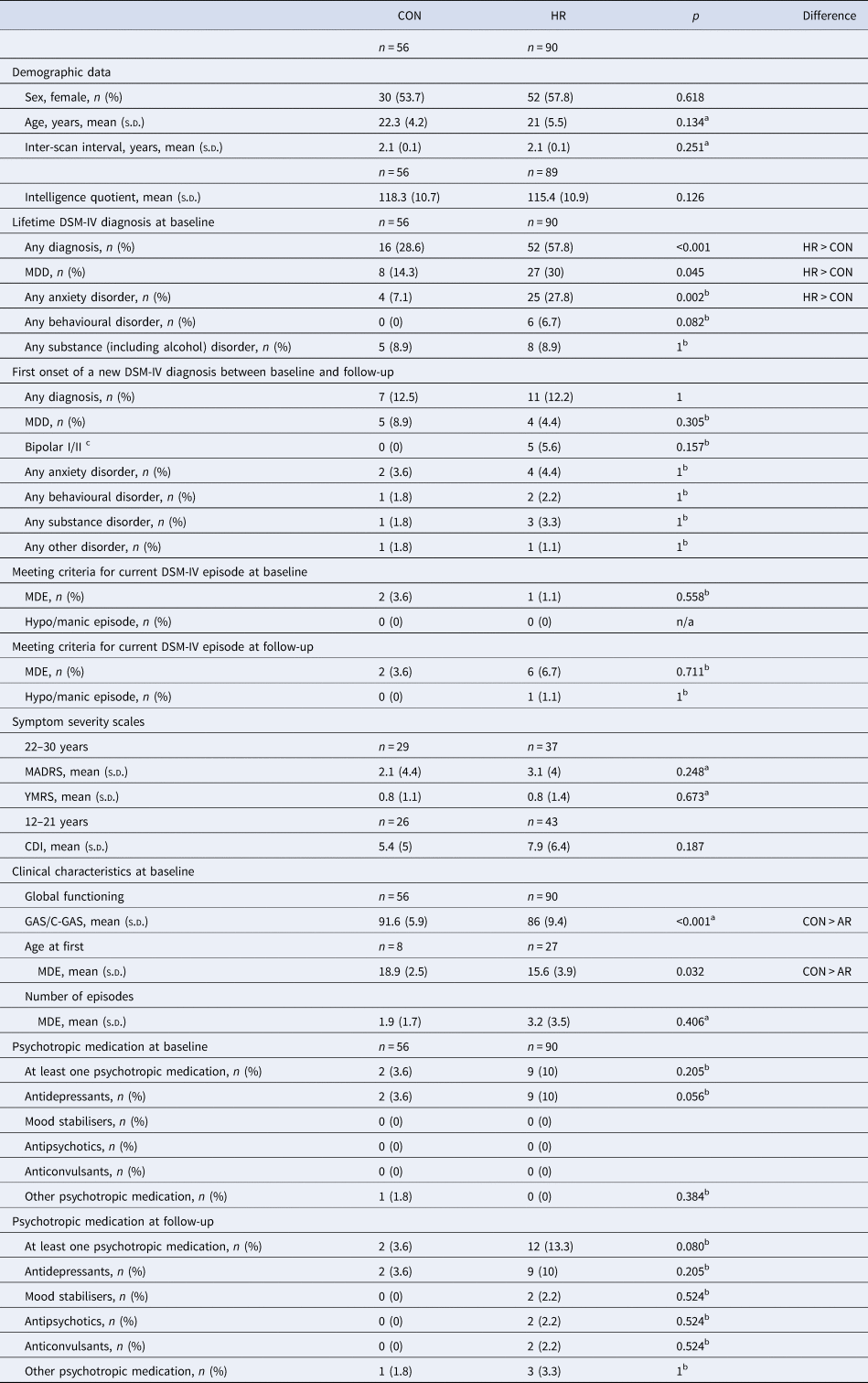
Groups did not differ significantly in mean age, mean IQ, sex distribution or mean time between scans (Table 1). Lifetime occurrences of at least one major depressive episode, lifetime DSM-IV diagnosis or anxiety were significantly higher in the HR than the CON group, consistent with previous reports on high-risk populations (Birmaher et al., Reference Birmaher, Axelson, Monk, Kalas, Goldstein, Hickey and Brent2009; Nurnberger et al., Reference Nurnberger, McInnis, Reich, Kastelic, Wilcox, Glowinski and Gershon2011). Amongst the HR participants, nine participants experienced a first mood episode from baseline to follow-up (five had a first onset of a DSM-IV manic or hypomanic episode and four had new onset of a major depressive episode). At follow-up, 32 HR had a lifetime diagnosis of any mood episode and 56 HR had a lifetime diagnosis of any lifetime DSM-IV diagnosis. Further details of new DSM-IV diagnoses at follow-up are provided in Table 1.
Demographic and clinical data for control (CON) and high-risk (HR) groups
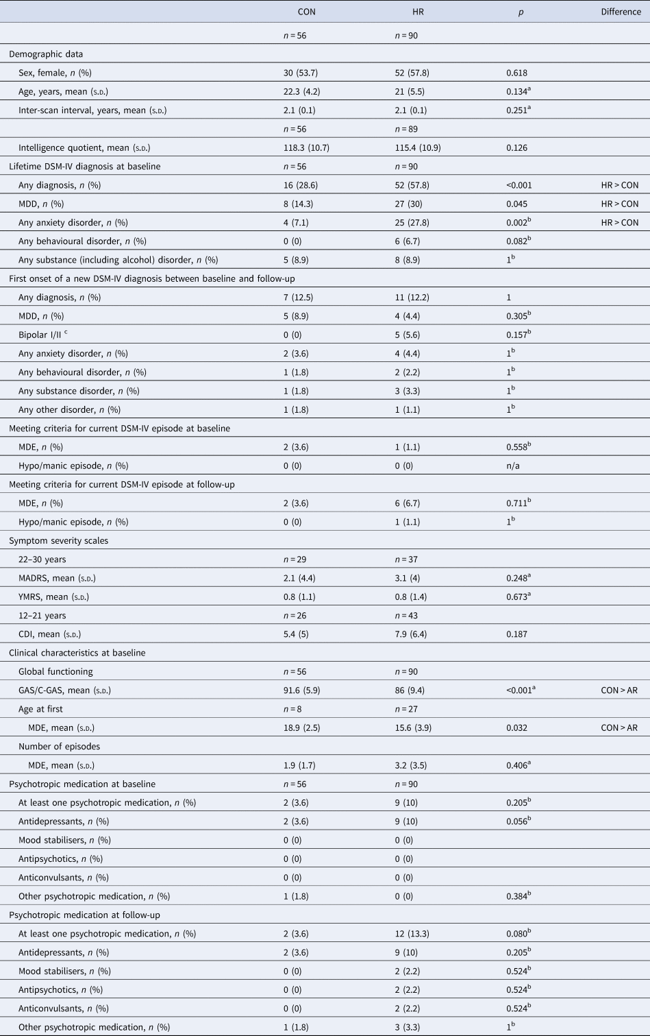
MADRS, Montgomery-Åsberg Depression Rating Scale; YMRS, Young Mania Rating Scale; CDI, Children's Depression Inventory: CON, controls; HR, high-risk; MDD, major depressive disorder; MDE, major depressive episode; GAS, Global Assessment Scale; C-GAS, Children's Global Assessment Scale; n/a, not applicable.
Diagnostic confidence rating ranges using the Best Estimate Methodology vary from 1 to 4, where 1 represents criteria not met for a diagnosis and 4 represents a definite diagnosis. All diagnoses listed here have a confidence rating of 3 or higher.
a Variable did not meet assumptions for parametric analysis. Mann–Whitney U was used.
b Variable did not meet assumptions for parametric analysis. Fisher's exact test was used.
c Five HR subjects experienced a first manic (two subjects) or hypomanic (three subjects) episode.
Structural imaging
Mean values of cortical and subcortical GM, subcortical WM volume, and cortical thickness and surface area are presented for group and time in online Supplementary Tables S2 and S3. Time effects are presented in online Supplementary Table S4.
Group by time interactions
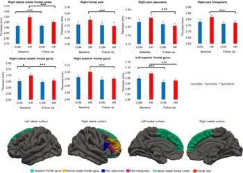
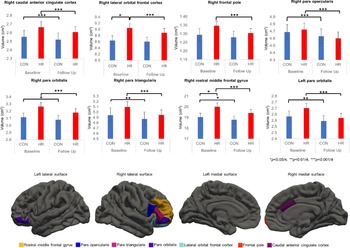
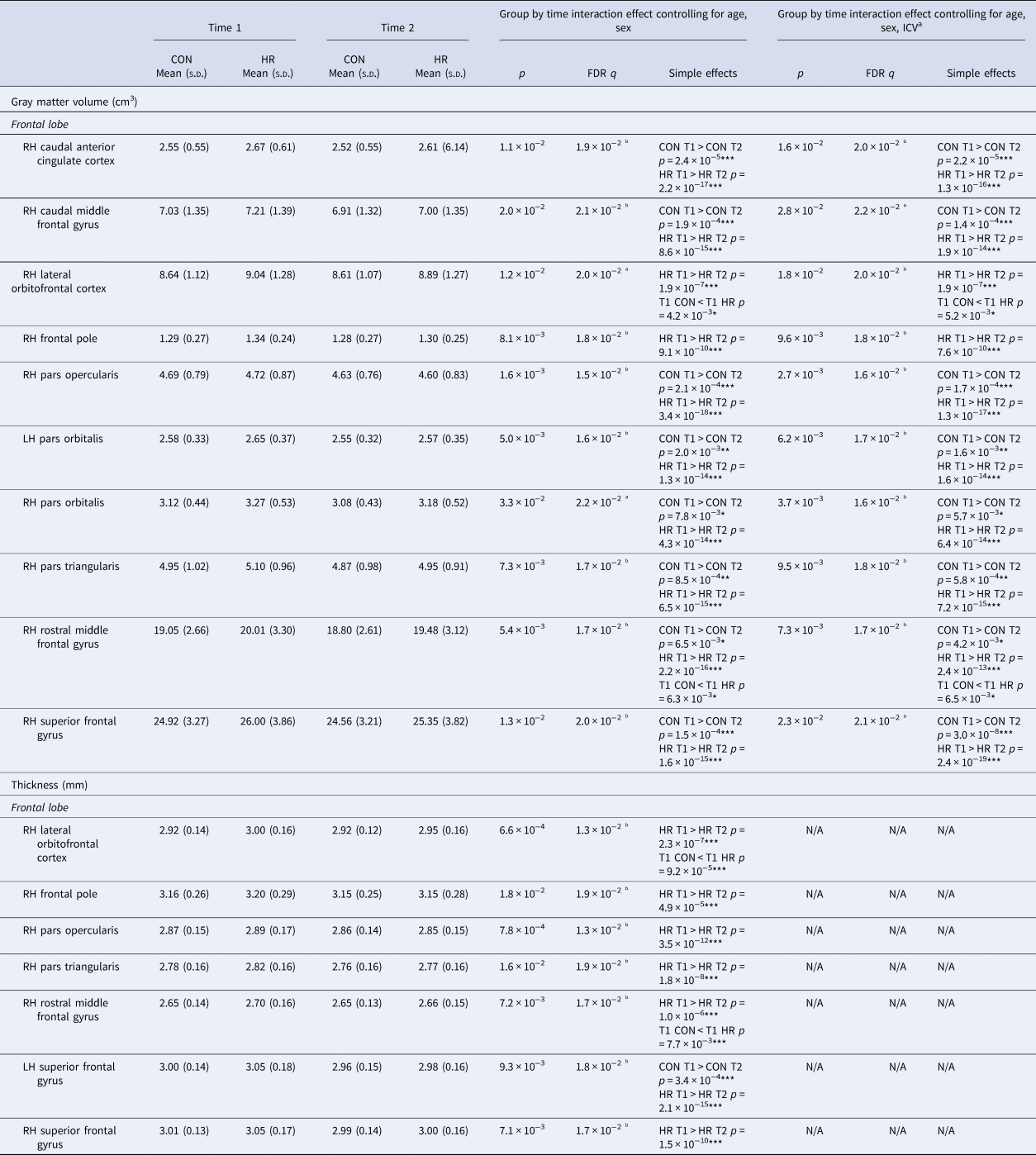
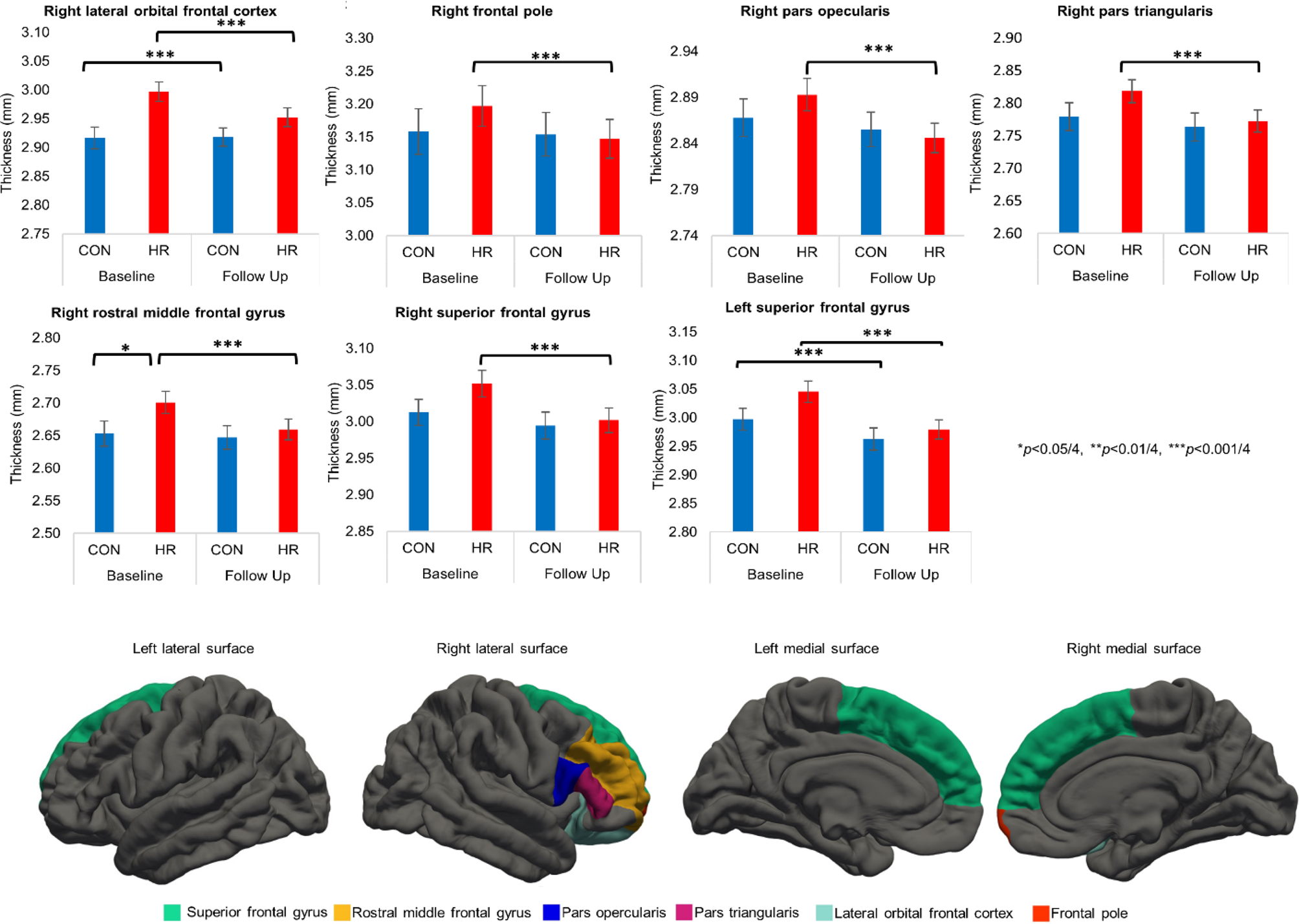
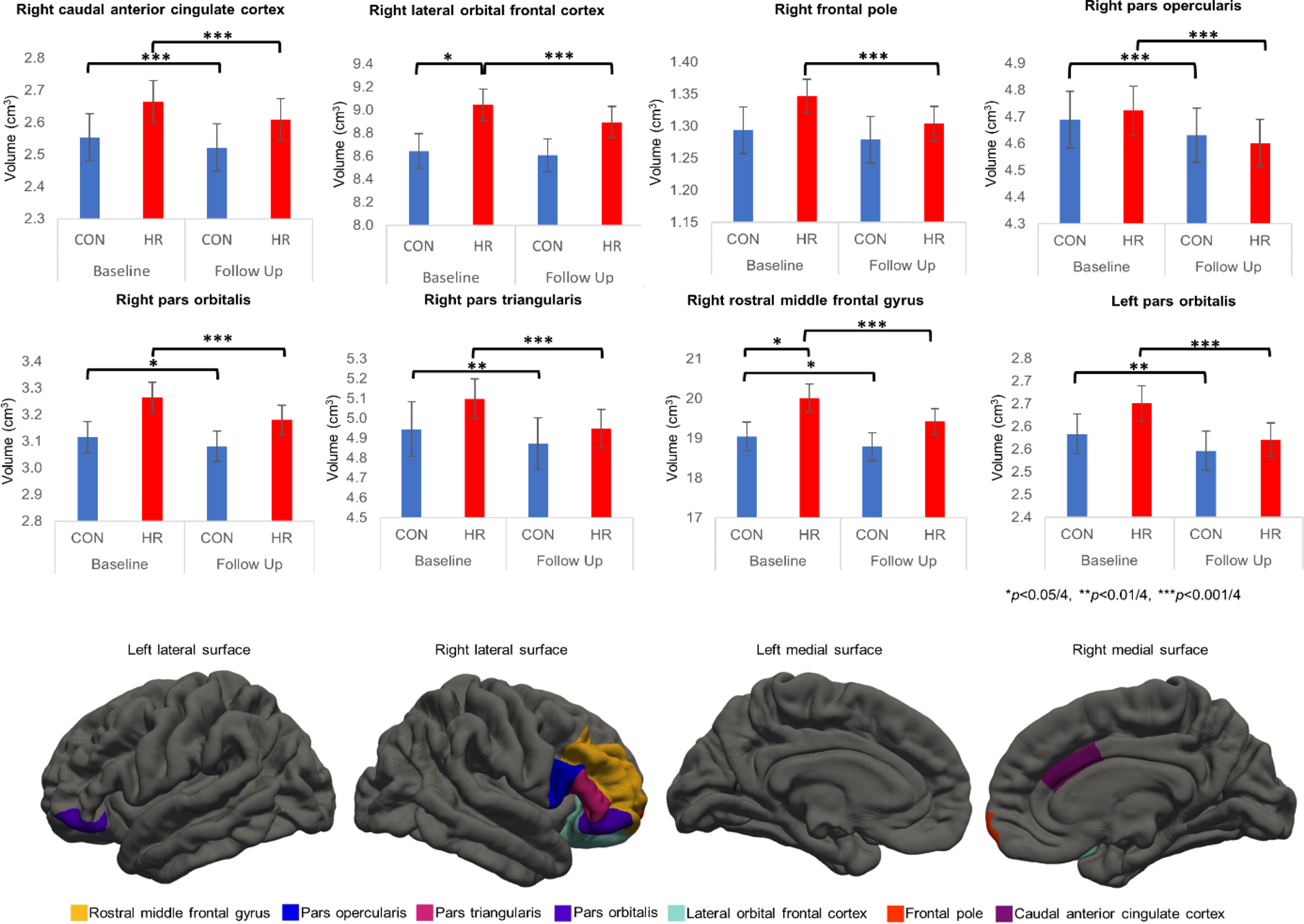
Group by time interactions are reported in Table 2 and Figs 1 and 2. After controlling for multiple comparisons, HR subjects showed accelerated cortical thinning and volumetric reduction over time (after ICV correction) in a number of right lateralised frontal regions: IFG (pars triangularis and pars opercularis), lateral orbitofrontal cortex, frontal pole and rostral middle frontal gyrus compared to controls. Additionally, the bilateral pars orbitalis region of the IFG and right caudal anterior cingulate cortex showed accelerated volume reductions (after ICV correction) in the HR group compared to controls, and there was a steeper rate of cortical thinning over time in the bilateral superior frontal gyrus in HR subjects. The right rostral middle frontal cortex and right lateral orbitofrontal cortex had larger volumes and the right rostral middle frontal cortex was thicker in HR subjects relative to controls for baseline only. Table 2 reports additional regions that survived prior to controlling for ICV; group by time effects that did not survive FDR correction are presented in online Supplementary Table S7.
Significant group by time interactions for gray matter thickness. Group by time interactions for gray matter thickness that survive FDR correction at a threshold of p < 0.05, with age and gender as covariates. Simple effects tests were tested against Bonferroni-adjusted significance levels (p < α/4). Values are means, with standard errors represented by vertical bars. Over time, HR subjects (n = 90) showed accelerated cortical thinning in a number of frontal regions relative to controls. CON (n = 56), controls; HR, high risk.
Significant group by time interactions for gray matter volume. Group by time interactions for gray matter volume that survive FDR correction at a threshold of p < 0.05, with age, gender and intracranial volume as covariates. Simple effects tests were tested against Bonferroni-adjusted significance levels (p < α/4). Values are means, with standard errors represented by vertical bars. Over time, HR subjects (n = 90) showed greater volume reductions relative to controls in widespread frontal regions compared to controls (n-56). CON, controls; HR, high-risk.
Group by time interactions that survived correction for multiple comparisons
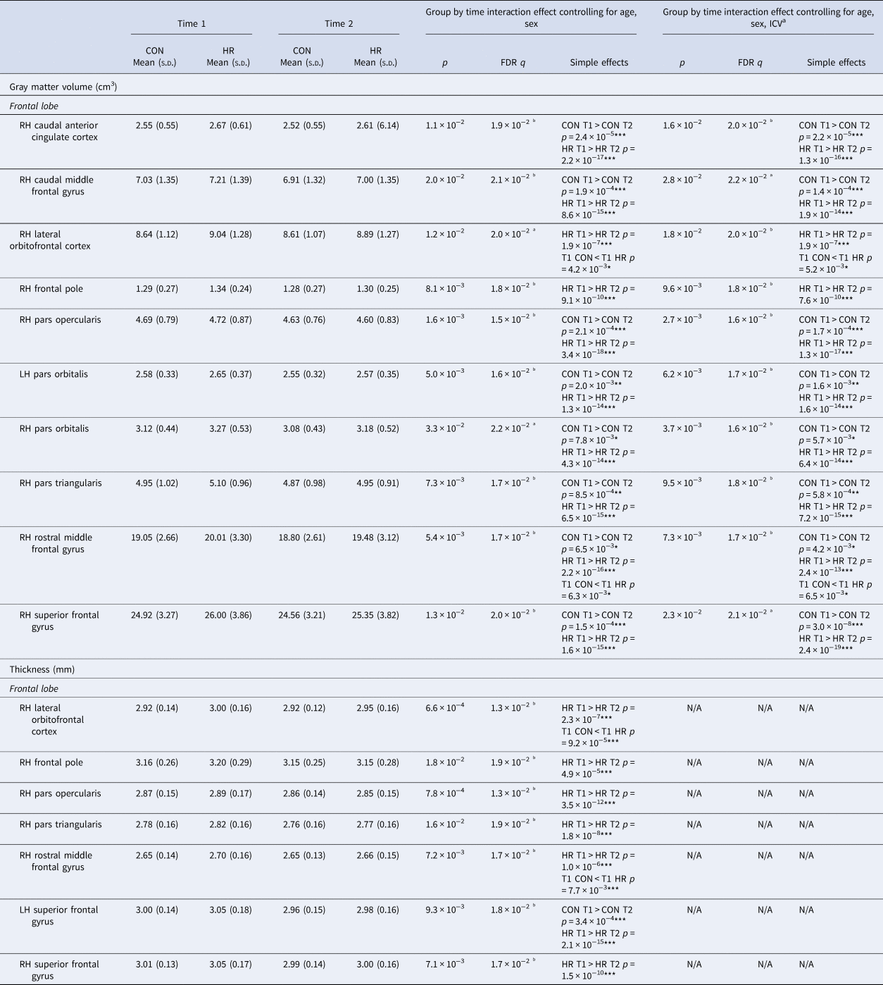
CON, controls; HR, high-risk; ICV, intracranial volume; LH, left hemisphere; RH, right hemisphere; N/A, not applicable.
Simple effects tests were tested against Bonferroni-adjusted significance levels (p < α/4). *p < 0.05/4, **p < 0.01/4, ***p < 0.001/4.
a Significant effects (p < 0.05) prior to controlling for multiple tests.
b Effects that survive FDR correction at a threshold of p < 0.05.
Group (across time) effects for ROIs that did not show a group by time interaction
Group effects are reported in online Supplementary Table S5. After correcting for multiple comparisons, we found that, independent of time, HR subjects had larger GM volumes (after ICV correction) in the right medial orbitofrontal cortex and accumbens area compared with CON subjects. Similarly, across time HR subjects had thicker left caudal anterior cingulate cortex compared with CON subjects. Online Supplementary Table S5 reports additional regions that survived prior to controlling for ICV; online Supplementary Table S6 reports group effects that did not survive correction for multiple tests.
Clinical sub-group results
Over time, all thickness and volume ROIs presented in Figs 1 and 2 revealed significant reductions in HR subjects without the new onset of a mood episode or new onset of any DSM-IV diagnosis. In HR subjects with the new onset of a mood episode (n = 9) or any DSM-IV diagnosis (n = 11), differences over time were not evident for some ROIs. As effect sizes for differences over time in these ROIs were similar for HR subjects without new diagnostic onsets compared to HR subjects with new onsets, it is likely that small sample sizes in HR subjects with new diagnostic onsets limited the ability to detect effects over time. Further details of these auxiliary analyses are provided in online Supplementary Results, Supplementary Tables S8 and S9, and Supplementary Figures S2–S4.
Auxiliary results
When current medications and mood episode; IQ; years of education; proband; lifetime alcohol use or dependence were included as predictors, all main effects of group and group by time interactions that survived correction for multiple comparisons remained significant (p < 0.05, FDR corrected). All ROIs that showed a main effect of group or a group by time interaction after correction for multiple comparisons (FDR corrected) remained significantly different after accounting for current mood state (MADRS or CDI score). When BMI was included as a predictor, four ROI group by time effects of volume survived prior to, but not after controlling for multiple tests and one ROI effect of thickness was not significant (see online Supplementary Results). However, BMI did not correlate these five ROIs (all ps>0.07 uncorrected). All other main effects of group and group by time interactions that survived correction for multiple comparisons remained significant (p < 0.05, FDR corrected) when BMI was included as a predictor.
Discussion
We studied longitudinally morphometric brain changes in young individuals at high genetic risk for BD. When compared to controls, HR subjects showed accelerated loss of both cortical thickness and volume in a number of right lateralised frontal regions, including IFG, lateral orbitofrontal cortex, frontal pole and rostral middle frontal gyrus. Accelerated volume reductions were seen in the left IFG and right caudal anterior cingulate cortex, and a steeper rate of cortical thinning in the bilateral superior frontal gyrus. There were no significant group by time interactions in surface area, suggesting that cortical thickness and not surface area was primarily responsible for the volume loss.
In a recent highly powered cross-sectional study of GM thickness and surface area from the international ENIGMA consortium, adults with established BD (n = 1837; average age 38 years) showed reduced cortical thickness in frontal regions including the left IFG, and rostral middle frontal cortex compared to controls (n = 2582; average age 35 years) (Hibar et al., Reference Hibar, Westlye, Doan, Jahanshad, Cheung, Ching and Mwangi2018). Although the direction of findings has been inconsistent in high-risk studies, frontal thickness and volume abnormalities have also been reported in individuals at risk of developing BD compared to controls (Hulshoff Pol et al., Reference Hulshoff Pol, van Baal, Schnack, Brans, van der Schot, Brouwer and Kahn2012; Nery et al., Reference Nery, Monkul and Lafer2013; Roberts et al., Reference Roberts, Lenroot, Frankland, Yeung, Gale, Wright and Mitchell2016). Frontal regions represent major hubs of executive control, and have a regulatory function over emotional processing, functions clearly aberrant in mood disorders (Townsend & Altshuler, Reference Townsend and Altshuler2012). More specifically, existing studies suggest that caudal regions of the anterior cingulate cortex are involved in appraisal and expression of emotion, whereas rostral portions of the middle frontal gyrus have a regulatory role with respect to limbic regions involved in generating emotional responses (Etkin, Egner, & Kalisch, Reference Etkin, Egner and Kalisch2011). The orbitofrontal cortex is understood to be involved in both the recognition of positive and negative reinforcers and the subsequent correction of behaviour (Rolls, Reference Rolls2004). The IFG, a region critical for response inhibition (Hampshire, Chamberlain, Monti, Duncan, & Owen, Reference Hampshire, Chamberlain, Monti, Duncan and Owen2010), in particular has been highlighted as a key region linked to the genetic vulnerability of BD (Hajek, Cullis, et al., Reference Hajek, Cullis, Novak, Kopecek, Blagdon, Propper and Alda2013). The structural changes that we report in these frontal regions may be related to activation differences in the prefrontal cortex seen in participants at-risk of developing BD using functional MRI during tasks of response inhibition (Roberts et al., Reference Roberts, Green, Breakspear, McCormack, Frankland, Wright and Mitchell2013) and emotional processing (Ladouceur et al., Reference Ladouceur, Diwadkar, White, Bass, Birmaher, Axelson and Phillips2013; Surguladze et al., Reference Surguladze, Marshall, Schulze, Hall, Walshe, Bramon and McDonald2010).
Our results are consistent with the negative association between duration of illness and IFG volume reported in cross-sectional studies (Hajek, Cullis, et al., Reference Hajek, Cullis, Novak, Kopecek, Blagdon, Propper and Alda2013; Matsuo et al., Reference Matsuo, Kopecek, Nicoletti, Hatch, Watanabe, Nery and Soares2012) and may suggest that illness-related plasticity could be involved. Our findings are also in keeping with longitudinal studies of BD patients that report accelerated frontal GM volume loss (Lim et al., Reference Lim, Baldessarini, Vieta, Yucel, Bora and Sim2013), and cortical thinning in inferior frontal cortices (Abé et al., Reference Abé, Liberg, Song, Bergen, Petrovic, Ekman and Landén2020) over time relative to controls.
Our finding of accelerated GM loss in widespread frontal regions in HR individuals suggests that the frontal differences previously observed in established BD are developing prior to the diagnosis of the disorder. For the most part, we found that abnormalities in the HR group were right lateralised. Our right lateralised findings may be related to a dominant role of the right hemisphere in emotional processing (Alves, Fukusima, & Aznar-Casanova, Reference Alves, Fukusima and Aznar-Casanova2008). Importantly, these findings were not explained by exposure to psychotropic drugs or current symptom severity.
It has been well-established that normal brain development is generally characterised by peak GM in childhood, followed by a steady decrease through the second and third decades, while WM generally expands until late adolescence, and is stable through adulthood before decelerating during old age (Mills et al., Reference Mills, Goddings, Herting, Meuwese, Blakemore, Crone and Sowell2016). However, peak GM and WM for specific cortical areas are typically reached at different ages (Tamnes et al., Reference Tamnes, Herting, Goddings, Meuwese, Blakemore, Dahl and Crone2017), with frontotemporal WM showing protracted maturation followed by a later decline (Yap et al., Reference Yap, Teh, Fusar-Poli, Sum, Kuswanto and Sim2013). A recent large-scale longitudinal study that replicated prior findings of cortical thinning during adolescent in a normal population also identified genes associated with cortical thinning (Parker et al., Reference Parker, Patel, Jackowski, Pan, Salum, Pausova and Paus2020). The authors report a considerable overlap between the genes involved in cortical thinning and the genes relevant to the aetiology of psychiatric disorders including BD.
Our group differences over time are likely to represent disturbances of normal brain development linked to increased genetic vulnerability to mood disorders. Speculatively, these findings might be viewed as maladaptive or of a neuroprotective nature. Our results are consistent with a pathological acceleration of the normal thinning, particularly of frontal thinning in this young age group. We hypothesise that molecular pathways underlying the development of normal cortical thinning overlap with those involved in the pathophysiology of BD. Findings with respect to the right rostral middle frontal and lateral orbitofrontal cortex, in particular, suggest a trajectory of an initial increase in GM thickness and volume at (or prior to) baseline followed by accelerated decline.
Exploratory sub-group analyses did not reveal group by time differences emerging in concert with the onset of a mood episode or ‘any’ DSM-IV diagnosis. Thus, we could not differentiate between those who could be viewed as ‘resilient’ over a 2-year period and those who were ‘vulnerable’, as defined by the subsequent onset of a disorder. We acknowledge that these exploratory sub-group analyses only have sufficient power to detect very strong differences between such resilient and vulnerable groups.
The direction of our findings is generally consistent with the Scottish Bipolar Family Study which reported significant decreases in right amygdala GM volume in high-risk individuals who were diagnosed with MDD 2 years after a second scan relative to both controls and high-risk individuals who remained well (Nickson et al., Reference Nickson, Chan, Papmeyer, Romaniuk, Macdonald, Stewart and Sussmann2016). However, it is noteworthy that this effect was only evident in that study at the region of interest, but not at the whole-brain level. Notably, both an earlier study from the same cohort that based group allocation on clinical diagnosis at the time of the second scan (Papmeyer et al., Reference Papmeyer, Sussmann, Stewart, Giles, Centola, Zannias and McIntosh2016) and our current study reported no significant between-group differences or between-group differences over time in a range of sub-cortical regions including the amygdala after controlling for multiple tests. We also hypothesise that abnormalities in frontal regions of the cortex are likely to represent trait markers of vulnerability to BD whereas sub-cortical abnormalities may only emerge during the course of the disorder.
In accord with the direction of our IFG findings, the Scottish group showed accelerated IFG thinning over time in high-risk individuals who remained well relative to controls (Papmeyer et al., Reference Papmeyer, Giles, Sussmann, Kielty, Stewart, Lawrie and McIntosh2015). Although controls did not differ from those high-risk individuals who were diagnosed with MDD by follow-up (n = 20), over time individuals with MDD showed left IFG thickening compared with high-risk individuals who remained well (n = 63) (Papmeyer et al., Reference Papmeyer, Giles, Sussmann, Kielty, Stewart, Lawrie and McIntosh2015). Our study had a larger number of high-risk individuals who were diagnosed with at least one depressive episode at follow-up (n = 31) but did not observe IFG differences between high-risk sub-groups. Further, the present study did not find IFG differences between HR subjects with (n = 32) v. without (n = 58) a mood episode by follow-up. Another study found that unaffected twins discordant for affective disorders showed greater IFG GM volume both at baseline and at 7-year follow-up (Macoveanu et al., Reference Macoveanu, Baaré, Madsen, Kessing, Siebner and Vinberg2018). However, given the relatively long inter-scan interval and the very limited sample of BD proband relatives in that study (n = 10), it is not possible to make direct comparisons with our current investigation.
Across the two time-points, high-risk subjects had larger GM volumes in the right medial orbitofrontal cortex and accumbens area and thicker left caudal anterior cingulate cortex compared with control subjects. Supporting our current finding, various brain regions have been reported to be significantly larger in those at high-risk of developing BD compared to controls in a number of prior studies (Bauer et al., Reference Bauer, Sanches, Suchting, Green, El Fangary, Zunta-Soares and Soares2014; Boccardi et al., Reference Boccardi, Almici, Bresciani, Caroli, Bonetti, Monchieri and Frisoni2010; Hajek et al., Reference Hajek, Gunde, Slaney, Propper, MacQueen, Duffy and Alda2009), including our own recent baseline cross-sectional study from the same cohort (Roberts et al., Reference Roberts, Lenroot, Frankland, Yeung, Gale, Wright and Mitchell2016). It should be noted that our two studies are not directly comparable as slightly different samples were used and the group findings reported in the present study were pooled across two time-points. These group differences remained stable over the 2-year inter-scan interval and likely occurred at some point prior to the baseline scan. Taken together, our findings suggest that differences may be progressive in some regions and stable in others.
Some limitations need to be addressed. As neuroimaging scans were only assessed over two time-points, only we could not access the modelling of non-linear trajectories. Sample numbers for exploratory HR sub-group analyses were small, limiting our ability to detect anything but very strong effects. It remains unknown whether HR participants without a DSM diagnosis will develop the new onset of affective and non-affective episodes. With all the current participants enrolled in an ongoing longitudinal study, it is expected that at least 10% of our HR individuals will ultimately develop BD over time. Ongoing neuroimaging of individuals over time will provide a clearer picture of whether there is continued accelerated decline in frontal regions and clarify whether such changes coincide with the onset of BD and other affective diagnoses. As genetic liability is not the only potential explanation of these findings, environmental risk factors such as chronic stress could also contribute to neural differences in high-risk populations.
As detailed in our Methods section, at baseline we did not exclude CON and HR participants with non-BD psychopathology. Our groups were distinguished and defined by the presence (HR) or absence (CON) of a first-degree relative with BD. In support of this genetic liability distinction, over the 2-year follow-up period, five participants in the HR group but none in the CON group developed a first-onset of hypo/mania. Further, as the ‘ecological’ approach which we employed reduces the likelihood of differences in symptom severity (such as depressive symptoms) between the CON and HR groups, the chances of demonstrating neuroimaging differences between the groups are thereby also reduced. As defining groups on the basis of family history are different from most neuroimaging studies in which groups are distinguished on the basis of diagnosis v. no diagnosis, we acknowledge that the results of other studies may not be directly comparable to the current report.
Finally, given that there is no standard practice for analysing neuroimaging data and reporting results we acknowledge that the results of other studies are not directly comparable. For example, differences in imaging parameters, such as field strength; FreeSurfer version; choice of region of interest v. whole-brain approach; or voxel-based v. atlas-based approach, may be attributed to the heterogeneity of findings in the literature. Given that volume is a gross measure of WM structure, methods such as diffusion imaging may reveal subtle microstructural differences that were not detected in the current study. Patterns of effects may also differ when examining other brain imaging modalities such as WM tracts and task-related or resting-state networks. Future studies are needed to provide better insight into multimodal and neurobiological processes mediating the increased risk of developing BD.
Conclusions
The present study confirmed that neurobiological differences over time are observable in individuals at increased risk of developing BD prior to the emergence of specific psychopathology. A steeper rate of GM loss was observed in right lateralised frontal regions, including the IFG, lateral orbitofrontal cortex, frontal pole and rostral middle frontal gyrus of high-risk individuals compared to controls. Structural brain development in high-risk individuals follows regionally specific complex trajectories, with a mix of differences that remained stable over time, baseline differences that normalised over time and different rates of progression over time in the absence of differences at either time-point. As abnormalities in cortical thickness were not always reflected as abnormalities in cortical volume, these results highlight the importance of assessing multiple complementary morphometric metrics. These findings provide new insights into the neurodevelopmental patterns associated with increased vulnerability for BD and may have future implications for early identification and intervention.
Supplementary material
The supplementary material for this article can be found at https://doi.org/10.1017/S0033291720003153
Acknowledgements
We are grateful to all participants and their families for their valuable contribution to this study.
Financial support
This study was funded by the Australian National Medical and Health Research Council [Program Grant 1037196 (PM and MB), Project Grant 1066177 (JMF, RL) and 1063960 (JMF, BJO)], the Lansdowne Foundation, Good Talk and the Keith Pettigrew Family Bequest (PM). We gratefully acknowledge the Janette Mary O'Neil Research Fellowship (JMF) and Betty Lynch (JMF) for supporting this work and our team.
Conflict of interest
None.