Introduction
The crystal chemistry of hydroxyperovskites has been reviewed by Mitchell et al. (Reference Mitchell, Welch and Chakmouradian2017). The key structural features are (1) the absence of a cavity A cation, (2) all oxygens form hydroxyl groups and (3) each oxygen is an H donor and acceptor. The absence of a cavity cation and the moderately strong hydrogen bonding leads to highly rotated octahedra.
There are two distinct groups: (1) single hydroxyperovskites with trivalent cations only and stoichiometry B(OH); (2) double hydroxyperovskites with stoichiometry BB′(OH)6, where B is a monovalent or divalent cation and B′ is a tetravalent or pentavalent cation. Polymorphism involving cubic and tetragonal structures has been observed in Ga(OH)3 and MnSn(OH)6. In double hydroxyperovskites B and B′ octahedra alternate.
Different hydrogen bonding configurations occur in hydroxyperovskites depending upon space group and octahedra tilt system. For example, cubic structures with space groups Pn  $\bar 3$ and Im
 $\bar 3$ have three equal in-phase tilts a + a + a + that lead to isolated 4-membered rings of ···O–H···O–H··· linkages. Crankshafts composed of hydrogen-bonded bridges occur in tetragonal, orthorhombic and monoclinic hydroxyperovskites in combination with isolated 4-membered O–H···O–H rings or zig-zag chains.
There are 15 known natural hydroxyperovskites: eight stannates, one antimonate, Fe(OH)3 (bernalite), In(OH)3 (dzhalindite), Ga(OH)3 (söhngeite), and three germanates (stottite, zincostottite and nancyrossite), all of which are exceptionally rare minerals. Only one hydroxyperovskite with B +B′5+(OH)6 stoichiometry is known: mopungite NaSb(OH)6.
Here, we report the discovery of a new double hydroxyperovskite and member of the stottite subgroup that is unusual in having five rather than the usual six H per formula: nancyrossite FeGeO6H5. The mineral honours Prof. Nancy L. Ross, mineral physicist and mineralogist at Virginia Polytechnic Institute, Blacksburg, Virginia, USA. Nancy Ross has made major contributions to our knowledge of the crystal chemistry, thermochemistry and crystallography of perovskites and related phases, including stottite. The mineral and name (symbol Ncy) have been approved by the International Mineralogical Association (IMA2024-033; Welch et al., Reference Welch, Najorka, Kleppe, Kampf and Spratt2024a).
Experimental methods
Sample
The sample containing stottite and nancyrossite was donated to MDW by the mineral collector and mineralogist William Pinch of Rochester, New York, USA. This sample, which comprises only loose crystals, also contains grains of sphalerite and galena and is from the germanium horizon at Level 30 of the Tsumeb mine, Namibia (19°15’S, 17°42ʹE), the type locality of stottite. In addition to broken crystals, stottite occurs in the sample as large cuboidal crystals up to 1 mm in diameter. Occasionally, larger crystals sometimes display stepped surfaces that are probably due to etching or growth phenomena. The new mineral described herein, nancyrossite, occurs as subhedral grains or fragments up to 0.3 mm maximum dimension, that do not display stepped surfaces.
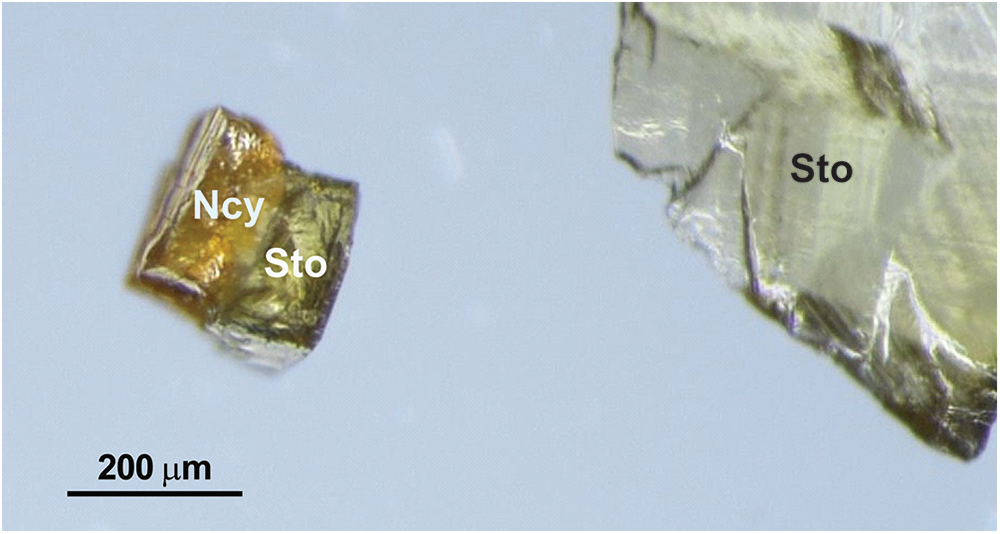
While selecting stottite crystals for a high-pressure study, numerous honey-coloured crystals were found that differed from the usual greenish yellow of stottite, as is shown in Fig. 1. These were screened by single-crystal X-ray diffraction (SCXRD) and found to have a much smaller unit cell than stottite, e.g. 396.76(2) Å3 and 396.66(2) Å3 versus 425.22(2) Å3. Structure determination by SCXRD indicated that the FeO6 octahedron of the honey-coloured phase was much smaller than that of stottite and was consistent with a dominantly ferric state for Fe. As such, this phase was recognised as a new mineral, and its formal description is given here.
Photomicrograph of a composite grain (left) comprising distinct nancyrossite (Ncy) and stottite (Sto) components; to the right of this composite grain is a larger single stottite crystal. SCXRD shows that the two crystals of the composite grain have structural continuity of their lattices whereby a and c axes are coincident. The outer surface of the composite grain shows growth steps also commonly seen in stottite and jeanbandyite, probably due to etching. The different colours of the two minerals are evident.

Three separate crystals were used in the characterisation of nancyrossite (crystals #7, #8 and #9). Crystals #7 and #8, for which crystal structures were determined, were used for analysis by electron microprobe and are mounted together on a glass slide. The full structure of crystal #9 is reported here. All three crystals are very similar and have almost identical structural and chemical data; they are deposited in the mineral collection of the Natural History Museum, London, UK, catalogue numbers BM2024,1, BM2024,2 and BM2024,3 for crystals #9 (holotype), #7 and #8, respectively.
Electron microprobe analysis
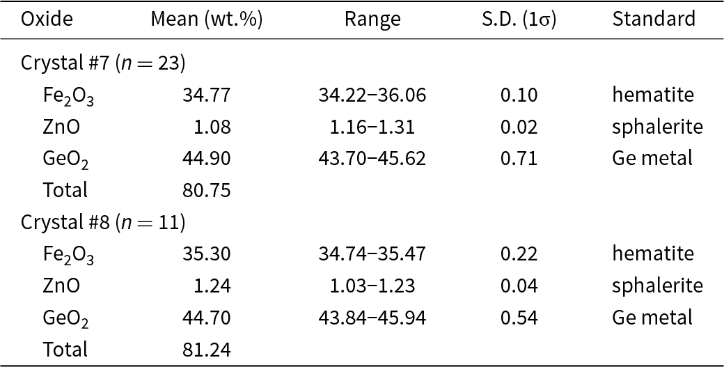
Two co-type crystals (#7 and #8) were mounted in epoxy and polished for electron microprobe analysis. Wavelength-dispersive analysis was done using a Cameca SX100 electron microprobe with an accelerating potential of 20 kV, a current of 20 nA and a 5 mm beam. Standards used are given in Table 1. A preliminary survey using energy-dispersive analysis indicated that the only metals present are Zn, Fe and Ge. Notably, Mn and Sn were below detection limits. The H content of nancyrossite could not be determined using chemical methods, because not enough material is available (five small crystals). Consequently, the H content is deduced from the crystal structure reported here. No suitable material was available for Mössbauer spectroscopy. Divalent and trivalent iron contents were deduced from the crystal structure, as described below.
Electron microprobe data for nancyrossite crystals #7 and #8.

Optical properties
The holotype crystal (#9) was used for determination of optical properties. The determination of RI values using a spindle stage was problematic because the refractive indices were above 1.8, the limit for accurate measurement in the stage’s optical cell due to the opacity (darkness) of the high-RI liquids needed. The optic axial angle (2V) was measured and determined using extinction data analysed with EXCALIBURW (Gunter et al., Reference Gunter, Bandli, Bloss, Evans, Su and Weaver2004). Conoscopic figures were consistent with the measured high 2V.
As is also the case with stottite (tetragonal, P42/n), nancyrossite displays anomalous biaxial optics. Jeanbandyite, cubic Pn  $\bar 3$, has anomalous uniaxial optics (Kampf, Reference Kampf1982). Birefringence values for β – α and γ – β were obtained using a Berek compensator. These values taken with those estimated using the Gladstone–Dale equation based upon the crystal structure determination of crystal #9 allows a meaningful description of the optical characteristics of nancyrossite.
Raman spectroscopy
Unpolarised Raman spectra were collected for crystals #8 (before mounting for electron microprobe) and #9. These crystals were placed on the culet of an ultra-low fluorescence diamond and spectra recorded over the range 100–3900 cm–1. The instrument used was the LabRAM HR800 (Horiba Jobin Yvon) at beamline I15, Diamond Light Source, UK. It was equipped with 1200g grating, a 20× objective, and an electrically cooled CCD detector. Excitation of each crystal used the 532 nm line of a 300 mW Torus laser focused down to a 10 μm spot on the sample and collected through a 75 mm confocal aperture using 180° back-scattering geometry. Laser power at the sample position varied between 3–4 mW; exposure times were 2× 120 s. The intrinsic resolution of the spectrometer is <1.0 cm–1 and calibrations are accurate to ±1 cm–1. The frequency of each Raman band was obtained by fitting Voigt line profiles using a least-squares algorithm.
X-ray diffraction
Quasi-powder X-ray diffraction data were collected for crystal #9 using a Gandolfi-type movement (φ and ω circles) on a Rapid II diffractometer (Rigaku Oxford Diffraction) operated at 45 kV and 36 mA, equipped with a curved image plate and graphite monochromatized CuKα radiation. Diffraction patterns were processed using the 2DP program (Rigaku-Oxford Diffraction) and the HighScore Plus program (Malvern Panalytical, Degen et al., Reference Degen, Sadki, Bron, König and Néner2014).
Single-crystal XRD was carried out using an Xcalibur four-circle single-crystal diffractometer (Rigaku-Oxford Diffraction) equipped with a 1K Eos area detector and graphite-monochromated MoKα radiation. Data collection and data reduction used the Crysalis program (©Rigaku Oxford Diffraction). Raw intensities were corrected for Lorentz, polarisation and absorption effects, and converted to structure factors. A multi-scan absorption correction was applied. Crystal structure determination used SHELX (Sheldrick, Reference Sheldrick2015), as detailed in the Results section.
Results
Chemical formula
Empirical formulae of nancyrossite crystals #7 and #8, calculated on the basis of six O2– + five H+, are: Fe3+1.01Zn0.03Ge0.98O6H5 (#7) and Fe3+1.01Zn0.04Ge0.98O6H5 (#8). The % H2O values shown in Table 1 were calculated for both average formulae for 2.5 H2O (5 H atoms per formula unit, apfu). The simplified formula is (Fe3+,Zn)GeO6H5. The ideal chemical formula is FeGeO6H5 which requires Fe2O3 34.80, GeO2 45.59, H2O 17.48, total 100 wt.%. The values for stottite, FeGe(OH)6, are: FeO 31.18, GeO2 45.39, H2O 23.42, total 100 wt.%. The empirical values are clearly closer to those of nancyrossite than stottite. The ferric state of Fe in nancyrossite is demonstrated by crystal structure determination and bond-valence calculations, as described below. The rationale for writing the ideal chemical formula as FeGeO6H5 is given in the Discussion section (‘Chemical formula’) with particular reference to jeanbandyite.
Optical properties
As noted above, the refractive indices of nancyrossite are very high and precluded direct determination using heavy liquids. The alternative approach outlined above gave the following properties: β – α = 0.005 (meas.), γ – β = 0.003 (meas); biaxial (–): α = 1.966 (calc.), β = 1.971 (calc.), γ = 1.974 (calc.); Gladstone-Dale n av = 1.97; 2Vmeas = 73.5(4)°, 2Vcalc = 75.4°; dispersion r < v slight; orientation X = c; non-pleochroic.
Raman spectroscopy
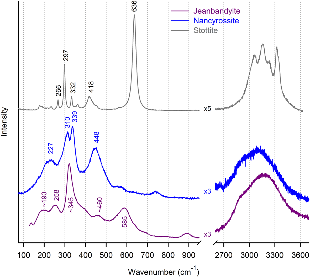
The Raman spectra of nancyrossite crystal #8 and jeanbandyite (Hingston Quarry, Cornwall, UK) are shown in Fig. 2. These spectra in the (OH)-stretching region are very similar, comprising two broad bands at ∼2850 and 3150 cm–1. The Raman spectrum of stottite from Tsumeb studied by Kleppe et al. (Reference Kleppe, Welch, Crichton and Jephcoat2012) is shown in Fig. 2 and comprises five resolved bands that lie within the envelope defined by the nancyrossite and jeanbandyite spectra. High Full Width at Half Maximum (FWHM) values for Raman and infrared bands spectra in the (OH)-stretching region is characteristic of hydroxyperovskites, e.g. 60–110 cm–1. Typical oxygen–oxygen donor–acceptor distances of hydroxyperovskites are 2.7–2.8 Å (see Table 7 below), implying weak hydrogen bonding. Hence, it is unlikely that the high FWHM values are due to significant anharmonicity of the O–H bond in these structures. The origin of the high FWHM values remains to be determined.
The Raman spectra of nancyrossite from Tsumeb (blue) compared with that of jeanbandyite from Hingston Quarry, Cornwall, UK (purple). Both spectra have two broad bands in the (OH)-stretching region 2700–3600 cm–1. At the top is the Raman spectrum of stottite from Tsumeb in which the five resolved OH bands lie within the same wavenumber range as the two broad bands of the nancyrossite and jeanbandyite spectra.

Powder X-ray diffraction
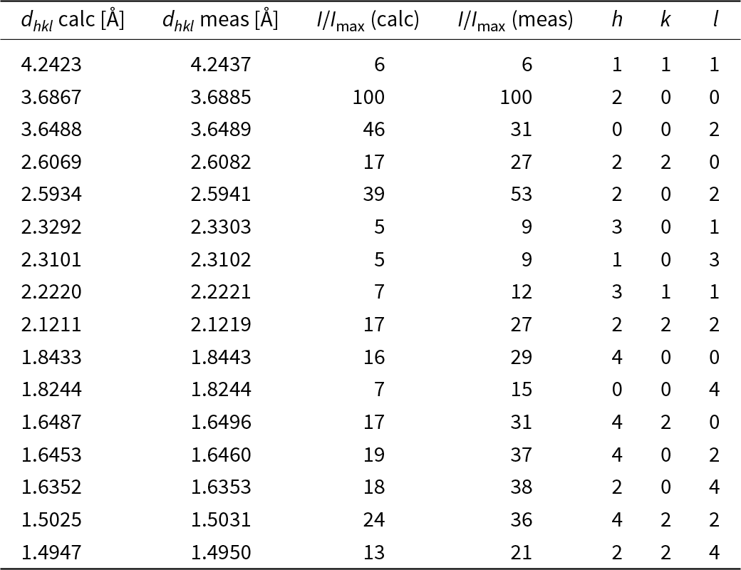
Calculated and measured dhkl values and their intensities are shown in Table 2. Refined unit cell parameters (tetragonal, space group P42/n) are a = 7.3770(1) Å, c = 7.2977(1) Å, V = 397.149(9) Å3; R(profile) = 0.019, wR(profile) = 0.030 and GoF =1.221.
Calculated and measured powder XRD data for nancyrossite from a quasi-random pattern of the holotype crystal collected using a Gandolfi-type movement with CuKα radiation. Only reflections with intensities above 5% are given.

Crystal structure: SCXRD
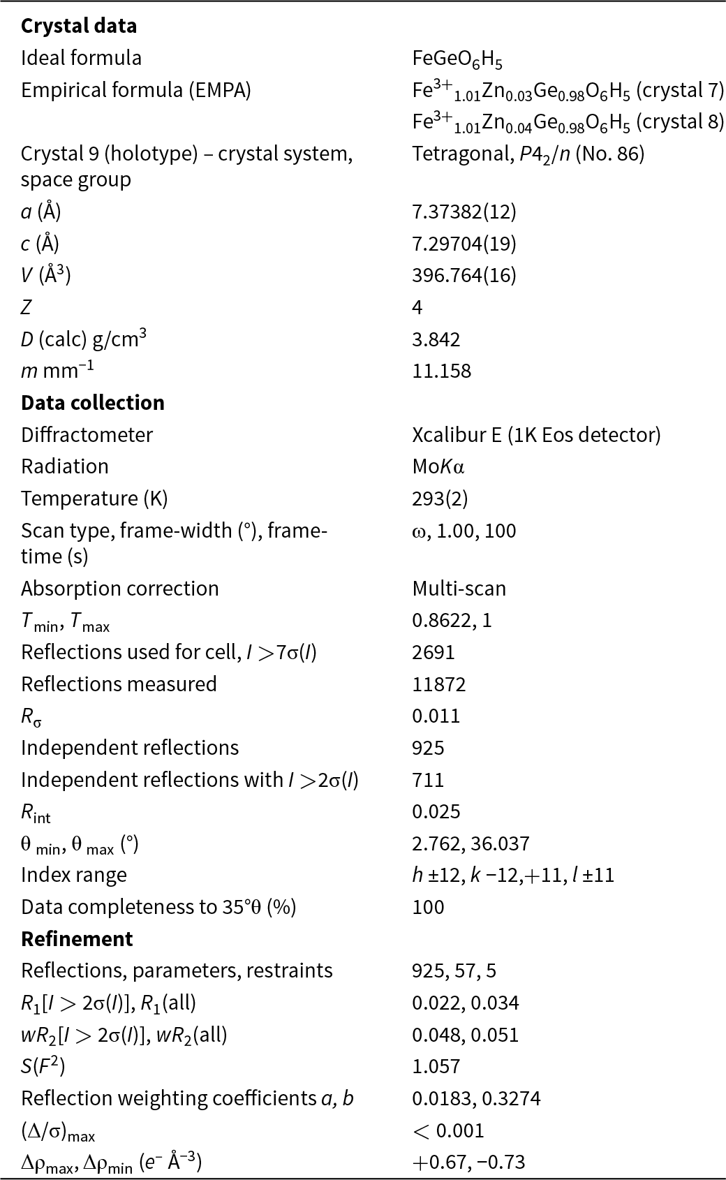
The structures of three nancyrossite crystals (#7, #8, #9) were determined in space group P42/n using SHELX (Sheldrick, Reference Sheldrick2015). The refined structures of all crystals are very similar. The structure of the holotype (#9) is reported here and is representative of the structures of all nancyrossite crystals studied. Only eight very weak violators of the n-glide and screw tetrad were present, all having I/σ(I) <0.6. Consequently, P42/n is considered the correct choice of space group. An attempt to refine in non-centrosymmetric space group P42, described below, clearly indicated that a centrosymmetric structure is the correct choice, i.e. space group P42/n.
Neutral scattering factors for Fe, Ge, O and H were used (Wilson, Reference Wilson1992). Refinement of site scattering at the Fe site confirmed that it is fully occupied by Fe and so the occupancy was not refined thereafter (occ. = 100% Fe). Minor merohedral twinning (minor twin = 8%) was detected during the early stages of refinement and this was incorporated into the refined structure model. A soft restraint of 0.85 ±0.05 Å for the O–H bond was used for the refinement of H atom positions; the isotropic displacement parameters U iso, of all H atoms were linked and refined as a single variable.
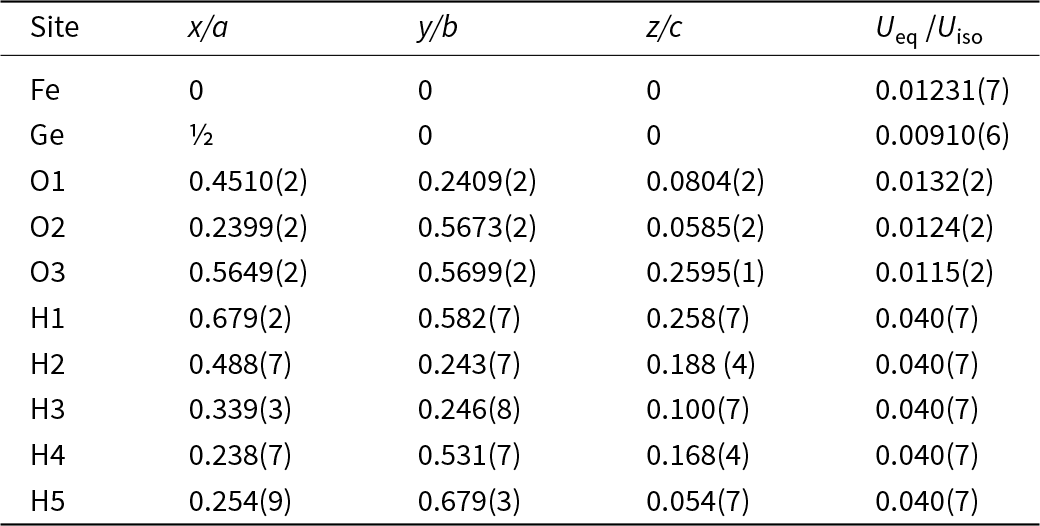
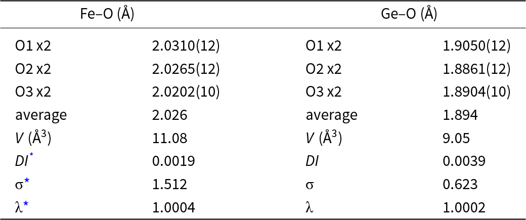
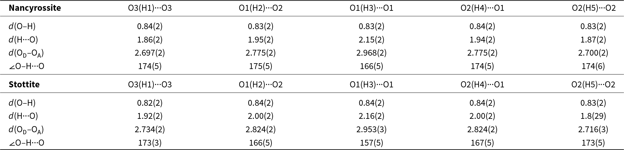
Results are summarised in Tables 3–8. A crystallographic information file (cif) for the holotype crystal (#9), including structure factors, is deposited as supplementary material (see below). A summary comparing unit cell parameters, refinement indices and geometric parameters for crystals #7, #8 and #9 is shown in Table 6.
Information relating to data collection and structure refinement of nancyrossite (crystal #9) at 293 K.

Atom coordinates and equivalent-isotropic displacement parameters (Å2) of nancyrossite (crystal 9). An overall U iso was refined for the five H atoms; all H sites are half-occupied.

Geometrical parameters for coordination polyhedra in nancyrossite.

* Distortion Index (DI), bond-angle variance (σ) and quadratic elongation (λ) defined by Robinson et al. (Reference Robinson, Gibbs and Ribbe1971).
Unit cell parameters, structure refinement parameters, bond-lengths and octahedral volumes for crystals #7, #8 and #9.

Hydrogen bonding in nancyrossite (crystal #9) and stottite. The values for stottite were obtained from a refinement of a crystal used in the study by Kleppe et al. (Reference Kleppe, Welch, Crichton and Jephcoat2012). Distances in Å, angles in degrees.

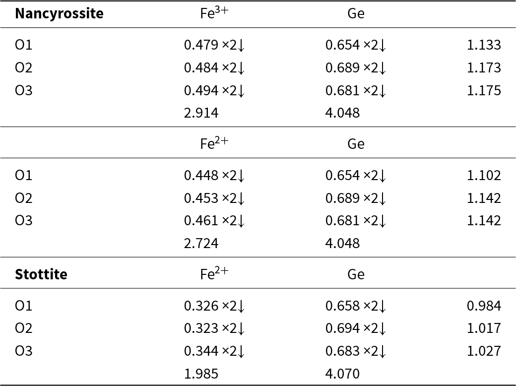
Bond valence calculations for nancyrossite and stottite. Data for stottite are calculated from a refinement of the same crystal as that studied by Kleppe et al. (Reference Kleppe, Welch, Crichton and Jephcoat2012). Bond valence parameters for Fe2+–O, Fe3+–O and Ge–O bonds were taken from Brese and O’Keeffe (Reference Brese and O’Keeffe1991).

It is important to realise that it is now possible to recognise partially occupied H sites using SCXRD when crystals of high quality are available, e.g. fully occupied versus half-occupied H sites (e.g. Lafuente et al., Reference Lafuente, Yang and Downs2015; Welch and Kleppe, Reference Welch and Kleppe2016; Welch et al., Reference Welch, Najorka and Wunder2024b). For example, in the early stages of refinement of the stottite (P42/n), the unmodelled residual electron-density in the difference-Fourier map given in the Q-list output by SHELX for a H-free model contains five maxima that correspond to one full and four partially occupied H sites that can be identified based upon O–H distance (0.8–0.9 Å for XRD) and O–H···O configuration (oxygen donor–acceptor distance and approximate O–H···O angle). Taken together with O–H distances, the highly directional O–H (non-bifurcated) hydrogen bonds of hydroxyperovskites allow plausible H positions to be checked and refined. This approach successfully located the five non-equivalent half-occupied sites of nancyrossite.
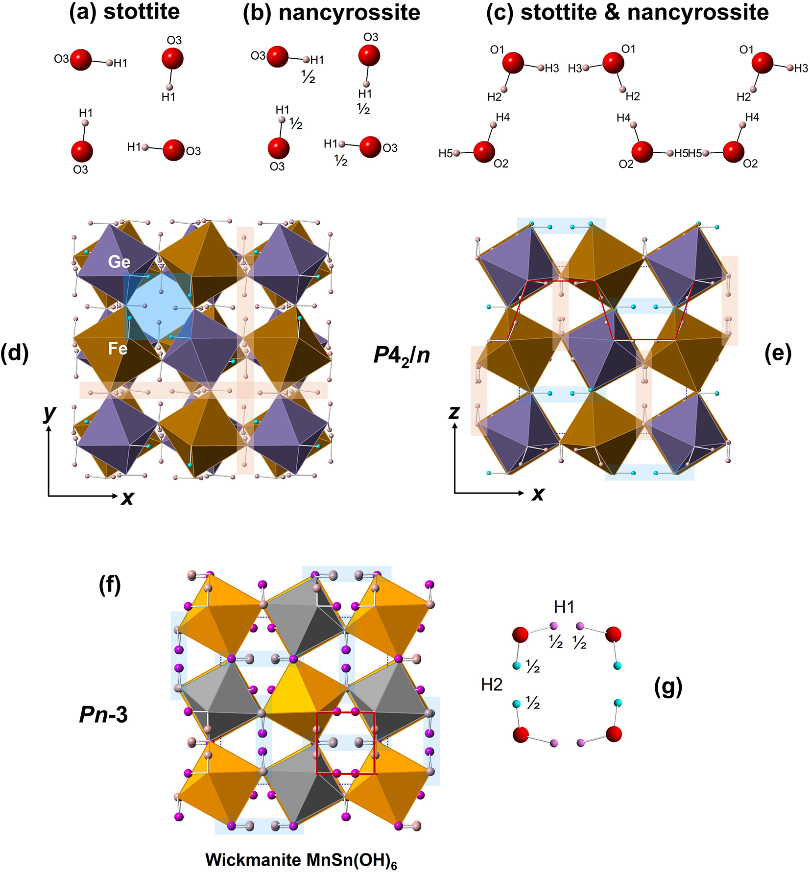
Iron, Ge and all five non-equivalent O atoms were found by direct methods and then refined by full-matrix least-squares. Plausible sites for all five H atoms were found (Table 4, Fig. 3). These sites are analogous to those of stottite except for the occupancy of the four-membered ring site H1, which appears to be half-occupied in nancyrossite (fully occupied in stottite). Hence, the five non-equivalent H sites in nancyrossite, all on general positions of multiplicity 8, are half occupied in the P42/n model. This result is consistent with 5 H atoms per formula unit (Z = 4): ¼ × (5 × ½ × 8) = 5 H apfu.
Structures of nancyrossite and wickmanite. Tetragonal structures such as nancyrossite and stottite have two in-phase and one out-of-phase (reverse) tilts of octahedra; cubic structures such as wickmanite and natanite have three equal in-phase tilts. These different tilt systems lead to different hydrogen bonding connectivities. Cubic structures have only isolated 4-membered rings of O–H···O bridges whereas tetragonal structures have O–H···O crankshafts and isolated O–H···O rings. (a) the ring of stottite with a fully occupied H1 site; (b) the ring of nancyrossite with a half-occupied H1 site; (c) the crankshaft shared by stottite and nancyrossite structures with four half-occupied non-equivalent H sites; (d) the nancyrossite structure projected onto (001) showing the reverse tilt parallel to [001]; (e) the nancyrossite structure projected onto (010) showing the in-phase tilts, and a single in-plane crankshaft shown as a red line; (f) the wickmanite structure (Pn  $\bar 3$) showing the in-phase tilts along all three axes; (g) the four-membered O–H ring of cubic hydroxyperovskites showing the two half-occupied non-equivalent H sites. Examples of the orientations of rings (blue) and crankshafts (brown) are shown. An example of an in-plane ring is shown for nancyrossite as a blue square in (d); an example of an in-plane ring in wickmanite is shown as an outlined red square in (f). Crankshafts extend parallel to the x and y axes of nancyrossite and stottite. The figures were created using Crystalmaker software.

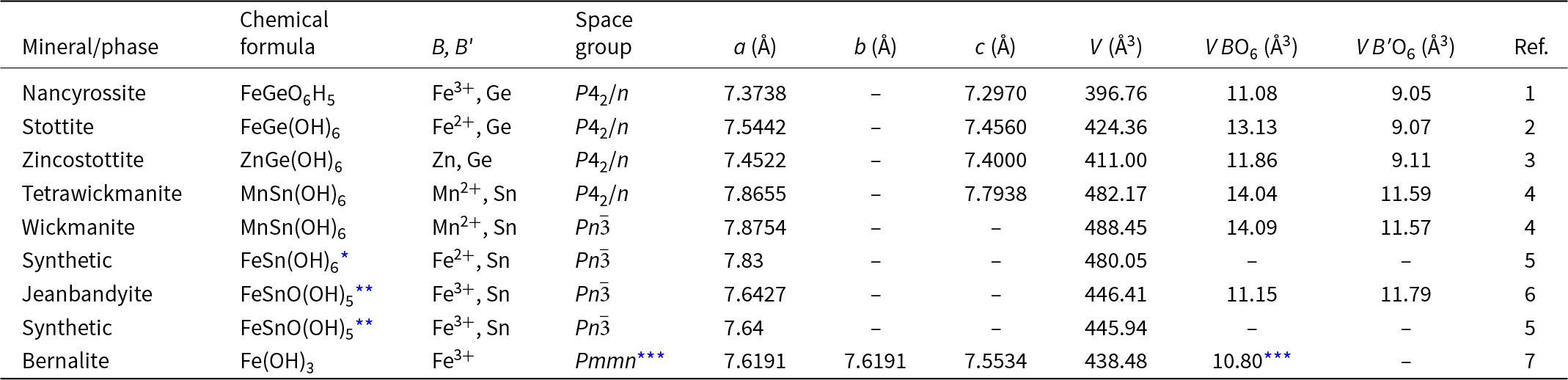
One of the two non-equivalent octahedra of nancyrossite has a volume of 9.05 Å3 and a geometry almost identical to that of the GeO6 octahedron of stottite (9.07 Å3). It is much smaller than the SnO6 octahedra of stannate hydroxyperovskites such as wickmanite MnSn(OH)6 (11.57Å3) and burtite CaSn(OH)6 (11.48 Å3). The other octahedron of nancyrossite is much smaller (11.08 Å3) than the Fe2+, Mn2+ and Mg octahedra of related hydroxyperovskites (Table 9); it is much closer to the value of the Fe3+O6 octahedron of bernalite Fe(OH)3 (10.8 Å3). Electron microprobe analysis (Table 1) confirms that the two octahedra are occupied by Fe and Ge. The small size of the Fe octahedron is consistent with occupancy by Fe3+ not Fe2+ (Table 7). Assuming a linear variation of the volume of the FeO6 octahedron with Fe2+/Fe3+ between stottite and bernalite, a value of 11.08 Å3 implies that the Fe site is 88% Fe3+. A calculation using OccQP (Wright et al., Reference Wright, Foley and Hughes2000), which uses site scattering values and bond valences, gives 0.897Fe3+:0.103Fe2+, in almost exact agreement with the result obtained using the bernalite/stottite V oct correlation.
Nancyrossite and related minerals.

References: (1) This proposal; (2) Kleppe et al. (Reference Kleppe, Welch, Crichton and Jephcoat2012); (3) Kampf et al. (Reference Kampf, Désor, Welch, Ma and Möhn2025); (4) Lafuente et al. (Reference Lafuente, Yang and Downs2015); (5) Nakayama et al. (Reference Nakayama, Kosuge and Kach1977); (6) Welch and Kampf (Reference Welch and Kampf2017); and (7) Welch et al. (Reference Welch, Crichton and Ross2005).
* A reliable structure for natanite is unavailable in the literature. Consequently, only the unit cell parameter and space group are shown here.
** Formula given here as it is reported by Nakayama et al. (Reference Nakayama, Kosuge and Kach1977) and Welch and Kampf (Reference Welch and Kampf2017).
*** Bernalite has only a single non-equivalent Fe3+ site (B = B′) and is metrically tetragonal but structurally orthorhombic.
Structure refinement of crystal #9 in non-centrosymmetric space group P42 (#77) was also attempted to test the robustness of the centrosymmetric (P42/n) model. Iron, Ge and all six non-equivalent oxygen positions appeared at the structure solution stage and stable refinement of these atoms was achieved. However, a Flack parameter of 0.46(12) and very high correlations between atoms related by a missing centre of symmetry clearly indicated that a centrosymmetric structure (i.e. P42/n) is the correct choice. Six plausible H sites were also apparent in the difference-Fourier maps at later stages of the refinement of the P42 structure. However, the refined overall U iso of these six sites was non-positive definite and it was not possible to refine the structure further.
A data collection at 100 K for a cotype crystal (MDW1) was made to see if any further information on H locations could be found. An excellent refinement in space group P42/n was obtained: R 1(all) = 0.026, wR 2(all) = 0.054 and GoF = 1.130. However, only three of the five H sites were located with certainty, all with half occupancy, i.e. the ring H site and two of the half-occupied crankshaft sites. It is, nonetheless, significant that the ring site refines more satisfactorily as half occupied in nancyrossite, unlike stottite for which full occupancy of this site is found consistently for different crystals (MDW, unpublished data).
Details of the hydrogen bonding in nancyrossite and stottite are shown in Table 7. In nancyrossite, the shortest OD–OA distances, 2.697 Å and 2.700 Å, are associated with the O(3)H(1)···O(3) of the ring and the O(2)H(5)···O(2) of the crankshaft, respectively. The longest OD–OA distance (2.968 Å) is O(1)H(3)···O(1) of the crankshaft. In stottite, the shortest OD–OA distances are for the O(3)H(1)···O(3) of the ring (2.734 Å) and O(2)H(5)···O(2) of the crankshaft (2.716 Å); the longest OD–OA distance is for O(1)H(3)···O(1) of the crankshaft (2.953 Å) as in nancyrossite distance (2.968 Å). Overall, there is close correspondence between OD–OA distances in nancyrossite and stottite.
Bond valence calculations
Bond valence sums (BVS) for Fe, Ge and O atoms of nancyrossite were calculated using the parameters of Brese and O’Keeffe (Reference Brese and O’Keeffe1991) and are shown in Table 8. These calculations confirm that Fe occurs as Fe3+ (BVS 2.91 valence units) rather than Fe2+ (2.72 vu). Oxygen bond valence sums range from 1.13 to 1.18 vu and indicate bonding to H. The same calculation for stottite is shown in Table 8 and it is clear that Fe is divalent (BVS 1.99 vu).
Discussion
Chemical formula
Crystal structure determination indicates that H atoms occur at positions that can be assigned to OH groups. However, writing the formula so as to include formal hydroxyl groups gives FeGeO(OH)5, as was done by Welch and Kampf (Reference Welch and Kampf2017) for jeanbandyite FeSnO(OH)5, and implies that one oxygen per formula is not bonded to H, which is not what is found by SCXRD, nor is it crystal-chemically reasonable as this oxygen will be very under-bonded (e.g. with a BVS of ∼ 1.2 vu). In the next section we apply the same reasoning to jeanbandyite. Given the association of H with oxygen as OH groups in nancyrossite as seen by XRD, we refer to this mineral as a hydroxyperovskite.
Relationship to other minerals
A summary of the crystallographic data for hydroxyperovskites related to nancyrossite is given in Table 9. Two minerals and one synthetic are particularly noteworthy with regard to nancyrossite: natanite, jeanbandyite and the synthetic analogue of the latter, reported by Nakayama et al. (Reference Nakayama, Kosuge and Kach1977) as FeSnO(OH)5. The reduction in unit cell volume for natanite → FeSnO(OH)5 is 33.64 Å3 (7%). The corresponding reduction for stottite → nancyrossite is 6.5% (27.60 Å3).
Using crystallographic and crystal-chemical arguments, Welch and Kampf (Reference Welch and Kampf2017) revised the formula of jeanbandyite from (Fe3+1-x, □x)(Sn1-y, □y)(OH)6 (□ = vacancy) reported by Kampf (Reference Kampf1982) to Fe3+xM 2+1–x Sn(OH)6-x (1 ≥ x ≥ 0.5) with an ideal end-member formula (x = 1) of Fe3+SnO(OH)5. However, this formula implies that one of the six oxygen atoms of the formula is not associated with H. The behaviour of H in nancyrossite has yet to be clarified. What is clear, however, is that the crystallographic data imply five H atoms per formula in the ideal structure. Until a model for H behaviour and distribution is available, we consider it to be more appropriate to write the chemical formula of jeanbandyite as FeSnO6H5 by analogy with what we propose here for nancyrossite, FeGeO6H5.
The Pn structure of hydroxyperovskites differs from that of tetragonal varieties by having different Glazer octahedral-tilt systems (Pn  $\bar 3$ = a +a +a +, P42/n = a +a +c –) that lead to very different hydrogen-bonding topologies (Mitchell et al., Reference Mitchell, Welch and Chakmouradian2017). The H-bonding arrangement of the cubic structure consists only of isolated four-membered rings, whereas the tetragonal structure has alternating (002) layers of four-membered rings and crankshafts, as shown in Fig. 3. Hence, the cubic topology (a +a +a +) of jeanbandyite reported by Kampf (Reference Kampf1982) and confirmed by Welch and Kampf (Reference Welch and Kampf2017) is correct as it cannot be confused with a tetragonal topology that has a reversed tilt (a+a+c–). Welch and Kampf (Reference Welch and Kampf2017) demonstrated that refinement of the crystal structure of jeanbandyite in space group P42/n fails to produce a satisfactory structure.
Zincostottite, ideally ZnGe(OH)6, presumed to be from the same Ge-rich horizon at Tsumeb as nancyrossite, was recently approved as a new mineral (Kampf et al., Reference Kampf, Désor, Welch, Ma and Möhn2025). The empirical formula reported is Zn0.77Fe3+0.23Ge1.00O6H5.77, with all iron in the ferric state and a corresponding H deficiency of 0.23 H apfu relative to the ideal formula. As such, this empirical formula can be considered a solid solution between zincostottite and nancyrossite.
The probable deprotonated site in nancyrossite is suggested by a comparison with jeanbandyite. The latter has the Pn  $\bar 3$ topology with only isolated rings, each having two non-equivalent H sites that, in the average structure found by diffraction methods, are both half occupied. The H sites of neighbouring rings are separated by a minimum distance of ∼2.7 Å. Whether or not this distance is too far for H interactions between adjacent rings (hopping?) is unknown. The significance of partial occupancy of H sites in hydroxyperovskites remains to be determined.
Single-crystal neutron diffraction (SCND) allows the accurate determination of occupancies of H sites. It is now possible to carry out SCND experiments on crystals with a minimum dimension of 0.1 mm. High quality crystals of nancyrossite larger than this dimension are available, and we intend to do SCND in the near future in order to locate the partially occupied H sites more accurately than is possible using SCXRD.
Implications
Synthetic hydrogen-doped perovskites are important materials for use as protonic conductors and electrolytic catalysts. Various protonation mechanisms have been used, such as the preparation of oxygen-deficient compositions followed by hydrolysis at oxygen vacancies, e.g. BaSc0.667W0.333O3 → BaSc0.8W0.2O2.8 → [BaSc0.8W0.2O2.8(OH)0.2 + 0.2 H+]. In contrast to such ‘hydrogen-doped’ structures which generate mobile protons, nancyrossite and jeanbandyite are already highly protonated and have vacant H sites that may facilitate proton migration or localised site hopping. Hydroxyperovskites are considered to be potentially significant electrolytic catalysts and photocatalysts (Evans et al., Reference Evans, Wu, Seshadri and Cheetham2020), with possible applications in fuel cells.
Stannate hydroxyperovskites present an opportunity to prepare synthetic samples of these materials as analogues of jeanbandyite FeSnO6H5, e.g. MnSnO6H5, CoSnO6H5 and NiSnO6H5. The solid solution behaviour of these materials which, in principle, could be used to tailor properties, is unexplored.
Recent studies of hydroxyperovskites have revealed interesting structural behaviour. For example, Welch and Kleppe (Reference Welch and Kleppe2016) found that natural Ga(OH)3 (söhngeite) transforms on heating from a P4 2/n structure to cubic Im  $\bar 3$ at ∼ 380 K. This transformation involves a major reconfiguration of the hydrogen bonding topology and may be driven by proton behaviour.
Compared with perovskites sensu stricto, hydroxyperovskites are relatively unexplored. For example, their solid solution behaviour is largely unknown. Stannate double hydroxyperovskites can be synthesised easily, e.g. CaCl2 + K2Sn(OH)6.3H2O → CaSn(OH)6 + 2KCl (removed by washing). Hence, there is scope for using synthetics to study solid solution behaviour.
With specific regard to hydroxyperovskites having partial H deficiencies, such as nancyrossite and jeanbandyite, the discovery of zincostottite with substantial (23 mol.%) nancyrossite component (Kampf et al., Reference Kampf, Désor, Welch, Ma and Möhn2025) indicates that solid solutions between BB′(OH)6 and BB′O6H5 double hydroxides could be synthesised to investigate the systematics of H behaviour in these materials.
Proton conduction has been reported for synthetic MgSn(OH)6 and CoSn(OH)6 powders at 298–500 K (Jena et al., Reference Jena, Kutty and Kutty2004). Above 523 K these compounds decompose to oxides. No attempts to measure proton conduction in natural hydroxyperovskites have been reported. Obtaining large crystals (>100 mm minimum dimension) of high quality, such as occur for nancyrossite and stottite, will allow the study of H conduction directly. Thus, there is considerable opportunity and motivation for exploring the crystal chemistry and properties of hydroxyperovskites.
Supplementary material
The supplementary material for this article can be found at https://doi.org/10.1180/mgm.2025.10.
Acknowledgements
MDW acknowledges the generosity of the late Bill Pinch of Rochester, New York State, in donating the superb sample from Tsumeb containing stottite and nancyrossite for research. We thank Callum Hatch at NHM, London for preparing the polished section of the single crystals for EMPA.
Competing interests
The authors declare none.