INTRODUCTION
The story of schistosome migration from the skin to the portal system unfolded over the decade between 1976 and 1986, much of it in the pages of ‘Parasitology’. The basic research on the normal parasite also impacted on more applied studies to determine the fate of challenge parasites in supposedly immune hosts, mostly laboratory mice. At the end of that period we knew a lot more about the elimination of larval parasites in the mouse model of schistosomiasis mansoni, which appeared to be due as much to the physical problems associated with a protracted migration as to immune-mediated killing. The personal view presented here is not intended as an exhaustive summary of the literature. Rather, it emphasizes the contributions and interactions that moved the story along, hopefully conveying some of the curiosity and excitement on one particular voyage of discovery.
BEFORE THE FLOOD
Investigations on schistosome migration began decades earlier. It was generally accepted that cercariae could penetrate host skin over any part of the body and the larvae subsequently travelled to the lungs. Beyond that organ the route was hazy, witness most textbook accounts of the life cycle where the issue is simply avoided – schistosomula reach the lungs, and then materialize later in the portal system. There had been speculation, but no hard data, that schistosomula might travel to the portal system via the systemic circulation (Yolles et al. Reference Yolles, Moore and Meleney1949). However, 2 papers dealing with lung-to-liver migration had proposed additional very different scenarios. Wilks, possibly influenced by the observation of petechiae on the pleural and liver surfaces, suggested that the route was via the pleural cavity, diaphragm and liver capsule, an idea put forward in some earlier studies; he reported “large numbers” of schistosomula in pleural washes to support his conclusions (Wilks, Reference Wilks1967). The third route, proposed for S. mattheei in sheep (Kruger et al. Reference Kruger, Heitman, Van Wyk and McCully1969) and improbable as it may seem, involved transit from skin to lungs in venous flow, and then a reverse migration through the right side of the heart, up the vena cava and hepatic vein to the liver, with parasites gaining access to the portal tract via the hepatic sinusoids. The supporting data were obtained by selective perfusion of clamped segments of the sheep vasculature and the enumeration of larvae recovered, between 5 and 11 days after exposure to tens of thousands of cercariae.
Even the timing of events in the skin was unclear, with some authors concluding that there was an extremely rapid transit of approximately 1 h, on the grounds that no larvae could be found in sections after that time (Standen, Reference Standen1953), whereas others suggested a stay of 3–4 days (Stirewalt, Reference Stirewalt1959). The technique for investigating skin residence, developed by Clegg and Smithers at the National Institute for Medical Research, Mill Hill, involved parasite recovery by mincing of tissue and its incubation in Hank's balanced salt solution (Clegg and Smithers, Reference Clegg and Smithers1968). Extracted schistosomula were classified as live or dead by dye exclusion and the proportions extrapolated to larvae that were not extracted. Approximately 34–44% non-viable schistosomula were reported in mice versus 13% for hamsters. The validity of these observations was reinforced by the recovery of more adult worms from hamsters than mice (66% vs 44%). Clegg and Smithers (Reference Clegg and Smithers1968) also concluded that the great majority of deaths occurred in the first 10 min, when the parasites were still in the epidermis. Rather surprisingly, they were unable to distinguish between live and dead parasites in skin sections!
MIGRATION IN NAIVE MICE
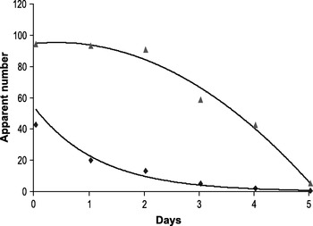
With these uncertainties about schistosome migration in her sights, Patricia Miller began her studies in York in the early 1970s. Her first experiment was a skin extraction time-course, from which she obtained a maximum value of 26% of penetrant cercariae 1 h after exposure, and a negative exponential recovery curve over a 6-day period (Fig. 1) (Miller and Wilson, Reference Miller and Wilson1978), apparently corroborating the earlier work on death in the skin (Clegg and Smithers, Reference Clegg and Smithers1968). However, there was a discrepancy between the apparent timing of parasite exit from the skin and arrival in the lungs, leading us to pose the question ‘where are the missing parasites’? We determined the actual residence time of schistosomula in the skin by exposing groups of animals to cercariae and then surgically excising the exposure site at 24-h intervals; parasites still resident in the skin at excision would not be recovered as adult worms at perfusion. The data provided estimates for t½ of stay in the skin of 88, 65 and 76 h for mice, hamsters and rats, respectively. Furthermore, the sigmoid form of the relationship with time, compared to the negative exponential obtained from tissue extraction showed that many viable parasites were present in the skin at the later sampling times (Fig. 1). Indeed, the declining efficiency of the skin recovery protocol between Days 0 and 4 should have been a wake-up call to researchers.
The timing of parasite migration through the skin (data re-plotted from Miller and Wilson, Reference Miller and Wilson1978). The profile of parasites recovered by mincing and incubation of the skin infection site (◆) with time post-exposure shows a negative exponential decline. In contrast, surgical excision of the site ( ) reveals that parasite exit follows a sigmoid pattern with a t½ stay of 88±23 h. The space between the two curves is a measure of the relative inefficiency of the extraction technique but nothing can be inferred about the absolute numbers of larvae present at each time-point.

The passage of parasites through the lungs was similarly monitored by the mincing and incubation technique, although its efficiency was now clearly questionable (Miller and Wilson, Reference Miller and Wilson1978). Peak recovery of schistosomula at Day 6 after exposure, amounted to only 23% of penetrant cercariae, versus 34% maturing as adult worms. The low numbers in the lungs could have been the result of death in the skin, and the discrepancy between lungs and liver to inefficient lung extraction, but there was another complicating factor. If the first parasites to reach the lungs left that organ before the last arrived from the skin (as indicated by the sigmoid exit curve) then the lung peak would never equate to skin numbers. This situation would take some years to resolve.
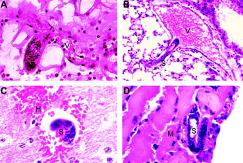
During these experimental studies we collaborated with the histopathologist, Dr Paul Wheater, of Nottingham University, to undertake a comprehensive appraisal of migrating schistosomes in mouse tissues (Fig. 2). For example, we reasoned that if schistosomula were traversing the diaphragm and liver capsule, they would surely be found in tissue sections. The study confirmed that skin migration occurred over a 5-day period (Wheater and Wilson, Reference Wheater and Wilson1979) with numbers following a sigmoid rather than negative exponential profile, corroborating the skin excision data while underlining the inadequacy of mincing and incubation to estimate parasite numbers. From parasite density in the skin, an approximate t½ for the duration of stay was estimated as 72 h. No evidence was found for the predicted dead or dying parasites; either they simply were not dying there or were rapidly cleared when they died. The histological study also indicated that the majority of penetrants remained in the epidermis or sebaceous glands for about 24 h. Over the next 6 days, small numbers could be located in the skin-draining lymph nodes, both beneath the capsule and in the hilar efferent lymphatics, so were obviously capable of traversing the nodes. Estimates of the relative density of schistosomula in the lungs revealed an asymmetrical profile with an early peak at Day 7 but a long tail out to the last sampling time at Day 20. Evidence for diaphragm transit was negligible, whereas schistosomula were found in systemic organs such as the brain and myocardium (Fig. 2), as well as the chambers of the left heart. Nevertheless, there was a time delay between numbers declining in the lungs and increasing in the portal tract. In the liver, schistosomula were found only in the hepatic artery and portal vein, supporting an arrival via splanchnic vessels and suggesting that the upstream end of the sinusoids must act as a ‘filter’. These histological observations strongly indicated that schistosomes travelled from the lungs to the portal system entirely within the vasculature, with the potential for multiple circuits. The problem was how to collect convincing experimental data that provided a dynamic rather than static picture. However, other opinions were current, with Lewis and Colley for example concluding on the basis of lung recoveries that there was no evidence for the recirculation of schistosomula, just a single pass through the lungs (Lewis and Colley, Reference Lewis and Colley1977).
Photomicrographs of schistosomula (S) in key locations during migration in the naïve mouse (after Wheater and Wilson, Reference Wheater and Wilson1979). (A) Penetrating a venule (V) in the dermis. (B) Emerging from a pulmonary venule into the main pulmonary vein (V) prior to passage to the left side of the heart. (C) In a cerebral blood vessel, surrounded by a micro-haemorrhage (H). (D) In a blood vessel of the myocardium (M). Images (B–D) provided the first hard evidence for the systemic phase of migration.

Around this time we began to pay more attention to the larval parasite itself. Ruth Lawson used an in vivo hamster cheek pouch preparation to document more precisely the kinetics of skin migration, capitalizing on the ability to track individual schistosomula for hours to days (Wilson and Lawson, Reference Wilson and Lawson1980). Penetrants ceased to move laterally in the epidemis within hours and remained external to its basement membrane for >40 h. About half the schistosomula had entered the dermis by 53 h and the majority left between 60 and 80 h by penetrating a blood vessel. This last was a laborious process (mean time ~8 h) that involved repeated application of the apical area to the vessel wall, until a hole developed through which the larva squeezed. An ex vivo study of the motility of schistosomula also charted the rhythmicity of the cycles of elongation and contraction (Wilson et al. Reference Wilson, Draskau, Miller and Lawson1978). Following arrival in the lungs, schistosomula increased up to 4-fold in length over a period of days with a commensurate decrease in diameter.
The light microscope observations on the cheek pouch were later complemented by ultrastructural studies showing that by the time schistosomula entered the dermis the acetabular glands and their contents had been lost (Crabtree and Wilson, Reference Crabtree and Wilson1985). These studies highlighted the role of the head gland as a potential source of lytic secretions to penetrate both the epidermal basement membrane and the wall of a venule. The importance of the muscular head capsule in squeezing the gland to aid secretion, and in providing traction for entry into the vessel lumen, was also recognized. A second study of the pulmonary phase of migration revealed the very tight fit of schistosomula within capillaries, with the implication that intravascular migration was a strenuous and protracted process (Crabtree and Wilson, Reference Crabtree and Wilson1986b). The very close apposition of parasite tegument and capillary endothelium also highlighted the possibilities for transcytosis of parasite antigens from vessel lumen to interstitial space.
A physiological investigation of the migrating schistosomulum ex vivo determined that the changes in morphology were not accompanied by a change in mass, i.e. they involved a re-differentiation of cells and tissues already present (Lawson and Wilson, Reference Lawson and Wilson1980). This suggested that the migrating schistosomulum was in a semi-quiescent metabolic state, consistent with the observation that no mitosis occurred until the portal system was reached (Clegg, Reference Clegg1965). After arrival in the liver, schistosomula contracted back to the size of skin worms. Blood feeding started after about 2 days, accompanied by a dramatic rise in both O2 consumption and lactic dehydrogenase activity that suggested a developmental switch was thrown. A final morphological study completed the picture when Jean Crabtree undertook a scanning electron microscope study of the developing parasite to tie in changes in surface morphology to the migration process (Crabtree and Wilson, Reference Crabtree and Wilson1980). The disappearance of the spines, which cover the whole cercaria, from the mid-body region of the developing skin schistosomulum was very noticeable. This left only small spinous regions at the extreme front and back; coupled with the observations on body rhythmicity, these changes suggested an adaptation for crawling through capillary beds in inch-worm fashion. The other salient feature was the enormous increase in surface area needed to accommodate the elongation of the schistosomulum body.
The discrepancy between the disappearance of schistosomula from the lungs and their accumulation in the liver prompted a further experimental study to investigate the question of how schistosomula travelled to the portal system (Miller and Wilson, Reference Miller and Wilson1980). If the diaphragm and liver capsule represented barriers of any kind then schistosomula should be retarded there but, unlike Wilks, we did not find them in pleural washes (Wilks, Reference Wilks1967). More plausibly they were traversing the capillary beds of systemic organs that, unlike the very elastic lungs, were not tissues from which they were readily extractable (although we did manage to recover small numbers from minced kidneys and spleen). We obtained the most convincing evidence for a systemic phase of migration by delivering ex vivo lung schistosomula to the left ventricle of the hamster heart. We first recovered them from the lungs 18 h later, thus demonstrating their ability to cross systemic capillary beds. Just over half delivered by this route matured in the portal system, the same proportion as when they were delivered directly back to the pulmonary vasculature. We also discovered that small numbers of ex vivo lung schistosomula, delivered to the portal vein, could negotiate the liver sinusoids and pass back to the lungs demonstrating that the hepatic ‘filter’ was not completely efficient, although 72% matured into adults. The filter effect may be due to the limiting vascular diameter of hepatic sinusoids being lower than that of systemic capillaries (Madden et al. Reference Madden, Paparo and Schwartz1968). These initial injection experiments provided the first indications that migration through the normal lung vasculature might be a tricky process for schistosomula. By 1980, we felt we had accumulated enough evidence to discard the trans-diaphragm hypothesis in favour of an entirely intravascular migration route but our observations still provided largely circumstantial evidence for a systemic phase of migration; we had to wait several years for the definitive proof.
MIGRATION AND ATTRITION IN MURINE MODELS OF IMMUNITY
Coincident with the early investigations on migration in York, other schistosome groups were following distinct but complementary lines of research. Most notably, what proved to be landmark studies on immunity to schistosomes in the mouse model were emerging from Murrel's group at the Naval Medical Research Institute in Bethesda, Maryland. David Dean demonstrated that mice harbouring a small bisexual primary infection developed strong ‘resistance’ to a cercarial challenge, in some groups reaching 100% (Dean et al. Reference Dean, Minard, Stirewalt, Vannier and Murrell1978b). However, neither male nor female single-sex infections elicited much protection, thus implicating the schistosome egg in the mechanism. A second study muddied the waters somewhat by showing that eggs alone could protect mice when injected intravenously to the lungs, but not via intra-peritoneal or subcutaneous routes (Dean et al. Reference Dean, Minard, Murrell and Vannier1978a). With hindsight it is easy to see how massive egg-induced pulmonary fibrosis could interfere with the intravascular migration of schistosomula.
Experiments from the same group, using gamma-irradiated cercariae to immunize mice, were more encouraging (Minard et al. Reference Minard, Dean, Jacobson, Vannier and Murrell1978a) and set the scene for much later work on immune mechanisms. Their relevance to this review is that a single exposure to 250–500 optimally irradiated cercariae would induce 70–80% protection against a normal cercarial challenge that persisted for at least 15 weeks. A follow-up study used tissue mincing and incubation to determine where both attenuated immunizing, and normal challenge parasites perished (Minard et al. Reference Minard, Dean, Vannier and Murrell1978b). It was apparent that reduced numbers of attenuated larvae reached the lungs as a consequence of irradiation, but large numbers of challenge parasites were recovered from that organ, with greater numbers present 10–15 days post-challenge in vaccinated mice than in naïve controls. In these data were hints of the upcoming controversy about the site of challenge parasite attrition. The radiation-attenuated vaccine was also developed by the Schistosomiasis group at the London School of Hygiene and Tropical Medicine (LSHTM), who explored the parameters of the model in a different mouse strain, confirming that a single percutaneous exposure of CBA mice to 500 cercariae could elicit 60% resistance against challenge (Bickle et al. Reference Bickle, Taylor, Doenhoff and Nelson1979). The Mill Hill group also published the results of protection experiments in mice vaccinated with irradiated cercariae (Miller and Smithers, Reference Miller and Smithers1980). Using tissue extraction methods, they concluded that the strong immunity induced “was largely effective in the skin”.
The belief that laboratory animals with a chronic infection became immune to subsequent challenge can be traced back to work by the Schistosomiasis group at Mill Hill on rhesus macaques (Smithers and Terry, Reference Smithers and Terry1969b). This was the ‘golden age’ of concomitant immunity whereby “invading schistosomula are destroyed by the immune response of the host while the adult worms that engendered this response are unaffected” (Smithers and Terry, Reference Smithers and Terry1969a). The concept was explored in mice in the early 1970s using parasite extraction from the lungs as a rapid assay of acquired immunity; a much reduced recovery of challenge parasites was recorded in mice with an 8 week or longer primary infection (Sher et al. Reference Sher, Mackenzie and Smithers1974). Furthermore, serum taken from mice with a 12–15 week primary infection was shown to confer partial resistance to challenge on naïve recipients – the classic passive transfer experiment – implicating antibodies as the protective agent (Sher et al. Reference Sher, Smithers and Mackenzie1975). These studies were followed up with a balance sheet of challenge parasite numbers in chronically infected and naïve mice, again using the mincing and incubation technique for the skin and lungs (Smithers and Gammage, Reference Smithers and Gammage1980). It was concluded that, as the same numbers of parasites could be recovered from the lungs and livers of naive animals, the missing 65% of penetrants must have died in the skin. In the chronically infected animals, further early death in the skin within the first 3 days was suggested, and a later phase between 6 and 14 days, either after the parasites had left the lungs or soon after arrival in the liver (were they still thinking in terms of a single pass through the lungs?). In parenthesis, we had reasoned that if 76% of parasites applied to the skin of a chronically infected animal died there, we should be able to find abundant evidence of their demise. We had previously failed to detect the projected dead parasites in the skin of naïve animals (Wheater and Wilson, Reference Wheater and Wilson1979) and we got a similar result with chronically infected mice; these ‘negative data’ were emphatically rejected by referees and never published.
In 1980 in York we began a collaboration with Quentin Bickle at LSHTM to investigate the migration and attrition of irradiated, immunizing cercariae and challenge parasites in mice, using quantitative histology. This truly laborious task, undertaken by Avis Mastin, required the identification of every schistosomulum in serial sections of complete lung lobes. The results were striking in that both irradiated and normal parasites arrived in the lungs in large numbers (57% and 48% of penetrants, respectively, were counted on day 7), but the irradiated parasites progressed no further and were still detectable in substantial numbers at Day 21 (Mastin et al. Reference Mastin, Bickle and Wilson1983). A similar observation was made about the fate of challenge larvae in control and vaccinated mice, with 52% and 63% of penetrants, respectively, in the lungs on Day 6. By viewing challenge schistosomula in situ in the lung sections we observed that inflammatory responses were present in vaccinated but not control mice. Over the next 15 years this observation had profound repercussions for studies on the immunological mechanism of challenge elimination in the irradiated vaccine model. An ultrastructural examination of the irradiated parasites during their extended stay in the lungs showed they had undergone the normal developmental changes associated with lung-stage migration (Mastin et al. Reference Mastin, Bickle and Wilson1985). However, at Days 13 and 21 the majority were lying free within the alveoli, with noticeable associated inflammation, and their ultimate fate appeared to be death by autophagy. Much later studies indicated that the radiation-induced lesion was a subtle effect on neuromuscular co-ordination that effectively prevented onward migration through the capillaries (Dillon et al. Reference Dillon, Feltwell, Skelton, Coulson, Wilson and Ivens2008; Harrop and Wilson, Reference Harrop and Wilson1993). Mastin's findings were later confirmed by the eminent pathologist Franz von Lichtenberg, working at the National Institutes of Health, Bethesda, with the conclusion that “the lung was a major site of parasite attrition for both immunising and challenge infections in the mouse irradiated vaccine model” (von Lichtenberg et al. Reference Von Lichtenberg, Correa-Oliveira and Sher1985).
Doubts about the validity of the ‘immunity’ exhibited by chronically infected mice surfaced around 1980. The group at the LSHTM noted an apparent correlation between the size of the mature worm burden, the lung, liver and gut egg burdens and the degree of protection, all incriminating the egg (Long et al. Reference Long, Harrison, Bickle, Bain, Nelson and Doenhoff1980). David Dean in Bethesda used mice with a parabiotic union to show that the protection elicited by irradiated cercariae could be transferred to naive recipients whereas that displayed by chronically infected mice could not; clearly the models differed in some fundamental way (Dean et al. Reference Dean, Bukowski and Clark1981). Against this background, combining the information we had generated about intravascular migration with the well-documented development of porta-caval shunts as a consequence of egg-induced pathology, I suggested that the immunity displayed by chronically infected mice might be an artefact of pathology (Wilson, Reference Wilson1980). The hypothesis predicted that schistosomula, arriving down the portal vessels from the intestinal capillary beds, could simply by-pass the hepatic filter to travel via the shunts back to the venous circulation and thence the lungs. The failure to trap schistosomula would thus appear as ‘immune-mediated’ attrition. The hypothesis was subsequently tested by the administration of ex-vivo lung schistosomula to the portal vein of mice with a chronic infection. Their recovery from the lungs, in proportion to the extent of porta-caval shunting, confirmed the effect of a ‘leaky hepatic portal system’ (Wilson et al. Reference Wilson, Coulson and McHugh1983). The relationship between the effects of S. mansoni infection on mouse vascular pathology were subsequently fully explored (McHugh et al. Reference McHugh, Coulson and Wilson1987a, Reference McHugh, Coulson and Wilsonb). The hypothesis and data proved controversial, but co-incidentally a congenitally leaky portal system was later found to be the explanation for the intrinsic immunity to schistosomes displayed by the majority of 129/Ola strain mice (Coulson and Wilson, Reference Coulson and Wilson1989), thereby sounding the death-knell of another model of protection.
To recapitulate the mouse studies, by 1982 the York group was convinced that migration from the skin to the portal system was entirely intravascular, with multiple circuits before chance indirect delivery to the portal system via the splanchnic vascular beds. They had found negligible evidence for parasite death in the skin of naïve or chronically infected animals. Conversely, the Mill Hill group put its faith in the tissue extraction results to come down in favour of early parasite death. The chronic infection model of immunity was still in vogue, although the Bethesda and LSHTM groups had produced data implicating the egg as the causative agent. The York group had yet to publish its findings on the leaky hepatic portal system, but there were conflicting data about whether the immunity could be passively transferred by serum or not. The radiation-attenuated vaccine model of protection did not suffer from limitations due to egg-induced pathology, and the protection could be transferred via a parabiotic union. Again, in this model, the proponents of tissue extraction believed that attrition occurred in the skin over the first 4 days after challenge. However, the York-LSHTM collaboration had also produced evidence, as yet unpublished but aired at meetings, that challenge parasite attrition occurred in the lungs. Could these conflicting views be reconciled?
AUTORADIOGRAPHIC TRACKING OF MIGRATING PARASITES
The breakthrough came with the demonstration by Jay Georgi at Cornell, NY, of the potential for autoradiographic tracking of schistosomula in the mammalian host (Georgi, Reference Georgi1982). The technique had its origins in the work of Niels Christensen at the Danish Bilharzia Laboratory to label developing cercariae by incubating the snail host with [75Se]-methionine, a gamma-emitting isotope (Christensen, Reference Christensen1977). Georgi took the logical step of preparing skin from the tails of mice exposed to labelled cercariae, and exposing it to X-ray film; each parasite appeared as a discrete dot so the whole skin population could be counted. Furthermore he showed that the parasites (more precisely their associated radioactivity) left the skin with a t½ of 91 h (cf. Patricia Miller's estimate of 88 h obtained from excision of abdominal skin). This was quickly followed by papers from the Bethesda group, in collaboration with Georgi, which extended the technique and applied it to longitudinal studies of migration. They demonstrated that lung tissues, gently compressed onto cardboard before autoradiography, would provide a very clear count of schistosomula present (Georgi et al. Reference Georgi, Dean and Chandiwanna1982). One of the first observations in naïve mice was that >90% of penetrants could be located as late as Day 7 when 86% of them were in the lungs (bad news for proponents of early death in the skin) (Mangold and Dean, Reference Mangold and Dean1983). Moreover, the technique was sufficiently sensitive to produce a profile of lung schistosomula numbers up to 14 days post-exposure (Georgi et al. Reference Georgi, Dean and Mangold1983). Applied to the skin, lungs and liver after challenge of mice with a chronic infection and naïve controls, the technique revealed that there was negligible death in the skin of both groups. In the controls, elimination occurred in lung and post-lung sites while in the infected animals, the extra attrition appeared to occur in the liver. The study also revealed that the lung mincing technique to recover schistosomula had a lower efficiency in previously-infected mice than naïve controls, thus invalidating its use as a comparator between the two groups.
Autoradiographic tracking was also used to determine the fate of attenuated vaccinating larvae, confirming Mastin's histological data (Mangold and Dean, Reference Mangold and Dean1984). A large proportion of 20 krad irradiated larvae migrated to the lungs where most were sequestered, with very few reaching the liver. Higher doses of radiation resulted in progressively fewer parasites reaching the lungs, with a lower level of protection elicited. Thus 85% of cercariae exposed to 90 krad remained in the skin. The study also showed that the technique could detect small numbers of labelled normal schistosomula in systemic organs such as kidney, brain, digestive and reproductive tracts – further proof for a systemic phase of migration.
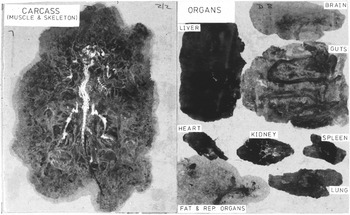
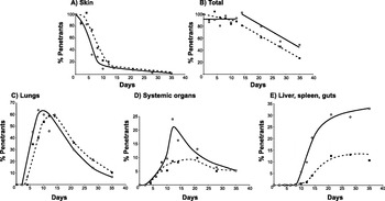
Autographic tracking was clearly a ‘must have’ technology and a short spell with Dean and Mangold in Bethesda in 1983 resulted in its transfer to York. Small additions to the technique such as intensifying screens and film exposure at −80°C increased the sensitivity of detection out to Day 35 worms. The entire mouse pelt was laid out, organ compression was standardized with a steel roller, and the muscles were squeezed from the eviscerated and de-brained carcass with a hydraulic press (Wilson et al. Reference Wilson, Coulson and Dixon1986). The result was a 2D version of the mouse (Fig. 3) in which every parasite could be located and counted. For the first time we were able to construct an accurate balance sheet from which to deduce the pattern of migration and site(s) of attrition, in naive mice and those exposed to the irradiated cercaria vaccine (Fig. 4). The results were clear cut, with a rapid decline in the numbers of parasites in the skin of naïve mice from Day 4 onwards and their commensurate appearance in the lungs (a small proportion of parasites lingered in the skin out to day 35). The lung peak was at day 8 and the systemic organ peak at Day 12 (24% of the Day 0 parasite total). The distribution of parasites between the various systemic organs was in excellent agreement with data for the fractional distribution of cardiac output in the mouse. Migration to the splanchnic organs and hence the liver occurred between Days 7 and 21, at which time parasites were still present and presumably trapped in pulmonary and systemic locations as there was no further recruitment to the portal population. Most striking, no decline in the total number of parasites was recorded until after Day 14, whereupon it proceeded at a steady rate out to Day 35; 16% of the Day 0 total was still detectable in non-splanchnic locations at this time. In many respects the migration of challenge parasites in mice vaccinated with irradiated cercariae proved identical to that in the naïve controls. There was a slower departure from the skin and a later peak in the lungs (Day 14). However, the main difference was that many fewer parasites reached the systemic and splanchnic organs (44% and 25% of normal values). Again, there was no early parasite attrition, the bulk occurring in the lungs at the same rate as in naive mice, but beginning 7 days earlier. Clearly, parasite death in both naive and vaccinated animals was a protracted process, not an acute lethal hit!
Mouse dissected for autoradiographic tracking. Muscles were squeezed from the eviscerated, debrained carcass under a hydraulic press, whilst individual organs were gently compressed onto absorbent card with a steel roller. The dried preparation was exposed to X-ray film to detect the number of parasites present in all organs, permitting an accurate balance sheet to be drawn up.

Balance sheet of parasite numbers in normal mice (▪ - - - ▪) and mice previously vaccinated with irradiated cercariae (○——○) (re-plotted from Wilson et al. Reference Wilson, Coulson and Dixon1986). Parasites leave the skin (A) of vaccinated mice with an approximate time delay of 1 day and there is a corresponding later rise in lung numbers (C). However, the lung peak (C) in the two groups at around 60% of penetrants, is similar. (N.B. As the first schistosomula leave the lungs before the last arrive from the skin, the peak never reaches skin numbers.) The total number of parasites detected (B) does not begin to decline before day 14 when ~10% remain in the skin, confirming the lack of death in that tissue. The real difference between the two mouse groups is revealed by the numbers in the systemic organs (minus splanchnic beds; D), where the peak in vaccinated animals is <50% that in normal mice. A similar difference is evident in the numbers detected in the splanchnic organs (liver, spleen, guts; E). The balance sheet of total numbers thus reveals that the lungs present the greatest obstacle to migration success, with the effect of the irradiated vaccine being to trap a greater proportion in that organ. Parasite attrition is a slow process (B) occurring over about 20 days, consistent with a mechanism of blocked migration, not cytotoxic killing.

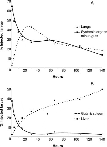
In parallel with the balance sheet experiments we investigated the kinetics of migration through the different vascular beds by administering labelled ex vivo lung schistosomula to naïve mice, and then tracking their movements by autoradiography (Fig. 5). The mean transit time through the lungs (corrected for returning parasites) was 30–35 h after femoral vein injection. Mean transit through systemic and intestinal capillary beds was 16 h and 6·5 h, respectively, after left ventricle injection, with distribution to systemic organs again in proportion to cardiac output, and both considerably more rapid than through the lungs that seemed to present a special obstacle. Transit time between organs was negligible, presumably passively with blood flow. It would thus appear that 2–3 circuits of the systemic-pulmonary vasculature taking >50 h each, would be sufficient to deliver the entire portal population.
Kinetics of migration through vascular capillary beds (re-plotted from Wilson and Coulson, Reference Wilson, Coulson and Dixon1986). Delivery of ex-vivo lung schistosomula to the left ventricle allows their rate of migration through different systemic capillary beds to be determined. The larvae are distributed instantaneously in a 0·33:0·67 ratio between splanchnic beds (B) and other systemic organs (A). Schistosomula traversed the intestinal capillary beds in a mean time of 6·5 h, to begin accumulating in the pre-sinusoidal vessels of the liver (B). The mean migration time through other systemic organs was 16 h, with numbers rising in the lungs to 40% of injected larvae (A) by about 25 h. With an estimated lung transit of 30–35 h (data not shown) there is a hint of a second wave of parasites passing through the systemic organs at around 65 h. The differences in transit time underline the obstacle that the lungs appear to present to successful migration and also explain why a greater proportion of schistosomula is present in the lungs, relative to the systemic organs, at a given time.

Two pieces of work rounded off the saga. In collaboration with Dean and Mangold we used the schistosomulum injection techniques and autoradiographic tracking, to clarify the site of challenge parasite elimination in vaccinated mice (Mangold et al. Reference Mangold, Dean, Coulson and Wilson1986). These data demonstrated that the lung-stage schistosomulum, believed insusceptible to in vitro cytotoxic killing mechanisms (Dessein et al. Reference Dessein, Samuelson, Butterworth, Hogan, Sherry, Vadas and David1981), was highly susceptible to elimination in mice vaccinated with irradiated cercariae, but only if reintroduced to the pulmonary or systemic organ circulation (the latter allowing the majority to return to the lungs), not into the portal vein. It was concluded that challenge elimination must occur in the lungs (and perhaps systemic organs) of vaccinated mice. It was indeed a slow process, since more than 7 days had to elapse after injection before a decline in parasite numbers, in vaccinated animals relative to controls, could be detected.
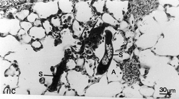
These data were complimented by Jean Crabtree's ultrastructural examination of the fate of challenge parasites in the lungs of normal and vaccinated mice (Crabtree and Wilson, Reference Crabtree and Wilson1986a). As previously observed (Mastin et al. Reference Mastin, Bickle and Wilson1983), from day 11 onwards an increasing proportion of parasites in naïve controls had left the blood vessels and were in the alveoli, where they attracted an inflammatory response, most likely the result of non-specific damage. This provides a plausible explanation of why more than half of penetrant cercariae fail to mature in the mouse. During passage through the lungs they rupture the blood vessels and enter the alveoli from which they cannot return (Fig. 6). In the vaccinated animals the pattern of localization was similar BUT the still-intravascular parasites attracted inflammatory foci comprising predominantly lymphocytes and macrophages. In some instances the foci disrupted the vascular highway while in others the plug of inflammatory cells appeared simply to block progress. Nearly all parasites were intact, despite the inflammation, a testament to their ability to resist oxidative attack.
Passage through the lungs is hazardous. Light micrograph of the lungs in a naive mouse 11 days post-infection showing an extended schistosomulum (S) half way into an alveolar space (A), from which re-entry to a blood vessel appears problematic (Crabtree and Wilson, 1986 a).

EPILOGUE
This was effectively the end of the saga. The attention of researchers turned towards investigations of the immunological mechanisms, and a search for the protective antigens that is still ongoing. Later publications were mostly directed towards more specialist immunology and molecular biology journals and the action moved away from ‘Parasitology’. We were left with a clear demonstration that schistosome migration from the skin to the portal system is entirely intravascular with minimal death in the skin, but obvious problems in lung transit. There was a conviction that the supposed ‘immunity’ developing in hosts with chronic bisexual infections was a product of egg-induced pathology. Lastly, in the irradiated vaccine model, we had a robust demonstration that high levels of protection could be elicited, and it was the lung, not the skin schistosomulum that was the target. This model is still referred to as the gold-standard, the yardstick against which the levels of protection elicited by recombinant antigens are compared, and its validity has been reinforced by its proven efficacy in primate models (Eberl et al. Reference Eberl, Langermans, Frost, Vervenne, Van Dam, Deelder, Thomas, Coulson and Wilson2001; Kariuki et al. Reference Kariuki, Farah, Yole, Mwenda, Van Dam, Deelder, Wilson and Coulson2004).
What of other schistosome species, hosts and models of immunity? There were a couple of studies with labelled S. haematobium and S. japonicum cercariae in mice. In the former, the lungs of naïve animals appeared to present an even greater obstacle to onward migration than for S. mansoni (Georgi et al. Reference Georgi, Wade and Dean1986). In the latter, it was concluded that the lungs and liver were the major sites of attrition both in naïve mice and animals vaccinated with irradiated cercariae (Laxer and Tuazon, Reference Laxer and Tuazon1992). Autoradiographic tracking was also shown to be feasible in rats (Knopf et al. Reference Knopf, Cioli, Mangold and Dean1986) and even baboons (Wilson et al. Reference Wilson, Coulson, Sturrock and Reid1990). Sadly, [75Se]-methionine, which made such a difference to our understanding of the dynamics of migration and attrition in the mouse, was withdrawn around 1990 so we do not presently have the option to find out. It can only be hoped that modern imaging methods will provide a replacement for autoradiographic tracking, with the resolution capable of detecting a 100×30 μm schistosomulum in an intact or compressed organ.
My thanks to Dr Patricia Coulson for a critical reading of the manuscript and to Drs Patricia Miller, Ruth Lawson, Avis Mastin, Simon McHugh and Professor Jean Crabtree whose studies in York contributed so much to our understanding of schistosome migration and attrition.