Implications
Hairless areas, scabs, wounds and swellings are common findings in cubicle housed dairy cows. They reflect conflicts between the cows and their housing environment and potentially impair cow welfare. The locations on the cow where these alterations are frequently found are the carpal joints, hocks and the hip area. Alterations at the hip area should consequently gain more attention in future research. Alterations at the inner and outer side of the hock as well as different types of alterations at the carpal joints should be investigated separately because they seem to have different causes.
Introduction
Integumentary alterations are a common finding in dairy cattle (e.g. Mülleder and Waiblinger, Reference Mülleder and Waiblinger2004; Veissier et al., Reference Veissier, Capdeville and Delval2004; Kielland et al., Reference Kielland, Ruud, Zanella and Østerhås2009). They reflect an impairment of the cows’ welfare as wounds and swellings may be painful, and alterations including hairless areas reflect repeated conflicts of the cow with its environment. So far, most studies focused on integument alterations in the hock region (e.g. Rodenburg et al., Reference Rodenburg, House and Anderson1994; Lombard et al., Reference Lombard, Tucker, von Keyserlingk, Kopral and Weary2010; Potterton et al., Reference Potterton, Green, Harris, Millar, Whay and Huxley2011a and Reference Potterton, Green, Millar, Brignell, Harris, Whay and Huxley2011b) and some additionally on the carpal joint (e.g. Spycher et al., Reference Spycher, Regula, Wechsler and Danuser2002; Fulwider et al., Reference Fulwider, Grandin, Garrick, Engle, Lamm, Dalsted and Rollin2007; Norring et al., Reference Norring, Manninen, de Passillé, Rushen, Munksgaard and Saloniemi2008). Some recent studies also looked at prevalences and possible influencing factors at the neck (Kielland et al., Reference Kielland, Bøe, Zanella and Østerås2010; Zaffino Heyerhoff et al., Reference Zaffino Heyerhoff, LeBlanc, DeVries, Nash, Gibbons, Orsei, Barkema, Solano, Rushen, de Passillé and Haley2014), the hip bone region and hind leg apart from hock and knee (Rouha-Mülleder et al., Reference Rouha-Mülleder, Palme and Waiblinger2010), as well as the hips, knees, fetlocks and thighs (Kielland et al., Reference Kielland, Ruud, Zanella and Østerhås2009). It is well known that lesions often occur at protruding parts of the cows’ body, for example, the lateral tarsal joint and the dorsal calcanei (Weary and Taszkun, Reference Weary and Taszkun2000), but to date no study is available examining the whole body and comparing prevalence values for the different body parts of dairy cows in cubicle housing.
For alterations in the hock area, it has been found that different factors may influence prevalence values for different specific locations at the hock (Weary and Taszkun, Reference Weary and Taszkun2000) or different types of alterations (Rutherford et al., Reference Rutherford, Langford, Jack, Sherwood, Lawrence and Haskell2008; Potterton et al., Reference Potterton, Green, Harris, Millar, Whay and Huxley2011a). We thus aimed to analyse possible correlations between prevalence values at different locations and of different types of alterations in order to provide a basis for future research on potential similar causes of related alterations.
In our investigation we examined 15 body locations of the dairy cow with the additional aim to ascertain the areas most affected by alterations in cubicle housing systems under a broad set of conditions (e.g. organic and conventional farming with dairy as well as dual purpose breeds). This may provide a basis for decisions about the relevance of further investigatory focus on those different body parts.
Animals, material and methods
Sample
Farms were selected with the help of the milk inspection boards and were required to have cubicle housing of Holstein Friesian or Fleckvieh cows and twice a day milking in a milking parlour. Farms could have complete indoor housing, access to an outdoor loafing area or pasture, but no turnout of cows for alpine grazing. For practical reasons, we only chose farms equipped with head lockers. We excluded farms with horned cows, rubber flooring in the alleys and acute outbreak of disease to reduce confounding. The original data set comprised 105 farms, visited in the winter housing period 2004/05. After exclusion of farms not fulfilling selection criteria and farms with missing values, 95 farms with a mean fat–protein-corrected yield of 8165 kg/year (range: 5773 to 10 146), a mean herd size of 59 milking cows (range: 28 to 156), and a cow/cubicle ratio of 0.97 (range: 0.62 to 1.28) remained; 51% of the farms had deep bedded cubicles, the other farms had raised cubicles with bare concrete, rubber mats or mattresses as cubicle base, with (29%) or without (20%) bedding material. The final data set consisted of three sub-data sets, one comprising 32 organic farms with Holstein Friesian cows distributed all over Germany, the second 32 conventional farms with Holstein Friesian cows in central Germany and the third 31 conventional farms with Fleckvieh cows in Austria.
On each farm, a random sample of 30 (unless there were fewer cows in the milking herd) to 52 cows was inspected. Sample size per farm was sufficient to estimate a within-herd prevalence of 50% with at least 10% precision and a confidence level of ⩾0.9. Cows were selected by marking cows in predefined random positions on the list of cows in milk at the time of farm visit if available, or alternatively by choosing/dismissing every xth cow in the parlour or feed rack. Non-lactating cows, cows from a breed other than Holstein Friesian or Fleckvieh, as well as cows in sick pens were excluded. Cows with an obvious acariasis were evaluated but data excluded from this analysis to reduce confounding. After data collection, cows with incomplete data on integument alterations were excluded, resulting in a sample size of 2922 cows for analysis.
Integument alterations
A total of 15 different sites of the cows’ body were inspected: the carpal joints, shoulders, flanks, hip bones, pin bones, knees, the medial, dorsal and lateral calcanei, medial and lateral tarsal joints and the rest of the hind legs, as well as the sacrum, back and dorsal neck. We differentiated hairless areas (H), scabs and wounds (W) and swellings (S). There was no minimum size defined, but all alterations detectable by visual inspection or palpation were included. Cows were inspected while fixed in the head lockers. A head torch was used for visual inspection if necessary.
Inter-assessor agreement
As data were collected by four different assessors, inter-assessor reliability testing took place before (on a total of 20 or 21 cows) and after (on a total of 40 to 69 cows) data collection on three to six farms. PABAK (Prevalence-adjusted bias-adjusted kappa; Byrt et al., Reference Byrt, Bishop and Carlin1993) was calculated for each assessor pair and each type of integument alteration separately, before and after data collection. For assessor testing, only the tarsal and carpal joints and calcanei were scored, as alterations were expected to be most prevalent on these body parts, making it most suitable for agreement testing. The score of each body part was treated as separate observation in the PABAK calculation. Testing six pairs before and after data collection resulted in 12 PABAK values based on 265 to 414 observations per assessor pair and alteration. Following the classifications of Fleiss et al. (Reference Fleiss, Levin and Paik2003) for κ-values we interpreted PABAKs ⩾0.75 as excellent, between 0.4 and 0.75 as fair to good and <0.4 as poor agreement. PABAKs ranged from 0.57 to 0.95. They were fair to good in 47% of all tests and excellent in 53% of all tests. The lowest mean PABAK per alteration type occurred for scabs and wounds after data collection (0.69), the highest mean PABAK was found for swellings before data collection (0.84).
Statistical methods
Two different measures were calculated: mean numbers of alterations per cow for descriptive statistics and herd level prevalences for cluster analysis. For bilateral body sites (all but neck, back and sacrum), numbers of alterations on both sides of the cow were added up to calculate mean numbers of alterations per cow. For herd level prevalences it was considered whether the specific alteration was present at least on one side of the cow or not. Herd prevalence values were calculated for all alterations and locations. If alterations at a specific location were present in at least 25% of the herds in at least one sub-data set the respective alteration and location was considered in the following cluster analysis.
The VARCLUS procedure (SAS 9.2) was used for hierarchical clustering of centroid components of the standardised prevalence values of alterations per location. The maximum number of clusters was defined to be equal to the number of variables. Different criteria exist to identify the optimal number of clusters in a given data set (Tibshirani et al., Reference Tibshirani, Walther and Hastie2001; Yan, Reference Yan2005) but in this study, the aim was to identify cluster patterns at different stages of clustering instead of identifying one optimal number of clusters. Relevant cluster stages were defined as stages resulting in a high increase in variance explained compared to the neighbouring splits (local maxima).
Results
Distribution of integumentary alterations
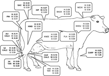
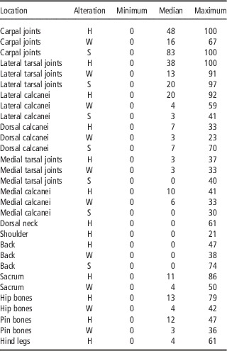
Highest mean numbers of alterations per cow occurred at the lower legs, specifically the carpal joints, the lateral tarsal joint and lateral calcanei, followed by sacrum, hips and pin bones (Figure 1). All types of alterations were found at all inspected locations. Similar to the distribution of mean numbers per cow, median herd prevalence values were highest for alterations at the carpal joint, lateral tarsal joint, lateral calcanei, hips, pin bones and sacrum (Table 1); 30 out of 45 combinations of alteration and location were present in at least 25% of the herds of at least one sub-data set (presented in Table 1) and were therefore used for cluster analysis.
Mean number of integument alterations per cow in a sample of 2922 dairy cows from 95 organic and conventional herds in Germany and Austria, means ⩾0.1 are bold. med=medial; dor=dorsal; lat=lateral; CARP=carpal joints; SHOU=shoulders; FLA=flanks; HIP=hip bones; PIN=pin bones; KNEE=knees; CALC=calcanei; TAR=tarsal joints; HIND=hind legs (apart from tarsus, calcaneus and knee); SAC=sacrum; BACK=back; NECK=dorsal neck; H=hairless areas; W=scabs and wounds; S=swellings.

Herd prevalence values (%) of hairless areas (H), scabs and wounds (W) and swellings (S) at different body locations of 2922 dairy cows

Clustering of alteration types and locations
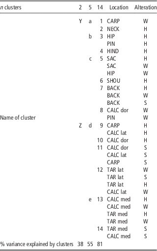
Converging was successful in all clustering steps. Three stages were identified which represented good numbers of clusters, that is, a comparably large increase of explained variance by splitting the last cluster was reached, but considerably less increase of explained variance by the following splittings. These stages represented two clusters, five clusters and 14 clusters (Table 2). The first split roughly divided the variables into one cluster with alterations at the lower legs (cluster Z, 16 members) and another cluster dominated by other body parts (cluster Y, 14 members). In the five cluster state, clusters had between 2 and 10 members. While cluster e represented all alterations at the medial tarsal joint and calcanei, cluster d was dominated by alterations at the lateral tarsal joint and lateral and dorsal calcanei and the carpal joint. Cluster c was dominated by alterations at the back and sacrum and cluster b exclusively represented hairless areas, namely at the hip and pin bones and hind legs. In the 14 cluster stage, some variables stood on their own, while the two largest clusters contained four variables which represented all alterations at the lateral tarsus and W at the lateral calcanei (cluster 12), and H and W at the medial tarsus and medial calcanei (cluster 13), respectively. S at these two locations formed a cluster on their own (cluster 14), while S at the dorsal and lateral calcanei and the carpal joints were combined in cluster 11. The first clustering stages thus represented mainly locations of alterations. With advanced splitting, types of alterations (hairless areas, scabs and wounds v. swellings) were differentiated.
Clusters resulting from hierarchical centroid clustering of herd prevalence values of integument alterations at different body parts of 2922 dairy cows in 95 herds in Germany and Austria

med=medial; dor=dorsal; lat=lateral; CARP=carpal joints; SHOU=shoulders; FLA=flanks; HIP=hip bones; PIN=pin bones; KNEE=knees; CALC=calcanei; TAR=tarsal joints; HIND=hind legs (apart from tarsus, calcaneus and knee); SAC=sacrum; BACK=back; NECK=dorsal neck; H=hairless areas; W=scabs and wounds; S=swellings.
Discussion
Inter-assessor agreement
Previous farm visits by a single observer had generated no concerns as to possible assessor bias in the detection of hairless areas, scabs, wounds and swellings. Assessor tests were therefore performed rather as a formality and only to compare inter-, but not intra-assessor agreement. Results and experience revealed that alteration detection was not as straightforward as it appeared. For instance, scabs are sometimes difficult to discriminate from dirt patches on carpal joints and hocks, though low agreement on this parameter might partly be due to the testing situation. When examining a cow only once, removing an encrustation to reveal whether it is dirt or a scab has no consequences, but with multiple assessors this changes the impression for those examining the cow afterwards. Therefore, it was decided not to remove any encrustations during the testing, which might be a reason for the lowest (but acceptable) mean PABAK of 0.69 found for W. A solution in future assessments could be to exclude joints with encrustations from the analysis for assessor agreement. However, this is no option for data collection on farm as exclusion of locations with encrustations may lead to underestimated prevalences.
Ideally, inter-assessor agreement should have been calculated separately for all alterations and locations. In the present study, this has been done for the alterations, but not for the locations. Furthermore, only the legs have been examined during assessor testing, as only the legs showed a good distribution of alterations on the training farms. The lack of the full scoring range is a very common finding when it comes to assessor training and testing on farm (personal experience), as frequently a farm has a specific ‘problem area’, which results in a specific alteration type or body location affected. Most other alterations and locations have a low prevalence, close or equal to zero. In these cases, inter-assessor agreement would be hard to judge, as there would be no variance in ratings at all. Not considering assessor agreement for each location separately might be problematic as Gibbons et al. (Reference Gibbons, Vasseur, Rushen and de Passillé2012) found differences in observer agreement at different locations, that is, better agreement on alterations at the tarsal joints and neck than at the carpal joints, which might, however, partly be due to slightly different definitions for carpal joint scores. In our study, consistent definitions were used for all locations. Despite this, when retrospectively looking at assessor agreement for the carpal joints, separately, it emerges in several instances that PABAK values were below the 0.4 threshold for acceptability for W and S. Consequently, the results for W and S at the carpal joints need to be treated with caution. On the other hand, herd prevalences of these alterations followed the same patterns (with lowest mean herd prevalences in the organic sub-data set) as alterations at the hock where agreement was sufficient, which speaks in favour of data quality. For H, agreement was always satisfactory.
Distribution of alterations
The mean numbers of alterations per location and cow as well as the median herd prevalence values found confirm that the most affected body parts are in the focus of recent research (e.g. Kielland et al., Reference Kielland, Ruud, Zanella and Østerhås2009; Husfeldt and Endres, Reference Husfeldt and Endres2012; Burow et al., Reference Burow, Thomsen, Rousing and Sørensen2013). Highest values, especially for W and S, were found at the carpal and tarsal joints and the calcanei.
The within-herd prevalences of the 30 alteration location combinations investigated in the cluster analysis showed a large range (Table 1). It was highest at the lateral tarsal (H 0% to 100%, S 0% to 97%, W 0% to 91%) and the carpal joints (0% to 100% for S and H, 0% to 67% for W). Despite the wide range of different conditions covered by the three sub-data sets, ranges within each sub-data set were in general close or equal to the range of the complete data set and comparable to other studies. Kielland et al. (Reference Kielland, Ruud, Zanella and Østerhås2009), for example, found a range of 0% to 100% for a combination of H, S and W at the carpal joints, with a mean herd prevalence of 35% for the left as well as for the right carpus, and Veissier et al. (Reference Veissier, Capdeville and Delval2004) a range of 0% to 100% for severe injuries, defined as open wounds or oedema, with a mean of 41%. Despite comparable prevalence ranges, mean prevalences reported are lower than median values in our study (S 83%, H 48%, W 16%), which might partly be due to the fact, that prevalences were given for each leg, separately (Kielland et al., Reference Kielland, Ruud, Zanella and Østerhås2009) or for severe injuries (Vessier et al., Reference Veissier, Capdeville and Delval2004). Differences in the scoring approach, for instance fixing cows in the feed rack for inspection, palpating additionally to visual inspection and using a head torch could be a further explanation. This is supported by Gibbons et al. (Reference Gibbons, Vasseur, Rushen and de Passillé2012) who found that accuracy of injury assessment depends on good lighting and the distance between cow and assessor. The median prevalence levels of lesions at the tarsal joints found in our sample seem in line with other recent publications. In Denmark as well as Canada, the combined prevalence for H, W and S at the hock was 47% (Burow et al., Reference Burow, Thomsen, Rousing and Sørensen2013; Zaffino Heyerhoff et al., Reference Zaffino Heyerhoff, LeBlanc, DeVries, Nash, Gibbons, Orsei, Barkema, Solano, Rushen, de Passillé and Haley2014). When looking at the different alterations at the tarsal joint, separately, much higher prevalences have been found in the United Kingdom for S (100%) as well as H (92%), but W (18%) was in a comparable range (Potterton et al., Reference Potterton, Green, Millar, Brignell, Harris, Whay and Huxley2011a). In a Norwegian study on the other hand, H (53%) and W (6%) were in a comparable range, but prevalence of S was considerably lower (1%) (Kielland et al., Reference Kielland, Ruud, Zanella and Østerhås2009). It remains unclear whether these discrepancies are due to country effects (e.g. the predominant breed ‘Norwegian Red’ in the Norwegian study), assessor effects or different definitions.
In accordance with Mülleder and Waiblinger (Reference Mülleder and Waiblinger2004) we found that W and H at the hips, sacrum and pin bone as well as H at the hind legs (apart from the joints) also occurred relatively frequently. Furthermore, attention should be given to S at the back, which usually occurred at the dorsal processes, and H at the dorsal neck as these alterations had high maximum prevalence values (back S 74%, neck H 61%) in our study and even higher maximum prevalence values of 96% (Gibbons et al., Reference Gibbons, Vasseur, Rushen and de Passillé2012) and 100% (Kielland et al., Reference Kielland, Bøe, Zanella and Østerås2010) for H at the neck have been reported in other studies.
Clustering
In the resulting clustering, location plays a more important role than type of alteration in most cases. Consequently, combining different alterations at one body location would make more sense than combining similar alterations at different body parts to conduct research regarding their causes. Only with more detailed clustering S were differentiated from H and W. H and W usually remained in the same cluster even with more detailed clustering, with a few exceptions: at the carpal joints, hips and pin bones, H were not in the same cluster as W. For carpal joints, W were in a different cluster already in the two clusters stage, together with H at the neck, which might reflect, for example, impacts of the neck rail or feed rack. For example, Mülleder and Waiblinger (Reference Mülleder and Waiblinger2004) found more alterations at the carpal joints on farms with feed bunk heights above 42 cm and Zaffino Heyerhoff et al. (Reference Zaffino Heyerhoff, LeBlanc, DeVries, Nash, Gibbons, Orsei, Barkema, Solano, Rushen, de Passillé and Haley2014) found that low feed rails increased the odds of neck injuries, but no feeding place properties were considered in the modelling process for the carpal lesions risk model in the same study. Nevertheless, Zaffino Heyerhoff et al. (Reference Zaffino Heyerhoff, LeBlanc, DeVries, Nash, Gibbons, Orsei, Barkema, Solano, Rushen, de Passillé and Haley2014) found odds for alterations of both the carpal joints and neck to increase with age of the animals. This might be attributed to the larger body size of older cows and thus less suitable dimensions of feed racks or cubicles.
Concerning H as well as S, the lateral and dorsal calcanei were more closely associated with the carpal joints than the rest of the hock area. They represent the protruding parts at the front and the rear of a lying cow, thus their alterations may both be caused by a too short lying area. This hypothesis is supported by the findings of Busato et al. (Reference Busato, Trachsel and Blum2000) who found fewer leg joint lesions on all legs with increasing stall length. This agrees with other studies reporting a positive effect of stall length or length of the lying area on the soundness of carpal joints (Köbrich, Reference Köbrich1993) and hock areas (Köbrich, Reference Köbrich1993; Weary and Taszkun, Reference Weary and Taszkun2000; Brenninkmeyer et al., Reference Brenninkmeyer, Dippel, Brinkmann, March, Winckler and Knierim2013), whereas Kielland et al. (Reference Kielland, Ruud, Zanella and Østerhås2009) found the opposite. The combination of W at the dorsal calcanei and the pin bones during clustering might have been influenced by the presence of a curb, which has been identified as a risk factor for lesions at the dorsal calcanei (Weary and Taszkun, Reference Weary and Taszkun2000). However, for W at the pin bones this hypothesis lacks confirmation.
The early separation of medial hock alterations from those on the lateral hocks during clustering suggests different underlying risk factors. In line with this, Brenninkmeyer and Winckler (Reference Brenninkmeyer and Winckler2012) did not find any common risk factors for the two neighbouring locations in their risk farm classification models: farms at risk of high lesion prevalences at the lateral hocks were mainly identified by the absence of a curb whereas for lesions at the medial hocks, milk yield and the proportion of skinny cows were most relevant. Milk yield might be an indicator of udder volume or hardness, leading to higher pressure on the inside of the legs when lying, and rubbing of the udder against the legs when walking. Alterations on the medial and lateral hocks should therefore be analysed separately in future studies.
The finding that clusters oftentimes consisted of the same or neighbouring locations and that H prevalence was always higher than W prevalence at the same location suggests that W develop from H over time (and partly recover to H when healing). Furthermore, it speaks in favour of the hypothesis, that underlying causes for H and W are the same, but need to be stronger or applied over a longer time period to cause the more severe alteration. On the other hand, S were oftentimes separated from H and W at the same location with more detailed clustering, which is in line with Potterton et al. (Reference Potterton, Green, Harris, Millar, Whay and Huxley2011a) who found several risk factors in common for H and W at the hocks, but only one common risk for H and S at the same location.
In addition, at the carpal joints and dorsal calcanei, results support the hypothesis of different underlying risks for S than for the other alterations. At these locations the prevalences of S were higher than the respective H prevalences. At the carpal joints, many different mechanisms apply: during the lying down process, high momentum acts on the carpal joints when the cow lowers its front. This may affect the joints to varying degrees, depending on the softness of the lying area and the cows’ height and BW and may be a cause of S. In line with this, Rushen et al. (Reference Rushen, Haley and de Passilé2007) found significantly more swollen carpal joints on concrete then on rubber mats and Brenninkmeyer and Winckler (Reference Brenninkmeyer and Winckler2012) identified the softness of the lying surface as common factor in two models differentiating farms at high risk of S at the carpal joints and dorsal calcanei from farms at a lower risk. In addition, in one of the two models the proportion of over-conditioned cows was another factor. In addition, in models for H, W and S together, cow height as well as hard lying surfaces have been identified as risks in one study (Kielland et al., Reference Kielland, Ruud, Zanella and Østerhås2009) and a higher body condition was identified as risk in another one (Mülleder and Waiblinger, Reference Mülleder and Waiblinger2004). In the case of inadequate cubicle measures, the cow then needs to crawl on the carpal joints to make space for her hind part to be lowered into the cubicle. In this situation, the structure of the lying surface might be detrimental. If it is for instance rough and abrasive (both rather on mats and mattresses than in bedded cubicles, but saw dust or dirt might be abrasive as well), it may lead to H and W. This mechanism does not only apply to lying down, but also to rising, if cubicle measures are not adequate. Several studies, identifying the influence of the length of the lying surface on carpal joint alterations, support this hypothesis (Köbrich Reference Köbrich1993, Mülleder and Waiblinger, Reference Mülleder and Waiblinger2004; Kielland et al., Reference Kielland, Ruud, Zanella and Østerhås2009). In addition, for W at the carpal joints, inadequate width of the cubicles and the absence of straw were the risk factors in common of two risk farm classification models (Brenninkmeyer and Winckler, Reference Brenninkmeyer and Winckler2012), and cleanliness, bedding frequency, softness and the length of the lying surface and the absence of a brisket board (enabling more crawling on the carpal joints) were risk factors in one of the classification models. An inadequate lying surface might furthermore affect the carpal joints while the cow is lying: if the surface is not deformable and the cow does not sink in, high constant pressure possibly leads to a sequence of alterations, from H to W and S. This would also affect all other protruding body parts that are in contact with the lying surface or other cubicle fittings. In line with this, it is a common finding that less alterations can be found in deep bedded cubicles with sand or straw (e.g. Schaub et al., Reference Schaub, Friedli and Wechser1999; Weary and Taszkun, Reference Weary and Taszkun2000; Spycher et al., Reference Spycher, Regula, Wechsler and Danuser2002; Kögler et al., Reference Kögler, Haidn, Herrmann and Reubold2004; Willen, Reference Willen2004; Rushen et al., Reference Rushen, Haley and de Passilé2007; Andreasen and Forkman, Reference Andreasen and Forkman2012) than on mats or concrete with no or minor bedding. The effect of high momentum during the lying down process as described for the carpal joints above (but in this case when lowering the hind end) could affect the dorsal calcanei if cubicles were too short (Brenninkmeyer and Winckler, Reference Brenninkmeyer and Winckler2012) and fitted with a curb (Weary and Taszkun, Reference Weary and Taszkun2000), although the curb may have a protective effect when looking at the complete hock area (Brenninkmeyer et al., Reference Brenninkmeyer, Dippel, Brinkmann, March, Winckler and Knierim2013). Further research is needed to investigate whether this could also inflict S at the back if cubicles are narrow and have inflexible cubicle partitions at a constraining height.
Conclusions
Integument alterations at hocks and carpal joints were found in high prevalences which justifies the focus of recent research on them. Some less affected body parts, as for example the sacrum, hip bones and pin bones, also have relevant alteration prevalences and should consequently gain more attention in future studies.
In causal studies, alteration types and their locations should not be merged unless it can be expected that they share the same influencing factors. Our results suggest that in most cases different types of alterations in the same location on the cows’ body are more closely associated than the same type of alteration in different locations. In particular, the medial side of the lower hind legs should be evaluated separately from the lateral side and H as well as S at the lateral and dorsal part of the calcanei might be combined with the same alterations at the carpal joints rather than with those at the neighbouring areas. Within locations, H and W prevalences correlate stronger with each other than with S prevalence in most cases, suggesting to investigate S separately in future causal studies.
The combination of large ranges of within-herd prevalences on the one hand and minimum values of zero, representing herds without any alteration of the respective type, on the other hand reveals a high potential for the further investigation of alteration causes on farm, and for actual improvement of cow welfare regarding these impairments.
Acknowledgements
The authors thank the participating farmers for their interest and generous hospitality, Annika Lucht and Jenni Humbert for their assistance with data collection and entry, and the milk recording agencies and breeding associations of Niederösterreich, Oberösterreich, Steiermark, Baden-Württemberg, Bayern, Nordrhein-Westfalen, Schleswig-Holstein, ZAR and VIT Verden for helping with farm selection and milk recording data. This study has been co-financed by the European Commission, within the 6th Framework Programme, Contract No. FOOD-CT-2004-506508. The text represents the authors’ views and does not necessarily represent a position of the European Commission who will not be liable for the use made of such information.