Introduction
Bovine viral diarrhea virus (BVDV) is considered one of the most important viruses infecting cattle worldwide. BVDV infections can cause multiple forms of disease with variable and complicated symptoms, such as growth retardation, persistent infection, hemorrhagic symptoms, respiratory and enteric infections, reproductive disease, and lethal mucosal disease (MD). This virus continues to baffle and batter the ruminant immune system.
Our knowledge continues to grow incrementally about BVDV and the immune system. Peterhans et al. (2003) provided an extensive review on BVDV and its effects on macrophages. Innate and adaptive immune responses to BVDV were reviewed by Brackenbury et al. (Reference Brackenbury, Carr and Charleston2003), and the immunosuppressive effects and the interaction of in vivo BVDV infection on interferon, antigen presentation and T-cell activation were reviewed. In our 2004 review of bovine immunity and BVDV (Chase et al., Reference Chase, Elmowalid and Yousif2004), innate and adaptive immunity (both cellular and humoral) were reviewed. In their 2010 review, Peterhans and Schweizer (Reference Peterhans and Schweizer2010) provided a great overview of common mechanisms that pestiviruses use to ‘outmaneuver’ their hosts, in particular the unique roles that the pestivirus-specific proteins, Npro and Erns, have with innate immunity. In 2013, we published an updated review examining the adaptive response to BVDV (Chase, Reference Chase2013). Also Peterhans and Schweizer (Reference Peterhans and Schweizer2013) published an exceptional review expanding on their 2010 review on the effect of Npro and Erns to include ‘tolerance of innate immunity’. In this review, we look at the latest information on immunological mechanisms that may contribute to immunosuppression and persistent infection, particularly as initiated by the innate immune system.
BVDV and the cells of the immune system
Does BVDV use the macrophage/dendritic cell as a ‘Lymphocyte Hitman’?
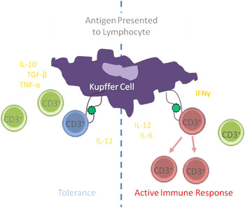
One of the early hematological hallmarks of BVDV is leukopenia, primarily lymphopenia, affecting T-helper cells, cytotoxic T cells and gamma–delta T cells in vivo (Bolin et al., Reference Bolin, McClurkin and Coria1985; Rebhun et al., Reference Rebhun, French, Perdrizet, Dubovi, Dill and Karcher1989; Gånheim et al., Reference Gånheim, Johannisson, Ohagen and Persson Waller2005). These peripheral blood lymphocytes undergo apoptosis in vitro (Ridpath et al., Reference Ridpath, Bendfeldt, Neill and Liebler-Tenorio2006), resulting in lymphoid depletion of B cells, T helper cells, cytotoxic T cells, and gamma–delta T cells to variable degrees depending on the virulence of the strain of BVDV (Ridpath et al., Reference Ridpath, Bendfeldt, Neill and Liebler-Tenorio2006; Chase, Reference Chase2013). B-cell levels in circulation are relatively normal (Gånheim et al., Reference Gånheim, Johannisson, Ohagen and Persson Waller2005) but B-cell depletion occurs extensively in the germinal centers of the lymph node (Liebler-Tenorio et al., Reference Liebler-Tenorio, Ridpath and Neill2002, Reference Liebler-Tenorio, Ridpath and Neill2003). Interestingly, there was no effect on circulating monocytes (Gånheim et al., Reference Gånheim, Johannisson, Ohagen and Persson Waller2005); although, in vitro infection of monocytes results in BVDV production (Brackenbury et al., Reference Brackenbury, Carr and Charleston2003; Rajput et al., 2014). Likewise, during maturation from monocytes to monocyte-derived dendritic cells (MDDC), monocytes cultured in the presence of IL-4 and GM-SCSF initially produce virus, but within 48 h after in vitro culture, the virus yield is reduced and ceases altogether by 120 h in culture (Rajput et al., 2014). Interestingly, monocyte-derived macrophages (MDM) also fail to produce progeny virus despite the presence of viral proteins and progeny viral RNA (Chase et al., Reference Chase, Elmowalid and Yousif2004; Rajput et al., 2014). There is no effect on MDDC or MDM viability following BVDV infection (Chase et al., Reference Chase, Elmowalid and Yousif2004; Rajput et al., 2014). In BVDV-infected lymphoid tissue, macrophages and dendritic cells (DCs) appear to be uninfected although they are surrounded by apoptotic lymphocytes (Liebler-Tenorio et al., Reference Liebler-Tenorio, Ridpath and Neill2002, Reference Liebler-Tenorio, Ridpath and Neill2003). The cytokines released by macrophages can vary, but have the potential to induce lymphoid apoptosis in certain circumstances; this mechanism may be employed by BVDV infection and the macrophage may be functioning in a ‘hitman’ capacity, eliminating lymphocytes that would otherwise be activated (Chase et al., Reference Chase, Elmowalid and Yousif2004; Sánchez-Cordón et al., Reference Sánchez-Cordón, NUNez, Salguero, Pedrera, Fernández de Marco and Gómez-Villamandos2005) (Fig. 1). Lymphoid depletion with macrophage and DC survival is also observed following infection with another pestivirus, classic swine fever virus (CSFV). Work with CSFV indicates that the likely cytokines involved in CSFV lymphoid apoptosis are interleukin 1 (IL-1), interleukin 6 (IL-6), and tumor necrosis factor alpha (TNF-α) (Choi et al., Reference Choi, Hwang and Chae2004; Sánchez-Cordón et al., Reference Sánchez-Cordón, NUNez, Salguero, Pedrera, Fernández de Marco and Gómez-Villamandos2005). Interestingly, studies with cultured macrophages infected with BVDV (Peterhans et al., Reference Peterhans, Jungi and Schweizer2003) or CSFV (Borca et al., Reference Borca, Gudmundsdottir, Fernández-Sainz, Holinka and Risatti2008) had increased IL-1 and IL-6 but did not have increased TNF-α production; however, in vivo infection with CSFV clearly demonstrated TNF-α at high levels in macrophages in both lymph nodes and spleen (Choi et al., Reference Choi, Hwang and Chae2004; Sánchez-Cordón et al., Reference Sánchez-Cordón, NUNez, Salguero, Pedrera, Fernández de Marco and Gómez-Villamandos2005). These macrophages were surrounded by apoptotic lymphocytes. Similar work needs to be done with BVDV.
B cells, T cells and plasma cells are all depleted by BVDV infection.

What about other innate ‘killers’ – neutrophils and natural killer cells?
Neutrophils
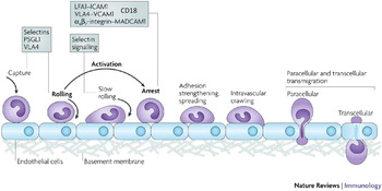
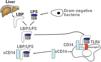
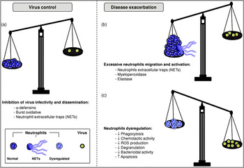
Probably no cell population has been more understudied for viral diseases than the neutrophils – the ultimate ‘bacteria killer’ (Gabriel et al., Reference Gabriel, Her and Ng2013). Neutrophils comprise from 20 to 70% of blood leukocytes depending on the age of animal (Menge et al., Reference Menge, Neufeld, Hirt, Schmeer, Bauerfeind, Baljer and Wieler1998). Although blood neutrophils decline following BVDV infection, the decrease is much less than that observed with lymphocytes (Roth et al., Reference Roth, Kaeberle and Griffith1981; Gånheim et al., Reference Gånheim, Johannisson, Ohagen and Persson Waller2005). BVDV causes a dramatic decrease in phagocytosis and killing that can last up 2 weeks following infection or vaccination (including two vaccine strains Singer and NADL) (Roth et al., Reference Roth, Kaeberle and Griffith1981; Roth and Kaeberle, Reference Roth and Kaeberle1983). Recently, we determined a direct in vitro effect of BVDV on bovine neutrophils (Thakur et al., Reference Thakur, Braun and Chase2014). Exposure of neutrophils to either non-cytopathic or cytopathic BVDV in vitro did not result in apoptosis but did decrease CD18 and CD62L (L-selectin) expression (Thakur et al., Reference Thakur, Braun and Chase2014). These two receptors are important for adhesion to endothelial cells, which is essential for neutrophil diapedesis (Fig. 2), indicating that BVDV could inhibit neutrophil migration. CD18 is the well-characterized leukocyte adhesion receptor associated with bovine leukocyte adhesion deficiency (BLAD) (Shuster et al., Reference Shuster, Kehrli, Ackermann and Gilbert1992). Curiously, neutrophils exposed to BVDV also had increased CD14 expression (Thakur et al., Reference Thakur, Braun and Chase2014). In contrast, CD14 was downregulated in BVDV-infected bovine macrophages in a strain-dependent manner (Chase et al., Reference Chase, Elmowalid and Yousif2004). CD14 is a polyspecific receptor, which recognizes predominately lipopolysaccharide (LPS) from the cell wall of Gram-negative bacteria (Fig. 3). CD14 is part of the toll-like receptor-4 (TLR-4) complex, which is an important receptor for pathogen recognition. Unlike monocytes/macrophages, neutrophil CD14 is not constitutively expressed but is induced by exposure to pathogen associated molecular patterns (PAMP) particularly LPS (Paape et al., Reference Paape, Bannerman, Zhao and Lee2003). Binding to neutrophil CD14 receptor results in production of the pro-inflammatory cytokines TNF-α and IL-1 (Finlay and Hancock, Reference Finlay and Hancock2004). Like monocytes/macrophages, neutrophil CD14 can also be secreted (sCD14; Sohn et al., Reference Sohn, Paape, Bannerman, Connor, Fetterer and Peters2007) and the sCD14 has been shown to downregulate the inflammatory response. Several papers examining the effect of BVDV on immunoresponsiveness have indicated that BVDV infection results in an early proinflammatory response with often a corresponding decrease in anti-inflammatory response which overlays quite remarkably to the increased expression of neutrophil CD14 and the shift to sCD14; therein, this may be a target of interest for future studies (Risalde et al., Reference Risalde, Gómez-Villamandos, Pedrera, Molina, Cerón, Martínez-Subiela and Sánchez-Cordón2011; Molina et al., Reference Molina, Risalde, Sánchez-Cordón, Romero-Palomo, Pedrera, Garfia and Gómez-Villamandos2014; Risalde et al., Reference Risalde, Molina, Sánchez-Cordón, Romero-Palomo, Pedrera and Gómez-Villamandos2014). Tempered inflammatory response and reduced diapedesis is likely to exacerbate any disease following viral infections, and is likely part of the immune dysregulation seen with BVDV (Fig. 4). Furthermore, while apoptosis was not noted in vitro, neutrophil decline has been observed in vivo (Roth et al., Reference Roth, Kaeberle and Griffith1981; Gånheim et al., Reference Gånheim, Johannisson, Ohagen and Persson Waller2005). CD14 expression on neutrophils may be recognized and serve as a target for induction of apoptosis by macrophages (Sladek and Rysanek, Reference Sladek and Rysanek2006). Future neutrophil research investigating the interaction and cytokine profile between BVDV-infected macrophages and neutrophils in vitro would be of great interest and potentially the ‘the lymphocyte hitman’ function of macrophages might be expanded to include neutrophils.
CD18 (Integrin) and L-selectins are essential for adhesion and extravasation of neutrophils. Adapted from Ley et al. (Reference Ley, Laudanna, Cybulsky and Nourshargh2007).

The interaction of LPS and CD14 in the neutrophil. Adapted from Zeldin et al. (Reference Zeldin, Eggleston, Chapman, Piedimonte, Renz and Peden2006). Environmental Health Perspectives, 620–626 open access.

Contradictory roles of neutrophils. Neutrophils can (a) control viral infections by releasing antimicrobial components and reactive oxygen species (ROS), and produce neutrophil extracellular traps (NETs) leading to the inhibition of virus infectivity, viral RNA degradation, and restrict virus dissemination. However, (b) excessive migration and overactivation of neutrophils can be detrimental to the host and exacerbate disease manifestation. (c) On the other hand, viruses can dysregulate neutrophil functions and alter phagocytosis, migration, ROS release, and degranulation. These often lead to secondary bacterial infections. Viruses can also induce apoptosis in neutrophils. Adapted from Gabriel et al. (Reference Gabriel, Her and Ng2013).

Natural killer (NK) cells
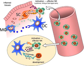
NK cells are an important cog of innate anti-viral defense, and their characteristics were well-summarized in an excellent review by Boysen and Storset (Reference Boysen and Storset2009) (Figs. 5–7). However, there are very little data on NK cell phenotype or function following viral infection particularly in cattle (Boysen and Storset, Reference Boysen and Storset2009). NK cells were first characterized in cattle in 2004 (Storset et al., Reference Storset, Kulberg, Berg, Boysen, Hope and Dissen2004). NK cells represent 1–10% of blood mononuclear cells (Storset et al., Reference Storset, Kulberg, Berg, Boysen, Hope and Dissen2004) and the percentage of NK cells in the blood is age dependent (Graham et al., Reference Graham, Thom, Howard, Boysen, Storset, Sopp and Hope2009). Like lymphocytes, NK cells are produced in the bone marrow and then populate lymphoid tissue after birth and following exposure to microbial populations (Fig. 5). Despite low relative numbers in the lymphoid tissue, it is estimated that there are 10X as many total NK cells in the lymphoid tissue as compared to the peripheral blood (Boysen et al., Reference Boysen, Gunnes, Pende, Valheim and Storset2008). NK distribution in lymph nodes varies depending on the type of lymph node with 2–4% NK cells in mucosal lymph nodes vs. 6–10% NK cells in spleen and non-mucosal lymph nodes (Boysen et al., Reference Boysen, Gunnes, Pende, Valheim and Storset2008). NK cells are present in the paracortical as well as in the medullary areas of bovine lymph nodes and are evenly distributed except in germinal centers where only a few NK cells are seen. In mucosal lymph nodes, lymphoid follicles are more numerous and large, while non-mucosal lymph nodes had smaller and fewer follicles. NK cells in lymphoid follicles can express interleukin-2 receptor (IL-2 receptor; CD25) and become lymphokine-activated killer cells (LAK) (Figures 6 and 7b), resulting in the killing of infected somatic cells. These cells also produce gamma interferon (IFN-γ), which further activates DC, T-helper cells and macrophages (Boysen et al., Reference Boysen, Gunnes, Pende, Valheim and Storset2008) (Figs. 5 and 8). Unlike other lymphocytes, NK immune suppression may result from a lack of mature NK cells rather than elimination of the population. NK cells are particularly dependent on IL-15 for their development and maturity (Endsley et al., Reference Endsley, Endsley and Estes2006; Lund et al., Reference Lund, Boysen, Dean, Davis, Park and Storset2012, Reference Lund, Boysen, Hope, Sjurseth and Storset2013). A recent study in cattle infected with highly acute 1373 BVDV strain had decreased levels of IL-15 in bronchial lymph nodes, suggesting lower NK activation (Palomares et al., Reference Palomares, Brock and Walz2014). We found that like macrophages and DCs (Chase et al., 2004; Rajput et al., Reference Rajput, Darweesh, Park, Braun, Mwangi, Young and Chase2014), BVDV infection of NK cells with either highly acute strain BVDV-2 1373 or mild strain BVDV-2 28508-5 had no effect on bovine NK cell viability in vivo (Darweesh, Reference Darweesh2013).
Body compartments for NK cell priming of human/murine NK cells. Upon significant microbial attack, primed NK cells re-enter the lymph nodes, where they are activated by DCs to produce interferon-gamma, which participate in the polarization of Th1 cells. In addition, primed NK cells are recruited into inflamed tissues, where they are activated by damaged/infected cells as well as accessory cells, triggering effector functions such as killing of damaged cells and release of cytokines. Unlike T-cells, these NK effector responses are not restricted by microbe-specific antigens. In individuals or species heavily exposed to microbes, this may be illustrative of a ‘steady state’ situation, and comparably more primed NK cells will be present in lymph nodes and circulation (DC, dendritic cell; MΦ, macrophage; SC, somatic cells). Adapted from Boysen and Storset (Reference Boysen and Storset2009).

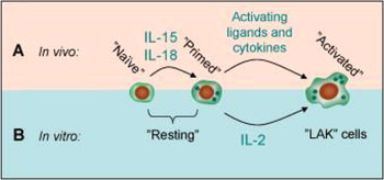
Relationship between NK cells in vivo and experimental LAK cells. (A) Recent evidence has shown that in the living organism, NK cells are initially naïve and poorly responsive, but following microbial exposure, priming by cytokines (IL-15 and IL-18) render the NK cells responsive. Primed NK cells become activated and fully functional in response to activating cytokines and ligands on the target cells. (B) ‘Resting’ isolated NK cells normally show poor or moderate cytotoxic capacity in in vitro assays, and it may not be clear whether these are naïve or primed. Stimulation of resting NK cells with IL-2 for several days result in LAK cells, which are strongly cytotoxic. Adapted from Boysen and Storset (Reference Boysen and Storset2009).

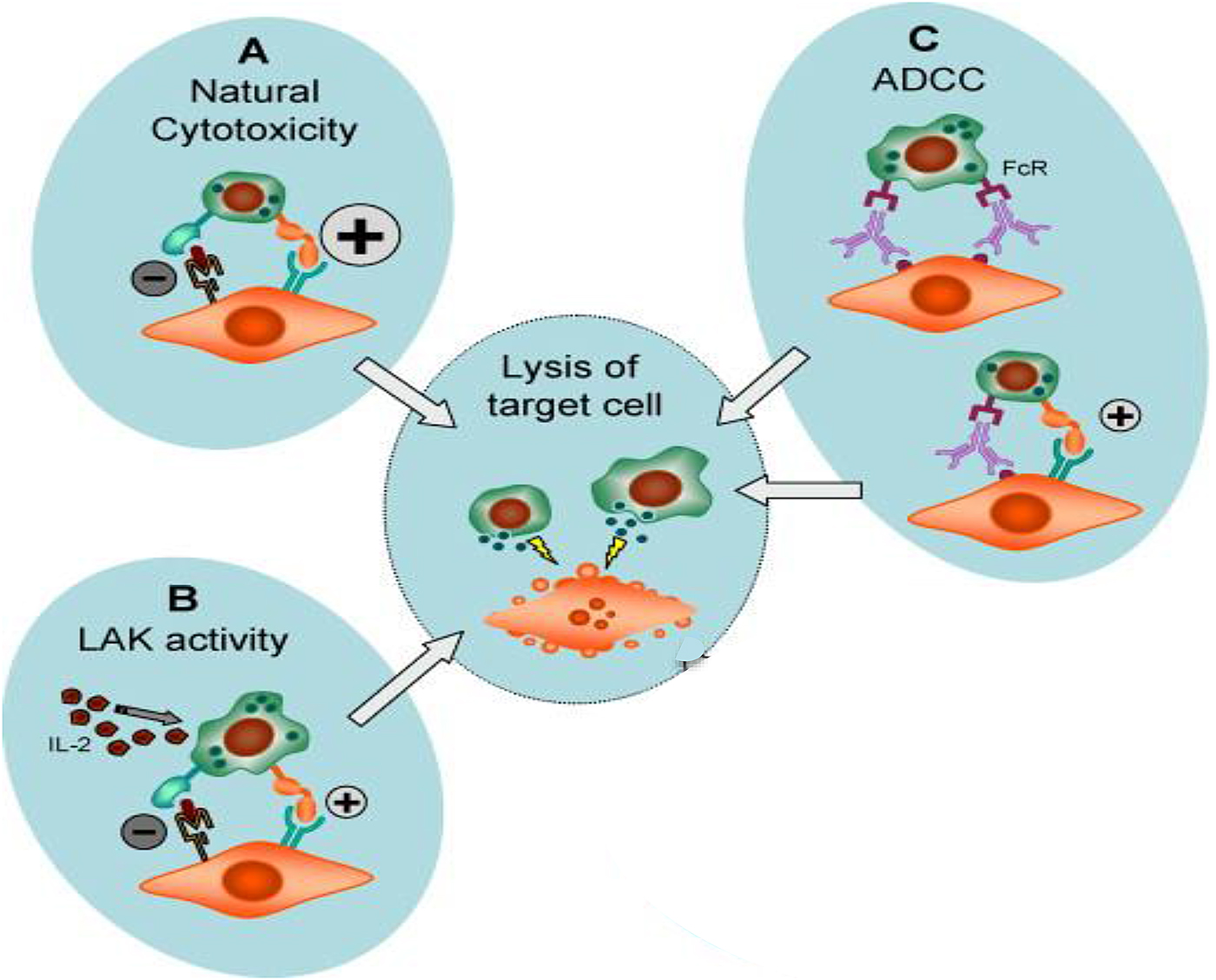
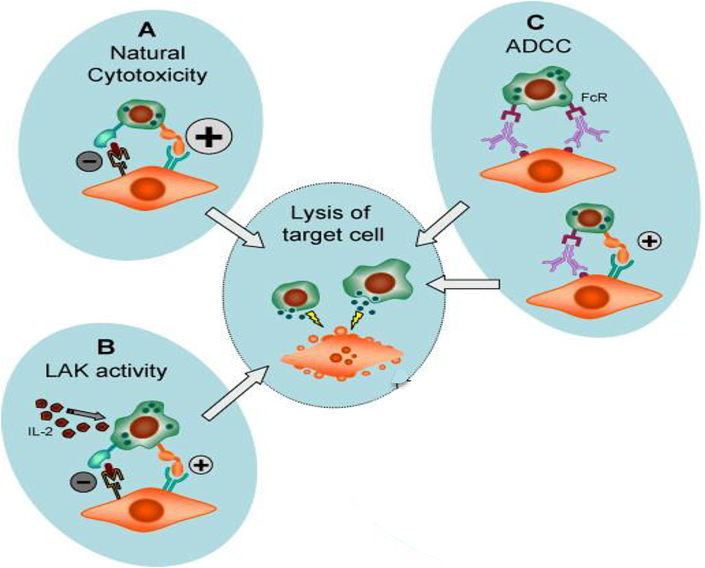
NK cytotoxic phenomena. The release of cytotoxic granula and subsequent lysis of a target cell may occur from different experimental settings: (A) natural cytotoxicity is the spontaneous lysis of a target cell by the NK cell. This results when inhibiting signals (−) to the NK cell are absent or insufficient to overrule activating (+) signals. (B) LAK activity is the result of pre-activation of the NK cell by cytokines, most often IL-2. (C) ADCC (antibody-dependent cell-mediated cytotoxicity) involved the cross-linking of Fcg receptors on the NK cell, by antibodies directed to structures on the target cell. This reaction may also involve other recognition events between NK cells and target cells. Adapted from Boysen and Storset (Reference Boysen and Storset2009).
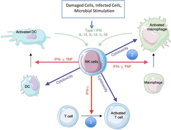
Cytokine production by NK cells. Several types of stimuli result in the NK cell production of cytokines such as IFN-γ and TNF. Such stimuli can be damaged or infected cells display altered cellular ligands and release cytokines like IFN-α/β, both recognized by NK cells. Microbial stimulation of accessory cells leads to their production of IL-12 and IL-18 that work in synergy to activate the NK cell. The cytokines released from NK cells trigger several mechanisms of cellular immunity, most importantly: (1) Assistance to DCs to produce ‘Th1’ cytokines, which promote cellular T-cell responses. (2) Prompting of macrophages to eliminate microbes (DC, dendritic cell). Adapted from Vivier et al. (Reference Vivier, Tomasello, Baratin, Walzer and Ugolini2008).

NK cells also present a new target for understanding the greater immunological dysfunction that results following BVDV infection. The formulation of the ‘missing self’ hypothesis by Ljunggren and Kärre (Reference Ljunggren and Kärre1990) early on, shaped perspectives regarding the importance of NK cells by suggesting that a principle function of NK cells is to eliminate cells that lack MHC class I (MHCI) or other ‘self-markers’ (Kärre et al., Reference Kärre, Ljunggren, Piontek and Kiessling1986). Such situations occur in cancer, viral infections and transplantation, indicating a prominent role for NK cells in these conditions. BVDV down-regulates MHCI in a strain-dependent manner (Chase et al., Reference Chase, Elmowalid and Yousif2004; Rajput et al., Reference Rajput, Darweesh, Park, Braun, Mwangi, Young and Chase2014), which is a compelling reason to look closer at the phenotypical impact of this change on NK cells.
In general, NK cell receptor biology is more complicated than other lymphocytes (Boysen and Storset, Reference Boysen and Storset2009). NK cells have a number of cell receptors; some that are activating (triggering) and some that are inhibitory (Moretta et al., Reference Moretta, Bottino, Vitale, Pende, Cantoni, Mingari, Biassoni and Moretta2001; Boysen and Storset, Reference Boysen and Storset2009) (Fig. 7a, b) and their activation is likewise variable (Figs. 7 and 8). The major NK phenotypic marker is the natural cytotoxicity activating (triggering) receptor (NCR1), which is also known as NKp46 (natural killer cell 46-kDa cell surface protein; CD335). The exact ligand for NKp46 is not specifically known, but is likely a tumor or viral protein (Boysen and Storset, Reference Boysen and Storset2009) (Fig. 7a). NK cells are also marked by granulysin; the antimicrobial protein contained in the NK and cytotoxic T-cells granules (Endsley et al., Reference Endsley, Furrer, Endsley, McIntosh, Maue, Waters, Lee and Estes2004) and the IgG Fc receptor IIIA (FcgRIIIA; CD16), which binds the constant region of the IgG molecule and is important for NK antibody-dependent cell-mediated cytotoxicity (ADCC) function (Fig. 7c).
There are phenotypic differences with adhesion marker CD2 (found also on T cells) between NK cells in circulation (CD2+) and those found in lymphoid tissue (CD2−) (Boysen et al., Reference Boysen, Olsen, Berg, Kulberg, Johansen and Storset2006; Lund et al., Reference Lund, Boysen, Hope, Sjurseth and Storset2013). In addition, bovine NK cells have another family of activation/inhibitory receptors, killer immunoglobulin-like receptors (KIRs) (Boysen and Storset, Reference Boysen and Storset2009). Although KIRs have been defined genetically in cattle, there is no functional work that has identified their roles in bovine NK activation/inhibition (Dobromylskyi and Ellis, Reference Dobromylskyj and Ellis2007). Likewise, other NK activating/inhibitory receptors have been identified in human and murine models; however, homologues have yet to be identified in ruminants (Boysen and Storset, Reference Boysen and Storset2009). Therefore, there is still much to characterize with respect to the normal NK phenotype, and also then further understanding the interaction between BVDV and NK cells.
Recently, the effect of BVDV strains on NK activation marker CD25, phenotype markers CD2, NKp46 and the granule protein, granulysin were investigated in IL-2 stimulated NK cells in vitro using the type cell depicted in Fig. 6. BVDV infection of NK cells with either highly virulent strain BVDV-2 1373 or mild strain BVDV-2 28508-5 decreased NKCD2+ and MHCI and had no significant effect on NK activation as measured by expression of CD25 (Darweesh, Reference Darweesh2013). BVDV-2 1373 strain increased the number of NKp46+ NK cells. Also both strains reduced NK cell killing, with BVDV-2 28508-5 mild strain having the largest reduction (Darweesh, Reference Darweesh2013). These studies indicate that BVDV infection resulted in NK phenotypic and activation changes that are strain dependent in vitro, and this may result in immune dysfunction in vivo. Therein, this work presents yet another immunological target that must be critically evaluated to gain insight into the BVDV immune dysfunction.
Persistence and innate immunity – a role for the liver and Kupffer cells?
For many years, veterinarians and researchers have worked under the assumption that BVDV persistent infection developed as a result of the immune incompetence of the fetus in the late first-early second trimester of pregnancy; however, many immune components are in place early in the second, if not the first trimester. Bovine fetuses have lymphoid development of the thymus, spleen and some peripheral nodes by the middle of the first trimester, day 42, day 55, and day 60, respectively (Schultz et al., Reference Schultz, Dunne and Heist1973). The gut-associated lymphoid tissue (GALT), however, was not noted in this study until later in the second trimester (175 days) in gestation. Germinal centers as well as some IgM in antigenically stimulated fetuses were present in the mid-first trimester as well at day 59, and IgG was in circulation by the mid-second trimester at 145 days (Schultz et al., Reference Schultz, Dunne and Heist1973). Bovine fetal immunocompetence is established around mid-second trimester of gestational (day 150) (Brock, Reference Brock2003). Understanding how BVDV evades and even alters these early immune parameters seems critical to gaining insight into the mechanism of the development of BVDV persistent infection.
The liver is an immunologically important organ with a particularly significant role in a developing fetus. The liver is the primary site of hematopoiesis and immune development at 100 days of gestation and studies have demonstrated that the liver, like a lymph node, is competent in primary activation of naive T cells (Bertolino et al., Reference Bertolino, McCaughan and Bowen2002; Bowen et al., Reference Bowen, Zen, Holz, Davis, McCaughan and Bertolino2004). Interestingly, the location where a lymphocyte is educated has tremendous implications on the outcome of its response to antigen presentation, because lymphocytes activated in the lymph node differentiate in distinctly different ways than those activated in the liver. Lymphocyte primary education in the hepatic tissues has been shown to provide a strong bias toward immune tolerance (Bowen et al., Reference Bowen, Zen, Holz, Davis, McCaughan and Bertolino2004). Overall, this prevailing response of the liver ensures the immune system does not react inappropriately to digestive antigens (Knolle and Gerken, Reference Knolle and Gerken2000). Early studies showed that intraportal application of antigens induced antigen-specific systemic tolerance (Cantor and Dumont, Reference Cantor and Dumont1967; Triger et al., Reference Triger, Cynamon and Wright1973; Li and Tian, Reference Li and Tian2013), suggesting that the immune response established in the liver can pilot the response in other immunological tissues as well.
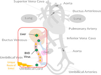
The structural architecture of the liver allows it a pivotal role in the balance of a host immune response (Knolle and Gerken, Reference Knolle and Gerken2000). Antigen-rich blood from the gastrointestinal tract in adults, and nutrient dense blood in the fetus, reaches the liver via the portal, or umbilical circulation, respectively, providing nutrients for digestion and storage (Fig. 9). The blood is then infused across a network of small diameter, high-pressure sinusoidal vessels. High resistance slows the flow significantly and allows maximal exposure to the non-parenchymal cells that comprise 20–40% of the liver. This population includes: endothelial cells, Kupffer cells (KCs), lymphocytes including B cells, and T-cell subsets: T regulatory, T helper, cytotoxic T cells, and DCs; gamma/delta cells, biliary cells and stellate cells.
Unique route of BVDV entry may be in part responsible for the development of BVDV persistent infection.

KCs are recognized as the liver's resident macrophages and have many macrophage-type functions, including cytokine secretion, MHC presentation, and microbial killing by production of oxygen-derived free radicals (Decker, Reference Decker1990, Reference Decker1998; Laskin and Pendino, Reference Laskin and Pendino1995). KCs exhibit both excitatory and tolerant measures in their interactions with leukocytes and are capable of secreting inflammatory mediators such as IL-1, IL-6, and TNF-α (Risalde et al., Reference Risalde, Gómez-Villamandos, Pedrera, Molina, Cerón, Martínez-Subiela and Sánchez-Cordón2011) as well as tolerogenic cytokines like IL-10 (Fig. 10) (Knolle et al., Reference Knolle, Schlaak, Uhrig, Kempf, Meyer zum and Gerken1995). The immune response of the KCs begins with the production of IL-12 and IL-18 in response to viral infection that activates local NK cells to produce IFN-γ. Following this activation, however, KCs release IL-10, which down regulates inflammatory mediators IL-6 and TNF-α and restores the more tolerant nature of the hepatic microenvironment (Tiegs and Lohse, Reference Tiegs and Lohse2010).
Immunological switch that KCs make between tolerance and active immune response.

KCs also mediate local and peripheral immune suppression for specific antigens by expressing a negative co-stimulator for T-cell activation alongside the antigen they are presenting on either MHC I or MHC II (Knolle and Gerken, Reference Knolle and Gerken2000). This negative co-stimulator is programmed cell death ligand 1 (PD-L1), which localizes circulating CD8+ and CD4+ T cells. CD8+ T cells in the liver begin to express cytotoxic lymphocyte antigen-4 (CTLA-4). CTLA-4 contributes to the suppression of immune response by inducing a T-regulatory cell profile (Tregs: CD4+CD25+FoxP3+) from naive and effector CD4+ T cells, converting them to Tregs, which induces tolerance (He et al., Reference He, Chen, Xu, Cai, Wu, Li and Chen2009). All of these responses, including portal tolerance, are lost if KCs are depleted in hepatic tissues (Roland et al., Reference Roland, Mangino, Duffy and Flye1993; Rai et al., Reference Rai, Loffreda, Karp, Yang, Lin and Diehl1997).
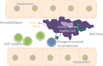
With Todd Hansen's group at Colorado State, as part of a larger study (Smirnova et al., Reference Smirnova, Webb, Bielefeldt-Ohmann, Van Campen, Antoniazzi, Morarie and Hansen2012, Reference Smirnova, Webb, McGill, Schaut, Bielefeldt-Ohmann, Van Campen, Sacco and Hansen2014), we analyzed fetal liver and isolated KCs from fetal calves whose dams were infected with BVDV at ~75 days of gestation followed by collection of fetuses at 82 and 89 days of gestation (7 and 14 days post-maternal infection). BVDV antigen was not detected in the fetuses at 82 days of gestation; however, BVDV was detected by immunohistochemistry in fetal liver at 89 days of gestation (14 days post-maternal infection) (Morarie et al., Reference Morarie, Braun, Smirnova, Hansen and Chase2012). Notably, within the fetal liver, only KCs were positive for BVDV antigen by IHC (Morarie et al., Reference Morarie, Braun, Smirnova, Hansen and Chase2012). The number of KCs was increased in the fetal liver at 89 days, and this is consistent with what was reported with acute BVDV liver infections (Risalde et al., Reference Risalde, Gómez-Villamandos, Pedrera, Molina, Cerón, Martínez-Subiela and Sánchez-Cordón2011). MHCI was expressed on 3X as many KCs in BVDV-infected calves (51 vs. 17%) as in control KCs from uninfected study fetuses at 89 days of gestation (Morarie et al., Reference Morarie, Braun, Smirnova, Hansen and Chase2012). MHC II was expressed on 2X as many BVDV-infected KCs (38 vs. 16%) as uninfected KCs from fetal calves (Morarie et al., Reference Morarie, Braun, Smirnova, Hansen and Chase2012). Cytokine analysis of supernatant collected from the BVDV-infected isolated KCs cultured for 24 h demonstrated lower levels of proinflammatory cytokines (TNF-α, IL-6, and IL-1β) than control KC supernatant (Morarie, Reference Morarie2012). There were also large aggregates of CD3+ lymphocytes noted in the fetal liver at gestational day 89 (Morarie, Reference Morarie2012). Taken together, this provides one plausible model for induction of tolerance of BVDV that results in BVDV PI and identifies yet another new target for research and better understanding into the development of persistent BVDV infection in calves (Fig. 11). BVDV-infected KCs in the tolerance phenotype may play a critical role in the forthcoming immunosuppression and persistent infection (Fig. 11). Once more, further in vitro study and analysis will be essential to unravel this mechanism in greater detail.
Model for the induction of BVDV tolerance by KCs.

Closing thoughts
BVDV continues to be a major problem for bovine health. With everything that we know about BVDV, there is still so much to learn. BVDV immune dysregulation can occur at many levels. New insight and perspectives have improved our understanding and shone light on several new areas to guide future research. The effect of BVDV infections on macrophages, NK cells, and neutrophils, and the mechanism of immune evasion in BVDV persistent infection, are targets that are critical to understanding BVDV pathogenesis. A greater insight in these innate immune responses will aid in the development of the next generation of BVDV prevention and control measures.