Cronobacter sakazakii, formerly Enterobacter sakazakii, is a gram-negative bacillus and an emerging pathogen in life-threatening infections, especially in infants (Iversen et al. Reference Iversen, Mullane, McCardel, Tal, Lehner, Fannin, Stephan and Joosten2008). C. sakazakii infection can lead to neonatal meningitis, sepsis, and necrotising enterocolitis, with a mortality rate between 40–80% (Liu et al. Reference Liu, Cai, Zhang, Gao, Yang, Zheng, Luo and Huang2006). Although powdered infant formula is the major vehicle for most Cronobacter infections, this bacterium is also isolated from milk, meat, plant food, and food ingredients including cereal, fruit and vegetables, herbs, spices, and marine products (Friedemann, Reference Friedemann2007; Chon et al. Reference Chon, Song, Kim, Hyeon and Seo2012).
Kefir is a type of fermented milk originating from the Caucasus Mountains in Eurasia. It is produced by the microbial activity of kefir ‘grains’ (starter cultures), which have a symbiosis containing over 50 species of lactic and acetic acid bacteria and yeast (Guzel-Seydim et al. Reference Guzel-Seydim, Kok-Tas and Greene2011; Kim et al. Reference Kim, Chon, Kim, Kim, Choi, Hwang and Seo2015a). In our previous study, we showed that kefir has antibacterial activity against C. sakazakii in pure culture and powdered infant formula (Kim et al. Reference Kim, Chon, Kang, Kim, Kim, Song and Seo2015b). Kefir supernatant not only inhibited the growth of C. sakazakii, but also killed vegetative cells present in powdered infant formula (Kim et al. Reference Kim, Chon, Kang, Kim, Kim, Song and Seo2015b). Although kefir contains multiple bacterial strains, information is limited regarding the relative contribution of single strain of lactic acid bacteria (LABs) on its antimicrobial activity.
Given this gap in knowledge, the current study was designed to achieve two objectives. First, the antimicrobial activity of supernatant derived from kefir-isolated LABs was evaluated and compared to that of a commercial probiotic strain. Second, the mechanisms underlying its antimicrobial activity against C. sakazakii were investigated.
Materials and methods
Bacterial strains used in this study
Lactobacillus kefiri DH5 and L. kefiranofaciens DH101 were isolated from kefir grains and used as test strains. As a control, Bifidobacterium longum BL720 was purchased from CHR. Hansen (Horsholm, Denmark) and used because this species was reported to have an antimicrobial activity against Escherichia coli O157:H7 (Ibrahim et al. Reference Ibrahim, Dharmavaram, Seo and Shahbazi2003). The three strains of bacteria were grown in MRS broth (Oxoid, Basingstoke, Hampshire, UK) at 37 °C for 72 h. The cultures were centrifuged and the supernatants filtered through a 0·45-μm syringe filter (Millipore, Bedford, MA, USA) for removing bacterial cell. The resultant sterile filtrates were used for evaluating the antimicrobial activity against C. sakazakii. C. sakazakii ATCC 29544 strain used in this study was originally obtained from the American Type Culture Collection (ATCC; Manassas, VA, USA). C. sakazakii ATCC 29544 was grown in nutrient broth (Oxoid) for 24 h at 37 °C.
Determination of antimicrobial activity of culture supernatant from kefir isolates
To determine the antimicrobial activity of kefir-isolated LAB, culture supernatants from B. longum 720, L. kefiri DH5, or L. kefiranofaciens DH101 were added to nutrient broth (100 µl of supernatant in 1 ml of broth, yielding a final concentration of 10% v/v) and each sample was then inoculated with a 50-μl suspension of C. sakazakii ATCC 29544 containing 6·33 ± 0·21 log CFU/ml. As described previously (Kim et al. Reference Kim, Chon, Kang, Kim, Kim, Song and Seo2015b), cultures were mixed and incubated at 37 °C, and optical density (595 nm) was measured for 24 h at 1-h intervals using a microplate reader (Multiskan FC; Thermo Fisher Scientific, Waltham, MA, USA). Based on the results, the strain with the greatest antimicrobial activity, L. kefiri DH5, was selected for the following experiments. In the follow-up studies of L. kefiri DH5-dependent antimicrobial activity, 100, 200, and 300 µl culture supernatants from this strain were added to 1 ml of nutrient broth, and each dilution was inoculated with 50-μl suspensions of C. sakazakii ATCC 29544 containing 6·21 ± 0·30 log CFU/ml. Growth curve analysis was conducted to evaluate the antimicrobial activity of these concentrates as described above. After incubating 6 and 24 h, viable C. sakazakii was also enumerated by plating 100 µl of the inoculum on Druggan–Forsythe–Iversen (DFI) agar and incubating for 24 h at 37 °C.
Measurement of the leakage of cytoplasmic materials
The cell membrane integrity of C. sakazakii was examined by measuring the release of cell constituents into the supernatant using methods described previously (Tiwari et al. Reference Tiwari, Behari and Sen2008; Guo et al. Reference Guo, Gai, Jiao, Wang, Zu and Fu2016). A total of 200 µl culture supernatants of L. kefiri DH5 were added to 1 ml of saline containing 8·21 ± 0·30 log CFU/ml of C. sakazakii ATCC 29544. Fresh MRS broth was used as a control. After incubation for 4 h at 37 °C, the samples were centrifuged at 2000 g for 10 min and analysed for nucleic acid and protein leakage. For nucleic acid leakage analysis, the absorbance at 260 nm (A260 nm) of the sample was determined with NanoDrop 2000 spectrophotometer (Thermo Fisher Scientific, Wilmington, DE, USA). Protein leakage analysis was performed using Bradford assay (Bio-Rad, Hercules, CA, USA) according to the manufacturer's guideline. For each sample, 20 µl of the supernatant was mixed in 230 µl of Bradford reagent. Optical density of the sample at 595 nm (OD595 nm) was measured after 5 min of incubation at room temperature.
Measurement of pH and titrable acidity of the supernatant
The pH of the sterile filtrate was determined using a pH meter model 205 (Testo, Lenzkirch, Germany) equipped with temperature compensation, and calibrated using pH 4·00 and 7·00 buffers. The titratable acidity was determined by the titration method using 0·1 N NaOH with phenolphthalein indicator (Sigma-Aldrich, St Louis, MO, USA) as until colour changed to rose pink.
Heat treatment and pH neutralisation
To address the mechanisms by which L. kefiri DH5-derived culture supernatants exerted antimicrobial activity, the supernatant was subjected to (i) heat treatment at 60 °C for 30 min or 100 °C for 10 min or (ii) neutralisation to pH 7·0 with 1 N NaOH. The 200 µl of heat-treated and neutralised supernatants were subjected to growth curve analysis as described above.
Statistical analysis
Data were analysed using SPSS version 18·0 (SPSS Inc., Chicago, IL, USA). Differences in optical density at 595 nm and the numbers of viable C. sakazakii cells after 6 and 24 h of treatment were analysed by analysis of variance (Tukey's method). The data on the leakage of cellular components was analysed by Student's t-test. A P < 0·05 was considered statistically significant.
Results and discussion
Antimicrobial activity of culture supernatant of kefir LAB isolates against C. sakazakii
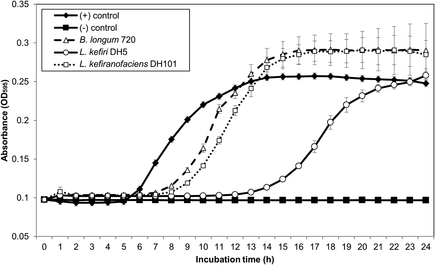
The addition of supernatants derived from cultures of L. kefiranofaciens DH101 and B. longum 720 prolonged the lag phase of C. sakazakii ATCC 29544 to 8 h, whereas the non-treated C. sakazakii cultures was 5 h (Fig. 1). Strikingly, L. kefiri DH5-derived culture supernatants prolonged the lag phase to 13–14 h, confirming that this bacterial strain was the most potent among those tested.
Suppression of C ronobacter sakazakii ATCC 29544 growth by lactic acid bacteria derived from kefir.

The pH values of the culture supernatants of L. kefiranofaciens DH101, L. kefiri DH5, and B. longum 720 were 4·39, 4·49, and 5·63, respectively. The titrable acidity of L. kefiranofaciens DH101, L. kefiri DH5, and B. longum 720 was 0·0568, 0·0468, and 0·0232, respectively, suggesting the culture supernatants of L. kefiri DH5 was more acidic than these of the other strains (P < 0·05).
There have been many reports on antimicrobial activity of LAB against pathogenic microorganisms to date (Hernandez et al. Reference Hernandez, Cardell and Zarate2005; Cizeikiene et al. Reference Cizeikiene, Juodeikienea, Paskeviciusb and Bartkienec2013; Jeong et al. Reference Jeong, Kim, Kang, Kim, Song, Kim and Seo2017). Indeed, although kefir's antimicrobial activity has been extensively investigated, only limited information is available on the effects of individual microorganisms present within this food product (Garrote et al. Reference Garrote, Abraham and De Antoni2000; Ulusoy et al. Reference Ulusoy, Olak, Hampikyan and Erkan2007). To the best of our knowledge, this is the first study to investigate the antimicrobial activity of kefir LAB isolates. Moreover, they are generally recognised as safe (GRAS), the application of LAB to control pathogens (instead of using antibiotics) is a favored strategy in both the food technology industry and in medicinal treatments of infectious diseases (Yang et al. Reference Yang, Fan, Jiang, Doucette and Fillmore2012).
L. kefiri and L. kefiranofaciens have been recognised as indicator microorganisms of kefir and reported to have pivotal roles in the beneficial health effects of kefir (CODEX Alimentarius Commission 2003; Chen et al. Reference Chen, Hsiao, Hong, Dai and Chen2012; Kim et al. Reference Kim, Chon, Kim, Yim, Kim and Seo2015c, Reference Kim, Kang, Jeong, Kim, Kim, Lee, Song and Seo2016). It is interesting that culture supernatnant of L. kefiri exerted stronger inhibition on C. sakazakii than that of L. kefiranofaciens, although the supernatnant of L. kefiranofaciens has lower pH and higher titrable acidity than that of L. kefiri. This can be explained that L. kefiranofaciens is homofermentative lactobacilli which mainly produces D-lactic acid homofermentatively, whereas L. kefiri is heterofermentative lactobacillli which produces balanced amount of D- and L-lactic acids, acetic acid, ethanol, and carbon dioxides (Kandler & Kunath, Reference Kandler and Kunath1983; Fujisawa et al. Reference Fujisawa, Adachi, Toba, Arihara and Mitsuoka1988). As the all of the metabolites of L. kefiri mentioned above could act as antimicrobial substances to the pathogenic bacteria, it could be postulated that synergistic effects of these compounds inhibited the growth of C. sakazakii more effectively than the metabolites of L. kefiranofaciens mostly composed of lactic acid (Kim et al. Reference Kim, Chon, Kang, Kim, Kim, Song and Seo2015b). It is also known that L. kefiri antagonises Clostridium difficile toxins and inhibits the adhesion of Salmonella Enteritidis to the intestinal epithelium (Golowczyc et al. Reference Golowczyc, Mobili, Garrote, Abraham and De Antoni2007; Carasi et al. Reference Carasi, Trejo, Pérez, De Antoni and Serradell2012). Together with our current observations, these data indicate that L. kefiri strains are good candidates for inclusion in novel probiotic formulas for controlling the foodborne pathogens in the dairy industry (Kim et al. Reference Kim, Jeong, Kang, Kim, Song and Seo2017).
Dose-dependent antimicrobial activity of L. kefiri DH5-derived supernatant
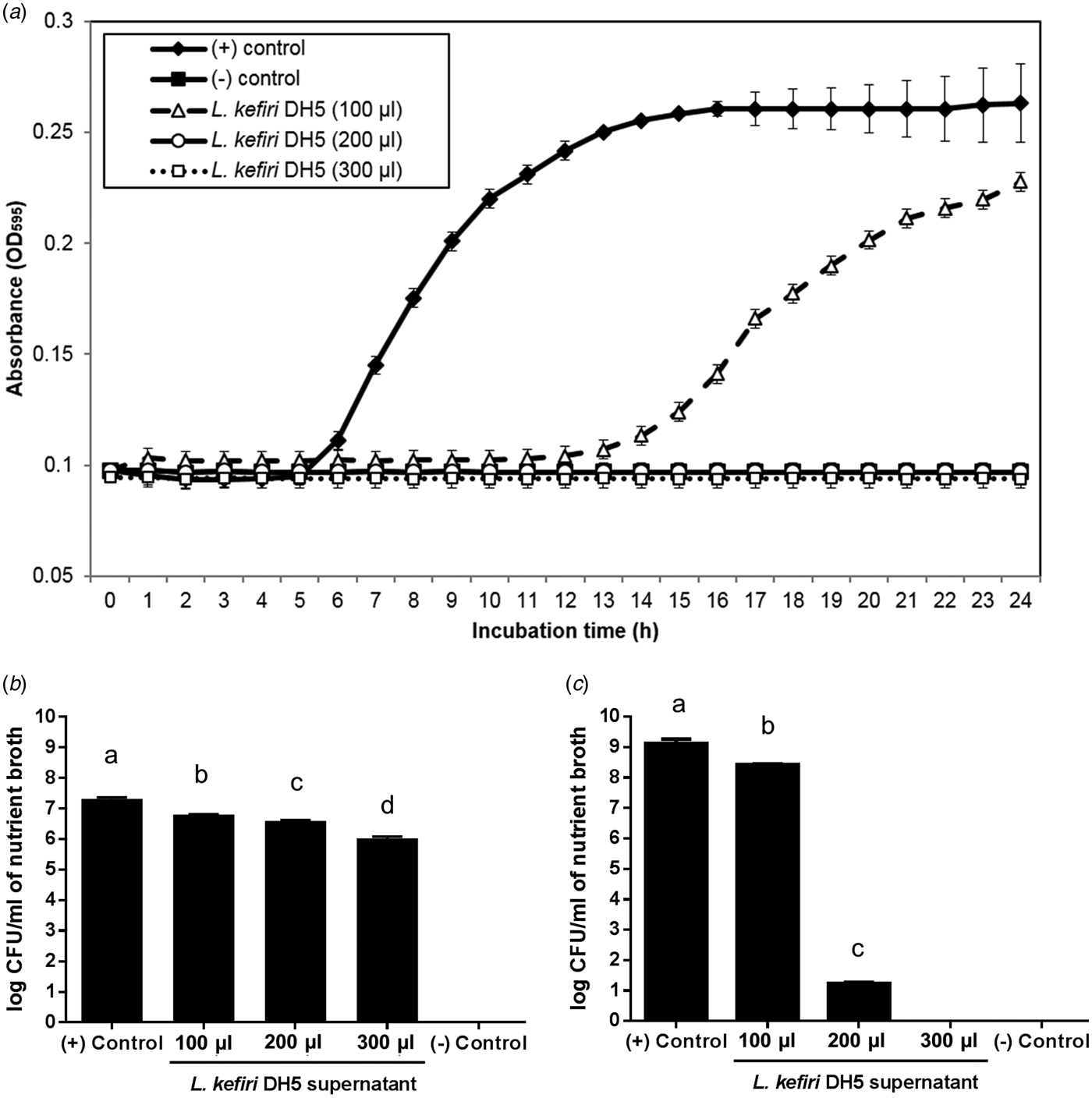
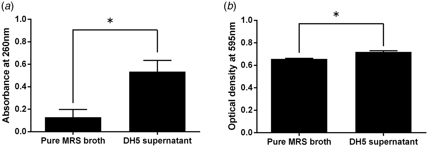
As the inhibitory effects of L. kefiri DH5 against C. sakazakii were superior to those of L. kefiranofaciens DH101, we chose the former strain for additional experiment on dose-effect relationship. Addition of 100 µl of supernatant derived from L. kefiri DH5 cultures slowed the growth of C. sakazakii when compared to the untreated control, whereas the growth was completely inhibited by 200 and 300 µl of supernatants (Fig. 2a). In plate-count enumeration, there was significant difference between the numbers of viable C. sakazakii cells in the control samples and samples with 100, 200, and 300 µl of L. kefiri DH5 supernatant after 6 h treatment (P < 0·05, Fig. 2b). After 24 h of treatment, the numbers of viable C. sakazakii cells in the control samples and samples with 100 and 200 µl of L. kefiri DH5 supernatant was significantly different (P < 0·05, Fig. 2c). No viable C. sakazakii cells were observed in the samples with 300 µl of L. kefiri DH5 supernatants after 24 h treatment (P < 0·05, Fig. 2c). Interestingly, the supernatant of L. kefiri DH5 induced the leakage of cytoplasmic materials (Fig. 3). The addition of 200 µl L. kefiri DH5 supernatant to the 1 ml of saline containing viable C. sakazakii cells resulted in disruption of the membrane integrity of C. sakazakii, which causes the leakage of nucleic acids (Fig. 3a) and proteins (Fig. 3b).
Dose-dependent antimicrobial activity of L. kefiri DH5 culture supernatant. (a) Growth curves of Cronobacter sakazakii ATCC 29544 grown in nutrient broth mixed with different volumes of the supernatants (100, 200, and 300 µl) from L. kefiri DH5 after incubating at 37 °C for 24 h. Numbers of viable Cronobacter sakazakii ATCC 29544 cells on Druggan–Forsythe–Iversen agar after (b) 6 h and (c) 24 h treatment.

The leakage of (a) nucleic acid and (b) protein of Cronobacter sakazakii ATCC 29544 by L. kefiri DH5-derived supernatant.

This dose-dependent antimicrobial activity is consistent with our previous findings that kefir supernatant solutions suppress growth of C. sakazakii cells in powdered infant formula (Kim et al. Reference Kim, Chon, Kang, Kim, Kim, Song and Seo2015b). In previous study, we found that all C. sakazakii cells were killed in powdered infant formula rehydrated with 30% kefir supernatant solution in both single strain and cocktail inoculum (Kim et al. Reference Kim, Chon, Kang, Kim, Kim, Song and Seo2015b). Considering the 10 and 20% addition of kefir supernatant solution reduced the number of viable C. sakazakii cells in powdered infant formula, the antimicrobial activity of kefir supernatant showed dose-dependent relationship (Kim et al. Reference Kim, Chon, Kang, Kim, Kim, Song and Seo2015b). The results of the present study are consistent with the previous study in that the culture supernatant of L. kefiri DH5 acted in a concentrate-dependent manner.
As C. sakazakii causes life-threatening infections in infants, countries including the United States, those in the European Union, and Korea, have established legal limits and adopted a zero tolerance policy regarding the presence of C. sakazakii in food (Huang et al. Reference Huang, Pang, Wang, Tang, Zhou, Zhang, Li, Tan, Li, Lin, Liu, Huang and Shi2015; MFDS, 2016; Sulaiman et al. Reference Sulaiman, Jacobs, Segars, Simpson and Kerdahi2016). In this sense, it is critical to determine whether the antimicrobial activity against C. sakazakii is bacteriostatic or bactericidal in nature. Our results indicate that metabolites derived from L. kefiri DH5 could be applied in the food industry to eliminate C. sakazakii from food products.
Effects of heat treatment and pH neutralisation on the antimicrobial activity of L. kefiri DH5-derived supernatant
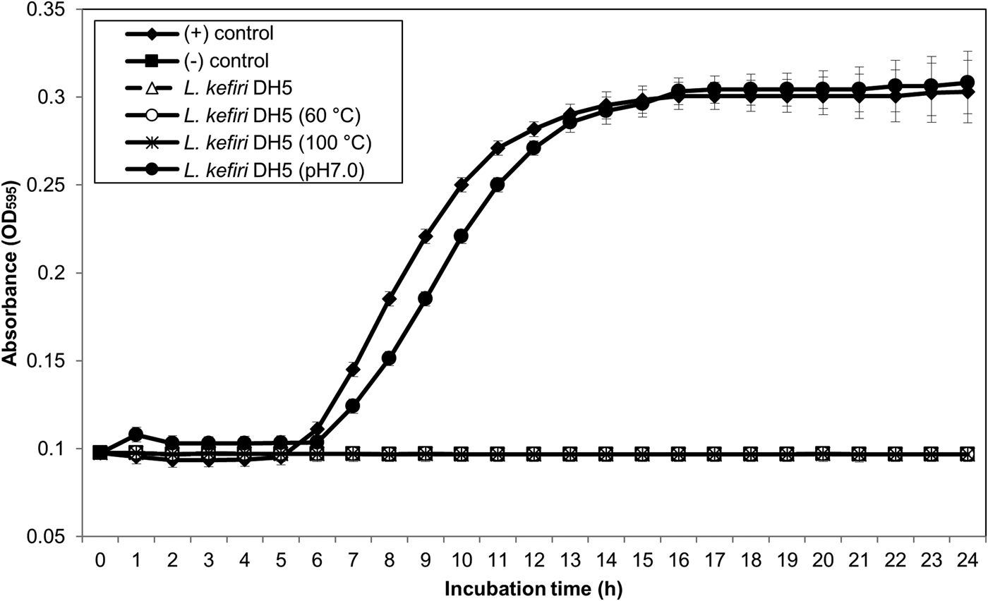
To address the mechanism by which L. kefiri DH5-derived culture supernatants exert antimicrobial activity, the supernatant was subjected to either heat treatment or pH neutralisation. While heat treatment had no effect, pH neutralisation significantly reduced the antimicrobial activity, as growth of C. sakazakii was observed from 6 h onwards (Fig. 4). These data indicate that organic acids produced by L. kefiri play a critical role in the antimicrobial effects of L. kefiri DH5-derived culture supernatants.
Effects of heat treatment and pH neutralisation on the antimicrobial activity of L. kefiri DH5-derived supernatant. Cronobacter sakazakii ATCC 29544 were grown in the presence of untreated L. kefiri DH5-derived supernatants, or in the presence of those subjected to heat treatment or pH neutralisation.

Metabolic products of LAB have strong antimicrobial effects against saprophytic and pathogenic bacteria (Schillinger, Reference Schillinger, Bills and Kung1990). Potential mechanisms underlying these effects include competition for nutrients and production of inhibitory compounds such as carbon dioxide, hydrogen peroxides, organic acids, diacetyl, and bacteriocins (Moraes et al. Reference Moraes, Perin, Ortolani, Yamazi, Vicosa and Nero2010). Garrote et al. (Reference Garrote, Abraham and De Antoni2000) reported that the inhibitory effects of kefir against gram-negative microorganisms such as pathogenic E. coli, Salmonella, and Shigella are mainly due to organic acids produced during fermentation processes. Our results are consistent with this, since C. sakazakii is gram-negative and belongs to the Enterobacteriaceae family. We infer that the antimicrobial activity of kefir against different types of pathogenic bacteria is due to the synergistic effects of several microorganisms present in this food product (Kim et al. Reference Kim, Chon, Kang, Kim, Kim, Song and Seo2015b).
This work was supported by the National Research Foundation of Korea (NRF) grant funded by the Korea government (MSIP) (No. 2015R1A2A2A01005017).