Interrupted aortic arch is defined as a complete luminal and anatomic discontinuity between the ascending and the descending aorta. This anomaly was first described by Steidele in 1778.Reference Steidele 1 More than 90% of infants with interrupted aortic arch die of circulatory failure within the first year of life if no intervention is performed. It is a very rare congenital malformation in young adults.Reference Ucak, Inan, Onan, Suleymanoglu, Temizkan and Yilmaz 2 – Reference Borgohain, Gupta, Grover and Gupta 4 Technically, conventional repair with end-to-end anastomosis through thoracotomy incision is a surgical challenge because of the extensive collateral circulation on the chest wall. Aortic repair through a mid-line sternotomy is an alternative to avoid injury to collateral vessels.Reference Korkmaz, Guden and Onan 3 In the recent years, hybrid procedures have been increasingly used in different types of congenital pathologies in adults. In this study, we report a successful hybrid approach for an adult who was diagnosed with interrupted aortic arch and a giant subclavian artery aneurysm.
Case report
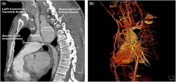
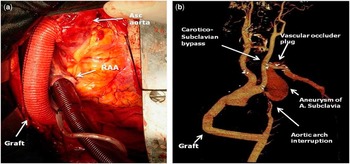
A 24-year-old man was referred to our hospital for evaluation of hypertension and pulse difference between the two arms. Peripheral pulses were palpable on both upper limbs, whereas pulses were diminished on the lower limbs compared with upper limbs. Blood pressures were 150/110 mmHg in the upper extremities and 80/60 mmHg in the lower extremities. Cardiac auscultation revealed a grade 3/6 systolic ejection murmur on the left sternal border. Chest X-ray showed a normal cardiac silhouette. Multislice CT angiography demonstrated interrupted aortic arch just below the left subclavian artery together with a giant aneurysm of the left subclavian artery measuring 4.49 cm in maximum diameter (Fig 1a). There was a massive collateral vessel circulation between the proximal and distal segments of the thoracic aorta (Fig 1b). This patient was referred for surgical intervention for repair of interrupted aortic arch and subclavian artery. Initially, a carotid–subclavian bypass was performed with a 7-mm polytetrafluoroethylene graft (W. L. Gore & Associates, Flagstaff, Arizona). Next, a single-staged extra-anatomic bypass (ventral aortic repair) through median sternotomy was performed. An 18-mm Dacron vascular graft (Hemashield gold-knitted double-velour vascular graft; Maquet, United States of America) was interposed between the ascending aorta and the descending thoracic aorta that was exposed through posterior pericardial incision. The neck of a giant aneurysm was ligated just below the left common carotid artery branch (Fig 2a). Furthermore, the proximal part of the subclavian artery close to the vertebral artery was sealed with a vascular plug by means of endovascular procedure.
( a ) Preoperative CT angiogram showing long-segment interrupted aortic arch type A with the left subclavian artery aneurysm (4.49 cm). ( b ) Extensive collateral development.

( a ) Operative view of the extra-anatomic ascending-to-descending aorta bypass graft (18-mm Dacron graft). ( b ) Postoperative CT angiography (three-dimensional reconstruction) shows the patent graft between the ascending and the descending aorta, carotico–subclavian bypass graft, and subclavian artery aneurysm excluded with vascular plug.

In the early postoperative period, CT angiography showed a patent graft (Fig 2b). The patient was discharged from the hospital without any complication on postoperative day 8.
Discussion
Interrupted aortic arch has been classified into three types – A, B, and C – based on the site of the aortic interruption by Celoria and Patton in 1959.Reference Celoria and Patton 5 In type A, interruption of the left aortic arc occurs distally to the origin of the left subclavian artery. In type B, interrupted left aortic arch, the interruption occurs distal to the origin of the left common carotid artery. In the type C, interrupted left aortic arch, the interruption occurs proximally to the origin of the left common carotid artery. If it is surgically untreated in the neonatal period, the median age of death is 4–10 days due to severe congestive heart failure, usually after physiological closure of the ductus arteriosus.Reference Ucak, Inan, Onan, Suleymanoglu, Temizkan and Yilmaz 2 Therefore, it is unusual to diagnose interrupted aortic arch in adults.
Clinically, there are differences between adult and neonatal forms of interrupted aortic arch. In the adult form, it is mostly reported as type A interruption without ventricular septal defect, whereas the neonatal form is frequently associated with ventricular septal defects and it is described as type B interruption.Reference Atalar, Robertson, Dyck and Ross 6 Isolated aortic arch interruption without a patent ductus arteriosus or aortopulmonary window is considered as a fatal condition during early stages of life.Reference Ucak, Inan, Onan, Suleymanoglu, Temizkan and Yilmaz 2 In the pathophysiology of isolated interrupted aortic arch in adulthood, a well-developed collateral circulation is necessary to enable survival. The presentation ranges from lack of symptoms to limb swelling with differential blood pressures in all extremities; however, collateral vessels may be subject to atrophy and atherosclerosis, which lead to other challenging problems such as rupture and bleeding.Reference Shirani and Soleymanzadeh 7
Differential diagnosis mostly includes interrupted aortic arch and aortic coarctation in adults. This is a rare case of interrupted aortic arch in an adult patient associated with subclavian aneurysm; however, it could be speculated that this anomaly was a real interrupted aortic arch or it was aortic coarctation with near atresia. In coarctation, angiographic images show that the arch retains its curvature, and the ascending aorta and the aortic arch have a normal or slightly enlarged calibre. In cases of interrupted aortic arch, the arch is typically of smaller calibre than normal, and the branches have a straighter course. During operation, we observed that the diameter of the aortic arch was progressively decreased. Therefore, we preferred an extra-anatomic bypass with ascending-to-descending aortic bypass rather than manipulating the aortic arch.
In adults, interrupted aortic arch has been traditionally repaired with graft interposition in a single stage using thoracotomy incision.Reference Ucak, Inan, Onan, Suleymanoglu, Temizkan and Yilmaz 2 , Reference Korkmaz, Guden and Onan 3 Anatomic repair is possible in type A and B interrupted aortic arch with graft interposition between the proximal and the distal segments of the interrupted aorta. In type A interrupted aortic arch, extra-anatomic repair can be performed with a subclavian artery-to-descending aorta bypass grafting. Ventral aortic repair is the preferred technique through a mid-line sternotomy incision in adult patients to avoid injury to the extensive collateral circulation on the chest wall. This approach can be preferred to reduce the risk of morbidity and mortality.Reference Mehrpooya, Eskandari and Salehi 8 , Reference Erkanli, Onan, Aktürk and Bakir 9 In this approach, anatomic or extra-anatomic repair necessitates the use of cardiopulmonary bypass; however, in case of a giant aneurysm of the subclavian artery, the management strategy is different from isolated cases of interrupted aortic arch. To the best of our knowledge, our patient is the first reported case on hybrid intervention for type A interrupted aortic arch with a left subclavian artery aneurysm in an adult. According to the size of the left subclavian artery aneurysm, we decided to perform hybrid intervention to decrease the risks of operation.
In conclusion, hybrid interventions can be considered in adults with interrupted aortic arch and subclavian artery aneurysm to decrease the mortality and morbidity of conventional operations.
Acknowledgement
None.
Financial Support
This research received no specific grant from any funding agency, commercial, or not-for-profit sectors.
Conflicts of Interest
None.
Ethical Standards
The patient was informed of the design and the purposes of the procedures according to Declaration of Helsinki Principles.