Introduction
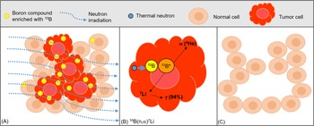
Boron neutron capture therapy (BNCT) is a tumour selective particle radiotherapy, based on the nuclear reaction known as boron neutron capture. It is considered a binary treatment, based on the administration of boron carriers incorporated preferentially by tumour cells, followed by irradiation with a thermal or epithermal neutron beam (RefsReference Coderre and Morris1–Reference Matsumoto3). The capture reaction between a 10B nucleus and a thermal neutron produces high-energy α-particles and recoiling 7Li nuclei. These particles have high linear energy transfer (LET), high relative biological efficacy and a short path length of approximately the diameter of one cell (<10 μm), circumscribing their damage to tumour cells, where 10B atoms are preferentially localised, sparing normal surrounding tissue (Fig. 1) (RefsReference Coderre and Morris1, Reference Barth4, Reference Hopewell5). In this sense, as BNCT involves biochemical rather than geometrical targeting, it would be potentially useful to treat all tumour cell populations, including undetectable micrometastases (Ref.Reference Pozzi6) and foci of malignant transformation in field-cancerised tissue (Ref.Reference Monti Hughes7).
Boron neutron capture therapy (BNCT). (A) Boron compounds enriched with the nonradioactive isotope 10B accumulate preferentially in tumour cells. Afterwards, the target tissue is exposed to a thermal or epithermal neutron irradiation (which becomes thermalised as it penetrates the tissue). The neutron component has little effect on tumour or normal tissue. (B) When 10B absorbs a thermal neutron (<0.5 eV) (11B*), rapidly disintegrates into an alpha (4He) particle (1.47 MeV) and a recoiled lithium nucleus (7Li) (0.84 MeV). Both deposit high energy along their short path, comparable with the diameter of one cell (<10 μm). A γ-ray of 0.48 MeV is emitted in 94% of the reactions. (C) Thus, only cells containing 10B (mostly tumour cells) are destroyed by the boron neutron capture reaction [10B(n,α)7Li], sparing normal surrounding cells (RefsReference Coderre and Morris1, Reference Suzuki2, Reference Dymova110).

BNCT is a mixed field irradiation, that consists of a boron dose component (the high-LET particles resulting from the boron neutron capture reaction) and a non-specific background dose. This background dose consists of low-LET gamma rays of the neutron beam and from the capture of a thermal neutron by a hydrogen atom, high-LET protons originated by the scattering of fast neutrons and high-LET protons from the capture of a thermal neutron by a nitrogen atom. As the background dose affects both normal and tumour tissue, BNCT protocols should ideally maximise the tumour selective boron radiation component (RefsReference Schwint8, Reference Schwint9).
Clinical studies of BNCT for glioblastoma multiforme, melanoma, recurrent head and neck tumours, lung and liver metastases, mesothelioma, extra-mammary Paget's disease have been performed or are underway in the United States, Japan, European Consortium, Sweden, Italy, Finland, Argentina and Taiwan (among others) employing mostly nuclear reactors as the neutron source (e.g. RefsReference González10–Reference Chen15). Extensive research has been carried out for the development of accelerator-based neutron sources that will enable in-hospital treatments, opening for more widespread clinical trials for different tumours (RefsReference Suzuki2, Reference Porra16). BNCT clinical trials for glioma and head and neck cancer have been recently performed in accelerator-based neutron sources in Japan (Ref.Reference Miyatake17). To date, the clinical results show therapeutic efficacy, associated with an improvement in patient quality of life and prolonged survival, with identified opportunities for improvement (Ref.Reference Schwint8).
For a successful BNCT, a sufficient number of 10B atoms (109 atoms/cell or 20–30 mg of 10B/g) must be delivered to the tumour, and enough thermal neutrons must be absorbed by the 10B atoms to sustain the lethal 10B(n,α)7Li reaction (RefsReference Matsumoto3, Reference Barth4). An ideal boron carrier should be non-toxic at therapeutic dose levels, should accumulate preferentially in tumour cells versus blood and normal tissue and should target all tumour cell populations (RefsReference Garabalino18, Reference Schwint and Trivillin19). Tumour cells poorly loaded with boron will be unresponsive to treatment, making homogeneous tumour boron targeting pivotal to therapeutic success. Higher tumour boron concentrations will allow for shorter irradiation times and a reduction in the background dose (Ref.Reference Schwint9). Boron localisation studies in different structures of the tumour and surrounding tissue are of importance to evaluate the potential efficacy of a boron compound and strategy (e.g. Ref.Reference Portu20).
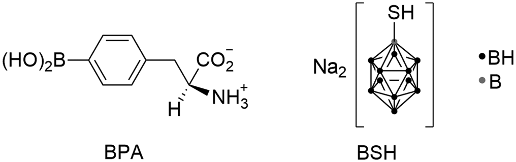
To date, two boron compounds have been significantly used in BNCT clinical trials: mercaptoundecahydrododecaborate-10B (BSH) and boronophenylalanine (BPA) (Fig. 2) (Ref.Reference Barth4). BSH is a water-soluble diffusive drug, principally used for malignant glioma. It does not cross the blood brain barrier (BBB) into normal brain, but accumulates in malignant brain tumours because of their disrupted BBB (Ref.Reference Ono21). However, BSH has poor membrane permeability and the accumulation of BSH in tumour cells is low (RefsReference Ono21, Reference Wada22). On the other hand, BPA has been used for several pathologies. It is an amino-acid derivative actively incorporated by tumour cells by the L-type amino acid transporter 1 (LAT1), and a small amount is taken up by LAT2. Tumour cells overexpress LAT1 versus normal cells, but LAT2 is expressed in both. However, since LAT2 transports less BPA than LAT1, a preferential accumulation of BPA is achieved in tumour cells (Ref.Reference Wada22).
Two boron compounds significantly used in BNCT clinical trials: BPA and BSH.

Despite their clinical use, both compounds exhibit several limitations. In this sense, new boron delivery agents are under development such as monoclonal antibodies (e.g. Ref.Reference Barth23), liposomes (e.g. Ref.Reference Heber24), nanoparticles (e.g. RefsReference Oleshkevich25, Reference Torresan26), boron cluster agents, carrier proteins conjugated with boron compounds (e.g. RefsReference Ban and Nakamura27, Reference Kawai28) and bimodal drugs (with anti-tumoural and BNCT effects) such as metallocarboranes (e.g. RefsReference Couto29, Reference Nuez-Martinez30). As an example, icosahedral boron clusters have shown the relevance of these compounds as exquisite frameworks in the design of novel drug candidates (RefsReference Endo31–Reference Poater33).
To date, tumour boron concentration in clinical trials has been estimated using empirical data models, depending on tumour/blood, tumour/normal tissue and normal tissue/blood concentration ratios. However, boron uptake and distribution in the tumour varies among patients. Imaging methods to monitor, in real time, boron uptake and distribution for each patient and each tumour would help to determine treatment indications and perform high-precision treatment planning (RefsReference Ferrari34–Reference Scott36). o-[18F]-Fluoro-l-p-boronophenylalanine ([18F]-l-FBPA), an18F-labelled radiopharmaceutical analogue of l-BPA, has been used as a positron emission tomography (PET) probe. However, this compound showed limitations (RefsReference Hiratsuka13, Reference Watanabe37). In this sense, huge efforts are devoted to the development of new theranostic compounds for BNCT (e.g. RefsReference Oleshkevich25, Reference Torresan26, Reference Sauerwein35, Reference Li38, Reference Kalot39). One example is the halogenated (most of them iodinated) carborane derivatives that were shown to provide nuclear imaging by PET-CT (positron emission tomography-computed tomography, 124I-, a positron emitter) and SPECT (single photon emission computed tomography, 125I-, a gamma emitter) (RefsReference Varadarajan40–Reference Teixidor, Viñas, Hosman and Eagling42).
However, if an ‘ideal’ boron carrier is identified as promising, many years of studies and very high costs will be necessary for it to be approved for treatment (Ref.Reference Schwint8). In this sense, the use of boron agents approved for their use in humans allows for a more direct and less costly extrapolation to a clinical environment. Optimising the delivery of BPA and BSH employing a bench to bedside approach will bridge the gap between research and clinical application (Ref.Reference Schwint8). Moreover, the understanding of the radiobiological mechanisms induced by BNCT will contribute to identify markers of cellular response to BNCT and may serve to modulate signalling pathways to enhance BNCT therapeutic efficacy and reduce associated toxicity (RefsReference Sato43, Reference Rodriguez44).
The aim of this review is to recapitulate examples of BNCT radiobiological studies for different pathologies and clinical scenarios, strategies to optimise boron targeting, enhance BNCT therapeutic effect and minimise radiotoxicity. It will also describe the radiobiological mechanisms induced by BNCT, and the importance of the detection of biomarkers to monitor and predict the therapeutic efficacy and toxicity of the therapy alone or combined with other strategies. Finally, it will briefly comment on the introduction of accelerator-based neutron sources in BNCT.
BNCT strategies for different pathologies
Translational research in adequate experimental models is necessary to optimise BNCT for different pathologies, particularly for those that respond poorly to standard therapies and would potentially benefit from more effective and selective therapies. Multiple radiobiological studies have been performed in different tumour models using different boron compounds, routes of compound administration and a range of administration strategies using nuclear reactors as the neutron source.
BNCT mediated by BPA or BSH has been widely explored in several animal models for different pathologies (e.g. RefsReference Pozzi6, Reference Kreimann45–Reference Andoh48). BPA and BSH are considered low molecular weight boron compounds, together with boric acid and GB-10 (decahydrodecaborate), which have been studied for many years as possible boron agents to be used in humans. Particularly, GB-10 was once approved for its use in patients by the U.S. Food and Drug Administration, but was not used in clinical trials (Ref.Reference Hawthorne and Lee49). Boric acid and GB-10 are both diffusive boron agents.
Different strategies trying to enhance the uptake of these boron drugs have been studied. For example, poly(vinyl alcohol)–BPA complex increased cellular uptake and slowed the untoward efflux in pancreatic adenocarcinoma and colon cancer models (Ref.Reference Nomoto50). Viral envelopes, emulsions, liposomes and ligands specific to overexpressed molecules (receptors and transporters) in cancer cells were also explored (e.g. RefsReference Fujii51–Reference Yanagie55).
Several studies demonstrated that the combined administration of these boron agents (BPA, BSH, GB-10 and boric acid) were proved to be superior to any single agent, helping to overcome the potential toxicity of higher doses of each of the compounds given alone (Ref.Reference Schwint8). For example, in glioma and squamous cell carcinoma (SCC) in vivo models, BNCT mediated by BPA + BSH induced the best survival and tumour control rates (RefsReference Ono56, Reference Barth57). In the hamster cheek pouch oral cancer model, the combination of the aberrant blood vessel boron targeting with GB-10 and tumour cell targeting with BPA, improved boron targeting homogeneity in the tumour and enhanced therapeutic efficacy at no additional cost in terms of toxicity (Ref.Reference Trivillin58). In the oral hamster precancer model, (BPA + GB-10)-BNCT induced a long-term partial inhibitory effect on tumour development from precancerous tissue with no normal tissue radiotoxicity and without exceeding precancerous tissue tolerance. The precancerous tissue surrounding tumours is more radiosensitive than normal mucosa and mimics field cancerised tissue in humans, the source of second primary tumours that cause therapeutic failure (Ref.Reference Monti Hughes7).
Another strategy that also profits from the benefits of combining boron compounds is sequential-BNCT (Seq-BNCT), which involves the sequential application of BPA-BNCT and GB-10-BNCT with an interval of 24–48 h between applications. The first application would reduce interstitial fluid pressure and induce cell death, favouring the intratumoural delivery of GB-10 in the second application, improving tumour boron targeting homogeneity. The brief interval between applications would avoid tumour re-population. A second application would favour targeting of tumour cell populations that failed to respond to the first application. In the hamster cheek pouch oral cancer model, Seq-BNCT significantly enhanced tumour response versus a dose-matched single application of (BPA + GB-10)-BNCT and did not exacerbate mucositis in precancerous tissue (Ref.Reference Molinari59).
Seq-BNCT would be useful in the case of patients requiring abbreviated treatment. For the case of patients that do not require abbreviated treatment, double applications of BNCT (full dose re-treatment) mediated by (GB-10 + BPA) could be employed. In the oral hamster precancer model, double BNCT enhanced tumour control and inhibited the development of second primary tumours from precancerous tissue. In terms of radiotoxicity, the second application of BNCT did not exacerbate the mucositis observed after the first treatment in precancerous tissue (e.g. RefsReference Monti Hughes60, Reference Monti Hughes61). A double application of BNCT would potentially serve to improve tumour control, to treat a local recurrence, to inhibit the development of second primary tumours from field cancerised tissue and/or treat second primary tumours that developed after the first BNCT treatment.
BNCT-induced mucositis affects patient quality of life and treatment. Moreover, BPA-BNCT-induced mucositis increased with the aggressiveness of the cancerisation protocol employed in the hamster cheek pouch (Ref.Reference Monti Hughes62). Histamine coupled to BPA-BNCT showed a reduction in BNCT-induced mucositis in precancerous tissue without jeopardising therapeutic efficacy (Ref.Reference Monti Hughes63). The use of radioprotectors would allow higher doses to be delivered to the tumour, protecting the surrounding dose-limiting tissues. The study of BNCT in different oral cancer patient scenarios would contribute to personalise BNCT for each patient condition (Ref.Reference Monti Hughes62).
Efforts have been made in the development of efficient 10B delivery vehicles and strategies that could target the tumour vasculature (RefsReference Koning64, Reference Kang65). Ono et al. (Ref.Reference Ono66) explored a selective inhibitor of tumour blood flow, flavone acetic acid (FAA) combined with BSH in an SCC model in mice. FAA would cause vascular collapse entrapping BSH in the tumour. FAA inhibited the clearance of BSH from the tumour but had little or no effect in normal tissue. High tumour levels of 10B enhanced sensitivity to thermal neutron irradiation. Another vascular targeting agent, the ZD6126 (N-acetylcochinol-O-phosphate), in combination with BNCT enhanced the sensitivity of the quiescent (Q) tumour cells compared with the total tumour cells in an SCC model in mice. Q cells are viable and clonogenic, but cease cell division in part because of hypoxia and depletion of nutrition in the tumour core. Therapies kill more efficiently proliferative rather than Q cells, the latter being responsible for recurrences after treatment (Ref.Reference Masunaga67). More recently, Yoneyama et al. (Ref.Reference Yoneyama68) studied BNCT after rapid, ultralow-dose administration of either tumour vasculature-specific annexin A1-targeting IFLLWQR (IF7)-conjugated 10BPA or 10BSH in bladder tumour-bearing mice, and demonstrated an enhancement in the therapeutic efficacy versus BPA and BSH alone. In the hamster cheek pouch oral cancer model, transient normalisation of defective blood vessels using thalidomide and administration of BPA in the normalisation window improved boron targeting homogeneity in tumours, enhancing tumour response (Ref.Reference Molinari69). Thalidomide treatment was combined with Seq-BNCT and achieved, in the hamster cheek pouch oral cancer model, 100% tumour response with 87% complete tumour remission, with no normal tissue toxicity and no cases of severe mucositis in dose-limiting precancerous tissue (Ref.Reference Molinari70).
Further seeking to favour boron delivery and distribution in tumours, electroporation associated with the administration of GB-10 improved gross boron uptake and microdistribution of GB-10 in the model of oral cancer in hamster (Ref.Reference Garabalino71). Ono et al. (Ref.Reference Ono21) also showed that BNCT and electroporation could be clinically useful. Sonoporation enhanced the efficiency of BNCT for oral SCC in nude mice by modulating the microlocalisation of BPA and BSH in tumours and increasing their intracellular levels (Ref.Reference Yamatomo72). The combination of low dose gamma irradiation with BNCT increased BPA accumulation in tumour and the tumour/normal tissue and tumour/blood boron concentration ratios, enhancing BNCT efficacy and extending the overall survival rate in an orthotopic human oral SCC animal model (Ref.Reference Lin73).
Particularly for glioblastoma, examples of techniques used to improve boron accumulation in brain tumours are BBB disruption by a hyperosmotic agent such as mannitol, intracarotid administration of the boron compound and convection enhanced delivery (RefsReference Barth74, Reference Futamura75). Slow infusions of BPA would improve boron targeting of infiltrating tumour cells in the brain (Ref.Reference Joel76). Neutron irradiation under continuous BPA infusion contributed to solve the problem of heterogeneous distribution of BPA (Ref.Reference Ono77). In an in vivo model for primary brain tumours and melanoma metastatic brain tumours, an X-ray boost enhanced the BNCT therapeutic effect (Ref.Reference Barth78). Gene therapy combined with BNCT increased cancer cell-killing effect by manipulating LAT1 expression in human glioma cells. This strategy could be useful in those tumours with low LAT1 expression (Ref.Reference Ohnishi79). Preloading with (l-3,4-dihydroxyphenylalanine) increased BPA uptake in glioma cells and, oral preloading with high-dose l-phenylalanine, reduced the accumulation of BPA in normal brain relative to tumour tissue, protecting normal brain (RefsReference Capuani80, Reference Watanabe81). Glioma stem cell-like cells may take up BPA and be amenable to targeting by BNCT (e.g. Ref.Reference Kondo82). In this sense, future studies on strategies to increase the boron uptake in these stem cells will help to overcome resistance and avoid recurrences.
The role of radiosensitisers in combination with BNCT and other particle therapies has been explored (e.g. RefsReference Kinashi83, Reference Qi84). For example, sodium butyrate, a histone deacetylase inhibitor, increased tumour boron concentration and acted as a radiosensitiser of BPA-BNCT for poorly differentiated thyroid carcinoma (Ref.Reference Perona85). Photosensitisers were also explored to combine photodynamic therapy and BNCT (RefsReference Ban and Nakamura27, Reference Conway-Kenny86). Tatebe et al. (Ref.Reference Tatebe87) explored the combined effect of rapamycin (mTOR – mammalian target of rapamycin – inhibitor) on the radiosensitivity of human oral cancer (SAS) cells treated with BPA-BNCT. Rapamycin inhibits the PI3K/AKT/mTOR pathway, inducing tumour growth inhibition. In this study, rapamycin inhibited BPA uptake in tumour cells and thus reduced their sensitivity to BNCT, suggesting the need for careful attention and precaution when two strategies are combined.
BNCT is a possible lung cancer treatment (e.g. RefsReference Bakeine88, Reference Farías89). BNCT therapeutic effect was demonstrated on lung metastases from colon carcinoma and clear cell sarcoma in animal models, with no toxicity in normal tissue (RefsReference Andoh90, Reference Trivillin91). BPA-BNCT with nicotinamide (a hypoxia-releasing agent) or bevacizumab (a vascular endothelial growth factor inhibitor) enhanced the reduction in lung metastases (Ref.Reference Masunaga92). In another study, BSH-BNCT combined with a hypoxic cytotoxin, tirapazamine, with or without mild temperature hyperthermia (that improves tumour oxygenation) improved local tumour control, whereas BPA-BNCT in combination with both tirapazamine and mild temperature hyperthermia as well as nicotinamide reduced the number of lung metastases (Ref.Reference Masunaga93). It was postulated that control of the chronic hypoxia-rich Q cell population in the primary solid tumour has the potential to impact the control of local tumours as a whole and that control of the acute hypoxia-rich total tumour cell population in the primary solid tumour has the potential to impact the control of lung metastases (Ref.Reference Masunaga92).
The combination of BNCT and immunotherapy is another attractive strategy to explore, as immunomodulatory effects were associated with BNCT (Ref.Reference Trivillin94). When ionising radiation is applied to a primary tumour, it can induce immunogenic cell death that may in turn trigger a cytotoxic immune response against the primary tumour and its metastasis. The abscopal effect is an ‘out-of-field’ inhibitory effect on tumour growth, first described associated with standard radiotherapy. In bladder tumour bearing mice, BNCT induced an immune response by the host against tumour cells (Ref.Reference Yoneyama68). Trivillin et al. (Ref.Reference Trivillin94) described, for the first time, the abscopal effect of BPA-BNCT in an ectopic model of colon cancer in BDIX rats. In the same model, Trivillin et al. (Ref.Reference Trivillin95) demonstrated that this immune response could be enhanced by the combination of BNCT with Bacillus Calmette–Guerin (BCG), an immune stimulator. They showed an induction of a powerful abscopal response and regional effects in metastasised lymph nodes. The combination of BCG and BNCT might act as an ‘anti-tumour vaccine’ where BNCT would trigger the process of tumour antigen generation and BCG would promote antigen presentation in an inflammatory microenvironment (Ref.Reference Trivillin95).
The effect of BNCT on immune cells was also studied. Kinashi et al. (Ref.Reference Kinashi96) studied the cytotoxic adverse effect of BNCT in peripheral T-lymphocytes of head and neck and brain tumour-treated patients. They reported that BNCT induced lower micronuclei induction in peripheral T-lymphocytes versus radioiodine treatment used for thyroid cancer. BNCT delivers a high dose to tumour causing little effect on the rest of the body. Aromando et al. (Ref.Reference Aromando97) observed, in the hamster cheek pouch oral cancer model, a significant reduction in the total number of mast cells in the tumour after BPA-BNCT suggesting a complementary mechanism of action of BNCT in tumour remission. Mast cell activation is associated with an increase in tumour cell proliferation. Kahn et al. (Ref.Reference Khan98) observed that boron-rich liposomes were assimilated by peripheral blood mononuclear cells. After neutron irradiation, these cells were modified to an anti-tumour phenotype, contributing to the inhibition of tumour growth in a murine mammary model. Finally, a chronic autoimmune pathology was treated with boron neutron capture synovectomy (BNCS). Rheumatoid arthritis is characterised by the proliferation and inflammation of the synovium. BPA-BNCS and GB-10-BNCS treated successfully the pathological synovium in a model of antigen-induced arthritis in rabbits (Ref.Reference Trivillin99).
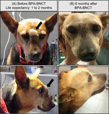
Radiobiological studies have contributed to clinical-veterinary BNCT studies. Biodistribution studies using BSH were performed in dogs with spontaneous brain tumours (Ref.Reference Kraft100). Another study showed a selective uptake of BPA by spontaneous undifferentiated thyroid carcinoma in dogs (Ref.Reference Dagrosa101). A comparison of the efficacy of BNCT and Gd-mediated neutron capture therapy for canine oral melanoma and osteosarcoma has been performed (Ref.Reference Mitin102) and the possible application of BSH-BNCT to spontaneous canine osteosarcoma has been studied (Ref.Reference Takeuchi103). Clinical-veterinary studies in cats with spontaneous SCCs and dogs with advanced head and neck cancer demonstrated that BPA-BNCT induced partial tumour control, with slight-moderate mucositis, excellent life quality and prolongation in the survival time estimated at recruitment (Fig. 3) (RefsReference Rao104–Reference Schwint106).
Example of a veterinary patient treated with BNCT. (A) Lucy had a spontaneous SCC in the nasal cavity and paranasal sinuses with bone destruction (arrow), with a life expectancy of 1–2 months. (B) Six months after BNCT mediated by BPA, Lucy exhibited a positive tumour response with optimum clinical signs and no protruding tumour mass. Computed tomography scans performed 1-month post treatment revealed a 50% reduction in tumour volume of the pretreatment tumour volume. Lucy was treated at the RA-6 nuclear reactor in Argentina (Ref.Reference Schwint106).

Radiobiological mechanisms induced by BNCT
The main target of radiation damage is DNA. Ionising radiation induces different types of DNA damage, being DNA double-strand breaks (DSBs) the most genotoxic lesions (Ref.Reference Zhou and Parsons107). Itwas demonstrated that DNA damage increases with LET of radiation. The high-LET particles released principally after the boron neutron capture reaction will induce a direct damage in DNA, causing irreparable DSBs. Instead, the low-LET radiation dose component induces mostly reparable DNA single-strand breaks (SSBs) by indirect damage (RefsReference Zhou and Parsons107–Reference Dymova110).
When DNA damage is detected, DNA damage response kinase ataxia-telangiectasia mutated and ataxia-telangiectasia and protein kinase rad3 (RAD3)-related are recruited, and their activation results in phosphorylated H2AX (H2A histone family member X, γH2AX), the activation of p53 and checkpoint kinase (CHK) CHK2 and CHK1. CHK2 activates G1/S checkpoint and CHK1 the G2/M checkpoint, inducing cell cycle arrest to allow the cell to repair the damaged DNA. Once DNA repair is finished, γH2AX is dephosphorylated (e.g. RefsReference Rodriguez44, Reference Kinashi111–Reference Chen114).
In higher eukaryotic cells, DSBs are repaired by the homologous recombination (HR) or the nonhomologous end-joining (NHEJ) mechanisms. NHEJ acts through DNA-PK, Ku70, Ku80 and DNA ligase 4 enzymes and HR acts through the products of Rad52 epistasis gene family (Rad51, Rad52, Rad54, Rad55, Rad57 and Rad58) (RefsReference Rodriguez44, Reference Kinashi111). HR is associated with DNA replication, as homologous DNA on a sister chromatid or chromosome acts as template to guide the broken strand repair. It is a relative accurate and error-free repair mechanism. On the other hand, NHEJ is less accurate and error-prone repair as broken compatible DNA ends are simply ligated with each other. NHEJ is the predominant pathway for DSB repair in mammalian species and it can function independently of the cell cycle phase, and particularly in G0/G1. Instead, HR is associated with the late S and G2 phase (RefsReference Rodriguez44, Reference Zhou and Parsons107). If DNA damage is not repaired, DNA damage-induced signalling pathways are activated and elicit cell cycle arrest and/or apoptosis, necrosis, autophagy or mitotic catastrophe (RefsReference Dymova110, Reference Chen114, Reference Fujita115).
Understanding the radiobiological mechanisms induced by BNCT will allow for the identification of cellular response markers to BNCT and possible signalling pathways that could be modulated to enhance therapeutic efficacy and reduce associated toxicity (e.g. RefsReference Sato43, Reference Rodriguez44).
BNCT-induced DNA damage was studied independently from repair mechanisms using strand break assays in DNA plasmids. These studies allow for the quantification of DSBs and SSBs induced by BNCT, and help to identify boron compounds that could effectively induce DSBs (RefsReference Okamoto108, Reference Jamsranjav116).
In vitro and in vivo studies analysed different parameters of DNA damage, as γ-H2AX foci, TP53BP1 foci (tumour suppressor gene p53-binding protein 1) and micronuclei induction. Studies in melanoma, glioblastoma and thyroid cancer cells showed that BNCT induced more DNA damage than γ-rays and X-rays (e.g. RefsReference Kinashi111, Reference Seki112, Reference Oliveira117, Reference Dagrosa118). Rodriguez et al. (Ref.Reference Rodriguez44) showed that γH2AX focus size was significantly larger in BPA-BNCT compared with neutrons or γ-irradiation, revealing high DSB complexity. They also observed that DNA damage and repair pathways were tissue specific. γH2AX focus pattern over time was different between thyroid cancer (WRO) and melanoma (MelJ) cell lines, and HR pathway was activated in WRO whereas in MelJ cell line both HR and NHEJ were induced. Kondo et al. (Ref.Reference Kondo119) studied the repair pathway in mouse embryonic fibroblast cell lines after BNCT, and found that DNA damage is partially repaired by ligase IV. Chen et al. (Ref.Reference Chen114) showed, in a hepatocellular carcinoma cell line, that BNCT mediated by boric acid arrested the cells at G2/M checkpoint and only the HR repair mechanism was activated. Kinashi et al. (Ref.Reference Kinashi111) demonstrated, in Ku80 mutant cell lines (xrs5), that a deficiency in the repair of DSBs enhanced BNCT sensitivity. Based on these results, possible strategies targeting the above-mentioned pathways (e.g. Ref.Reference Zhou and Parsons107) may potentiate BNCT efficacy.
BNCT-induced DNA damage was also studied in vivo. For example, Kondo et al. (Ref.Reference Kondo120) demonstrated, in a tumour brain model, that γH2AX foci remained at 24 h after BPA-BNCT versus γ-rays that were repaired by that time. Masutani et al. (Ref.Reference Masutani121) showed that, at 6 h after BPA-BNCT, there is an increase of γH2AX and HMGB1 (high mobility group box 1 protein) in a lymphosarcoma xenograft model. At 20 h after BNCT, persistent staining of both γH2AX and PAR [poly(ADP-ribose)] was observed. Accumulated PAR produced by poly(ADP-ribose) polymerase (a nuclear enzyme that signals the presence of DNA damage) triggers apoptosis inducing factor-dependent apoptosis or necrosis. Instead, at 20 h, HMGB1 becomes low suggesting a possible leakage of the cell. HMGB1 is a nuclear protein reported as a marker of necrosis and also one of the proteins in damage-associated molecular patterns which leaks out after cell damage and activates inflammatory responses. HMGB1 could serve as an early marker for BNCT, whereas persistent staining of γH2AX and PAR could be late markers of BNCT. Proteome analyses in human SCC cells after BPA-BNCT showed that early after BNCT, proteins functioning in endoplasmic reticulum, DNA repair and RNA processing showed dynamic changes. BNCT induced fragments of endoplasmic reticulum-localised lymphoid-restricted membrane protein that were also detected in a rat sarcoma graft model 24 h after BNCT, suggesting that this might be a possible hallmark of BNCT-induced cellular response (Ref.Reference Sato43). The characterisation of biomarkers to monitor DNA, cellular damage and molecular pathways activated after BNCT would contribute to the development of a personalised BNCT (RefsReference Ferrari34, Reference Sauerwein35, Reference Sato43). For example, in radiotherapy, a new concept that helped to personalise dose prescription is the genomic-adjusted radiation dose (GARD). GARD is a clinical model for genomic radiation dosing that could allow the individualisation of radiotherapy dose to tumour radiosensitivity and allows for the design of genomically guided clinical trials in radiation oncology (RefsReference Scott36, Reference Scott122). This precision concept could be worth exploring for BNCT, based on the actual and future findings on biomarkers of DNA damage and cellular response and considering tumour patient biology.
Although p53 is implicated in DNA damage and cell stress response, it is one of the most commonly inactivated tumour suppressors in human cancers, limiting the effectiveness of conventional therapy to treat tumours (RefsReference Seki112, Reference Fujita123, Reference Kinashi124). Seki et al. (Ref.Reference Seki112) demonstrated that BNCT-induced DNA damage did not depend on p53. In glioblastoma and oral SCC cells, although p53 mutant type cells are more resistant to BNCT, BPA-BNCT was able to induce apoptosis in p53-dependent and independent manners (RefsReference Dymova110, Reference Seki112). Kinashi et al. (Ref.Reference Kinashi124) performed BNCT combined with temozolomide (TMZ), an alkylating agent that produces DNA damage, which is repaired by methylguanine methyltransferase (MGMT). A172 (wild-type p53, low MGMT activity) was more radiosensitive to neutron irradiation than T98G (mutated-p53, high MGMT activity), but the difference was reduced when exposed to BPA-BNCT. In T98G cells, TMZ treatment did not enhance the cell-killing effect of neutron or γ-irradiation. However, T98G cells were more resistant to BPA-BNCT + TMZ, probably because of an increase in DNA repair activity of MGMT after TMZ. Taken together, BNCT has an advantage over conventional therapy when treating p53-mutated tumours. However, when combining with other therapies, these results suggest that tumour characterisation prior to BNCT would be useful to choose the best treatment for each tumour.
Several studies described BNCT-induced cell death mechanisms. For example, BNCT induced necrosis and apoptosis in melanoma cells by regulating Bcl-2/Bax expression (B-cell lymphoma-2, antiapoptotic factor/Bax, proapoptotic factor, respectively), releasing cytochrome c (an early marker of apoptosis) and increasing the levels of tumour necrosis factor receptor and cleaved caspases 3, 7, 8 and 9. None of these cellular markers was affected in melanocytes (RefsReference Faião-Flores125, Reference Faião-Flores126). In vivo BNCT studies also showed a decreased expression of Bcl-2 and increased expression of Bax. One day after BNCT, gene expression of caspases 3 and 8 was significantly elevated and cleaved caspases 8 and 9 were observed. Seven days after BNCT, cleaved caspases 3, 7, 8 and 9 were noted. BNCT first effect would be necrosis at the site of irradiation, with apoptosis occurring after 7 days (Ref.Reference Faião-Flores125).
In in vivo models of lymphoma, SCC, mammary carcinoma and sarcoma, BNCT induced apoptosis 6 h after treatment (RefsReference Masunaga127, Reference Masunaga128). BNCT mediated by boric acid also arrested hepatocarcinoma cells at G2/M checkpoint, with an induction of activated caspase 3 at 24 and 48 h after BNCT (Ref.Reference Chen114). Caspase 3 activation was also reported in the undifferentiated thyroid xenograft cancer model (Ref.Reference Dagrosa129). Perona et al. (RefsReference Perona85, Reference Perona130) showed that BNCT induced G2 arrest, necrosis and apoptosis at 24 and 48 h post-irradiation in undifferentiated thyroid carcinoma cells. BNCT induced apoptosis in glioblastoma cells, mediated by the Bcl-2/Bax pathway (RefsReference Seki112, Reference Wang131). BPA-BNCT induced G1 and G2 arrest in SAS cells with caspase 3 activation (RefsReference Sato43, Reference Lin73, Reference Fujita123, Reference Kamida132). In this cell line, apoptosis is associated with G1 arrest and functional p53 is required for the induction of apoptosis related to G1 arrest (RefsReference Fujita123, Reference Kamida132). In SAS/mutated-p53, the lack of G1 arrest and related apoptosis may contribute to cell death resistance (Ref.Reference Fujita123).
G2/M arrest and apoptosis via the mitochondrial pathway was also reported in glioma stem cells (GSCs) induced by BPA-BNCT (Ref.Reference Sun133). Cancer stem cells are considered to be highly resistant to DNA damage and part of the quiescent tumour population (Ref.Reference Masunaga134). In this sense, BNCT could offer an appreciable therapeutic advantage to prevent tumour recurrence, and may become a promising treatment in recurrent glioma, as X-ray and chemotherapy are not useful to treat GSCs (Ref.Reference Kondo82).
However, there are studies that showed that apoptosis not always has a significant role. Kamida et al. (Ref.Reference Kamida135) studied BNCT in a human oral SCC xenograft model. One day after BNCT, they observed tumour growth inhibition, reduction in DNA synthesis, enlargement and vacuolation of tumour cells, multinucleated cell formation and dense infiltration of inflammatory cells in the tumour stroma. Apoptosis was not significantly greater than that of untreated control, suggesting a post-mitotic cell death. In the hamster cheek pouch oral cancer model, after BPA-BNCT, vacuolation, necrotic foci within tumour parenchyma and the persistence of viable cells with evidence of damage were observed (Ref.Reference Kreimann45). Studies in this animal model showed that, one day after BNCT, BPA-BNCT induced G1 arrest and an inhibition in DNA synthesis with no evidence of apoptosis (Ref.Reference Aromando136). Same results were reported in an in vivo prostate cancer model (Ref.Reference Takahara46). Fujita et al. (Ref.Reference Fujita115) demonstrated that BNCT inhibited the growth of SAS/neo and SAS/mp53 tumours. However, recurrences were observed in the SAS/mp53 group. Histological examination revealed chromosomal condensation, micronucleation, nuclear segmentation and vacuolation. Notably, multinucleated giant cells appeared in SAS/mp53 tumours early after BNCT, suggesting mitotic catastrophe. Overall, BNCT induces different cell-cycle arrest and cell death depending on the cell line and tissue.
Tumour microenvironment is important in tumour progression (Ref.Reference Jing137). BNCT is capable of affecting the interaction of tumour cells with their microenvironment. BNCT decreased collagen synthesis in nearly 60% of melanoma cells without affecting normal cells. This alteration in the microenvironment could induce cell detachment of the extracellular matrix inducing a type of cell death called anoikis (RefsReference Faião-Flores125, Reference Faião-Flores126).
Hypoxia is a typical microenvironment feature in nearly all solid tumours, which promotes a more aggressive and metastatic phenotype (Ref.Reference Jing137). Wada et al. (Ref.Reference Wada22) showed that BPA uptake of human glioblastoma cells decreases when oxygen concentration decreases. They observed a greater proportion of cells in the G0/G1 phase after 72 h under hypoxic conditions, consistent with Yoshida et al. (Ref.Reference Yoshida138) that demonstrated that cells in the G2/M phase accumulate more 10B than cells in the G0/G1 phase. In the tumour, hypoxia can cause the tumour cells to enter a quiescent state and have less uptake capacity of 10B-carrier than aerobic cells, and consequently a significantly lower sensitivity to BNCT (RefsReference Masunaga127, Reference Masunaga139–Reference Masunaga141). This effect was stronger with BPA versus BSH, as BSH is a diffusion drug whereas BPA depends on the ability of the cells to take up BPA (RefsReference Masunaga93, Reference Masunaga and Ono140). In hypoxic tumour cells, the transcription factor hypoxia-inducible factor 1α (HIF-1α) accumulates and mediates an adaptive response to hypoxia (Ref. Reference Harada142). Harada et al. (Ref. Reference Harada142) demonstrated that LAT1 expression is controlled by HIF-1α, as they restored LAT1 expression in hypoxic cells using an HIF inhibitor [YC-1: 3-(5′-hydroxymethyl-2′-furyl)-1-benzylindazole] under hypoxic conditions. YC-1 administration to glioblastoma and oral SCC cell lines enhanced the cell-killing effect of BNCT in hypoxic cells. Sanada et al. (Ref.Reference Sanada143), in murine SCC cells, reported that hypoxia-treated cells incorporated less BPA and exhibited down-regulation of LAT1. On the contrary, in hypoxia-treated HIF-1α-deficient cells, BPA uptake and LAT1 expression were not decreased, and the survival was lower than wild-type cells. Additionally, more DNA damage was induced in HIF-1α-deficient cells than in wild-type cells. In this sense, hypoxia may decrease BPA-BNCT therapeutic effect, whereas the disruption of HIF-1α enhanced sensitivity to BPA-BNCT. Masunaga et al. (RefsReference Masunaga113, Reference Masunaga128) observed that boron compound uptake depends not only on oxygenation status but also on normal function of a p53. Intratumour distribution of BPA depends on the oxygen pressure in tumour tissues when tumours are p53 wild type. Instead, p53-mutated tumour cells’ uptake capacity was not dependent on oxygen pressure, resulting BNCT a very promising therapy for controlling p53-mutated tumours’ radioresistant to γ-rays (Ref.Reference Masunaga113).
One problem related to BNCT is its potential to induce mutations in normal cells without boron, which are near the boron-containing tumour cells. Kinashi et al. (RefsReference Kinashi144, Reference Kinashi145) studied the bystander effect of BSH-BNCT in CHO-K1 cells (Chinese hamster ovary cells K1). They reported that were principally point mutations, demonstrating a different mechanism than those induced by the direct neutron beam irradiation in BNCT, which induce total and partial deletions. They also found that long-lived radicals could be more important than short-lived radicals for the induction of mutations in bystander cells by BNCT, suggesting the use of long-lived radical scavengers to reduce the mutation rate in the surrounding tissues. Kinashi et al. (Ref.Reference Kinashi146) demonstrated that ascorbic acid scavenged long-lived radicals and apparently protect against mutagenicity of BNCT in CHO-K1 cells. Another study showed that BNCT could increase the metastatic capability of high-grade gliomas because of bystander effects in adjacent cells, and that the nuclear factor kappa B (NFκB) could be implicated in this bystander response (Ref.Reference Kondo147). As NFκB regulates cancer, inflammation and immune responses (Ref.Reference Tilborghs148), strategies that could target NFκB would enhance BNCT tumour control and minimise radiotoxicity. For example, inhibition of NFκB has the potential to reverse resistance to radiotherapy (Ref.Reference Tilborghs148).
Accelerator-based BNCT studies
The emerging trend is to perform BNCT studies and clinical trials in accelerator-based neutron sources (AB-BNCT). The knowledge gained from BNCT radiobiological studies in reactor-based neutron sources will pave the way in this new era. Accelerators are beneficial in terms of public acceptance; they involve less complex, less expensive and a more compact technology; they can be spectral tuned depending on the need of each patient and can be installed in hospitals (Ref.Reference Miyatake17). Several accelerators were installed and actually used for clinical trials or are under development around the World (e.g. RefsReference Suzuki2, Reference Porras149, Reference Cartelli150). These sources would expand the clinical BNCT services to more patients, and would conceivably help to make BNCT a standard treatment modality for various types of cancer (Ref.Reference Miyatake17).
Experimental studies and clinical trials demonstrated the safety and efficacy of AB-BNCT (e.g. RefsReference Miyatake17, Reference Sato151–Reference Kawabata154). Besides, new treatment methods combining proton beam sources and BNCT technology are under attention (RefsReference Matsumoto3, Reference Fukunaga155). Cirrone et al. (Ref.Reference Cirrone156) published the first experimental proof of proton boron capture therapy to enhance proton therapy effectiveness. Boron compounds for boron proton-capture enhanced proton therapy are currently under development (Ref.Reference Hideghéty157). AB-BNCT radiobiological studies in different models are necessary to explore potential differences and similarities between the radiobiology involved in reactor-based and accelerator-based BNCT, the mechanisms specifically involved in the effect of AB-BNCT and strategies to enhance AB-BNCT.
Concluding remarks
BNCT is a tumour selective particle therapy. It is a non-mutilating, organ preserving therapy that exerts a significant tumour control with good quality of life, as it is usually administered in a single session or at most two, in cases in which no other therapy could be useful (e.g. Ref.Reference Schwint106). Its therapeutic effect depends largely on boron biodistribution and microdistribution in the tumour. This review reports different strategies to enhance boron uptake and distribution in the tumour, increase BNCT therapeutic effect and minimise the induced side effects. Besides, this review describes that DSBs induced by BNCT are complex and cancer type specific as well as the elicited repair mechanisms and type of cell death induced. It also mentions the importance of BNCT theranostic strategies and the evaluation of biomarkers before and after BNCT, to monitor therapeutic and adverse effects and perform an early diagnosis of potential recurrences.
Radiobiological studies showed that BNCT could be broadened to other pathologies apart from those well reported to date. Several of the strategies employed in radiobiological studies are currently approved for their use in humans, an asset in terms of potential extrapolation to a clinical scenario. BNCT radiobiological studies would contribute to design novel, safe and effective clinical BNCT protocols for existing or new targets for BNCT.
Acknowledgements
The author thanks Dr Amanda E.Schwint and team colleagues, and acknowledges collaborations and International Society for Neutron Capture Therapy (https://isnct.net/). The author thanks Dr Lucía R.Fernandez for her kind assistance with Figure 2.
Financial support
This study was supported by CNEA, CONICET and PICT-2019-00523, FONCyT, Agencia Nacional de Promoción de la Investigación, el Desarrollo Tecnológico y la Innovación, Argentina.
Conflict of interest
The author declares that there are no conflicts of interest.
Ethical standards
When applicable, ethical approval was obtained by the authors of each of the cited papers.