The founders of paediatric cardiology lived and worked in the latter half of the 20th century, but grew the specialty from seeds planted in earlier times. In the 17th and 18th centuries, anatomists, and pathologists described mystifying cardiac abnormalities in infants, children, and young adults dying of cyanosis or cardiac failure.Reference Campbell1, Reference Rashkind2 Later, physicians working during the 19th, and the early part of the 20th, centuries compiled case reports and treatises about cardiac abnormalities, both congenital and acquired. The report on “la maladie bleue” by Étienne-Louis Arthur Fallot, published in 1888, is an example.Reference Fallot3 We did not include such specific publications, but rather chose to concentrate on broader texts. If available, we incorporated biographical information on the authors before summarising his or her work. We examined texts from 14 authors, beginning with “On Malformations of the Human Heart”, published in 1814 by John Richard Farré, and ending with the “Heart in Early Life”, written by George Alexander Sutherland, and appearing in 1914. These volumes, in English, French, German, and Spanish, and composed by clinicians, anatomists, morphologists, and pathologists, recorded fundamental facts and insights. Then, as now, congenital cardiac malformations engendered international interest.
John Richard Farré
Farré was born in 1775, on the island of Barbados. In 1792, he travelled to London for medical and surgical training at Guy’s Hospital. Farré briefly went back to Barbados before permanently returning to London. Later, he helped found English ophthalmology. His interests also included morbid anatomy, including cardiac malformations. With time, Farré diverted his attention from cardiac malformations, and developed a successful practice. Even so, he built an extensive collection of medical specimens, and became an early promoter of how pathology could complement clinical medicine. He died in 1862, at the age of 85.4, 5
His book published in 1814, and entitled “Pathological Researches. Essay I: On Malformations of the Human Heart”, was likely the earliest monograph that correlated multiple histories of patients with congenital cardiac disease with cardiopathological findings.Reference Farré6 Farré described the contents, “…nearly all the malformations of the heart, which have been published, are concisely stated or referred to [up to that time].” To judge from his introduction, he intended to write a second essay on pericarditis and carditis, and a third one on chronic carditis. He must not have completed them, as we were unable to find these titles after searching the internet and WorldCat.org (a global catalogue of library collections).
His curiosity about morbid anatomy was not simply academic. He recognized the importance of clinicopathologic correlations. Farré opined: “In investigating diseases by anatomy, the author chiefly proposes to contribute to the diagnostic part of medicine. The study of symptoms, without regard to the organic changes which give rise to them, leads to a confused knowledge of the genera, species, and varieties of internal diseases.”
He divided his monograph into two parts. The first part was devoted to conditions where “black blood” mingled with red blood. He further divided this part into categories of “Single Hearts,” in which he included functionally single ventricles with associated malformations, and “Imperfect Double Hearts”, which included atrial and ventricular septal defects, ventricular septal defects with associated abnormalities, pulmonary atresia, and transposition. His second part comprised “double hearts”, with stenoses of the arterial and atrioventricular valves. The double heart was defined on the basis of an otherwise normal heart with four chambers.
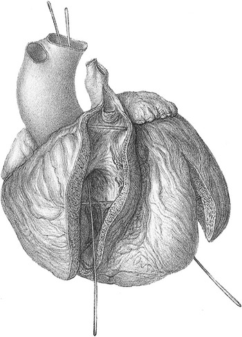
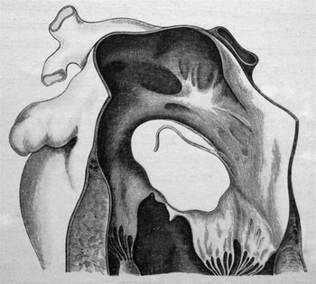
The description provided of a newborn dying at 4 days exemplified his insights. The cardiac anatomy was consistent with a common atrium, common atrioventricular valve, a solitary ventricle, and common arterial trunk. His splendid illustration (Fig. 1) shows the solitary ventricle giving rise to the common arterial trunk, and illustrates separate origin of the right and left pulmonary arteries from the posterior aspect of the trunk, the arrangement making up the second subset of specimens with common trunk identified by Collett and Edwards in their popular system for classificationReference Collett and Edwards7. Common trunk in association with a solitary ventricle, however, is exceedingly rare, and we are unaware of comparable descriptions of the lesion illustrated by Farré (Fig. 1). Hardly surprisingly, at 2 days, the patient had been in florid cardiac failure. Farré wrote, “His respiration, indeed, was remarkably quick…. The action of the diaphragm was unusual…it forcibly bent inwards…. The pulsations of the heart were too strong…the pulse at the wrist could not be felt, the skin was pallid and cold.”
The specimen illustrated by Farré showing a common arterial trunk. As can be seen, the truncal valve is supported by a complete muscular infundibulum, and there is no evidence of a ventricular septal defect. The apical trabeculations could be of right ventricular morphology, so the common valve might have been connected exclusively to a dominant right ventricle in presence of a postero-inferior left-sided incomplete left ventricle. Farré, however, described the ventricle as being a solitary structure.

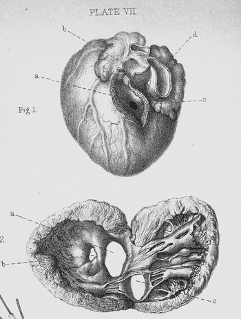
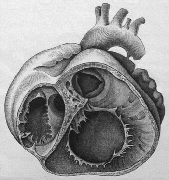
At autopsy, Farré postulated the physiology underpinning the clinical findings, describing that “The blood returning from the body and lungs being poured into one auricle, more completely mixed in the single ventricle, and from thence propelled into the common artery, it is obvious that the symptoms must vary in proportion to the size of the pulmonary branches which sprung from that artery.” These were remarkable conclusions for 1814. It was even 5 years before René-Théophile-Hyacinthe Laënnec invented the stethoscope.Reference Laennec8 Among the other cases he described was one with the anatomy we now recognise as being highlighted by Fallot as the commonest cause of “la maladie bleue” (Fig. 2). His illustration shows a probe passing directly into the aorta from the right ventricle, indicating the biventricular aortic origin, and it is clear that the pulmonary trunk is appreciably narrower than the aorta, with its origin guarded by a stenotic valve with 2 leaflets. The ventricular septal defect is shown with a muscular postero inferior rim.
The specimen illustrated by Farré in this figure has the unequivocal phenotypic morphology of the entity we now describe as tetralogy of Fallot. As we will see, this is but one of the several illustrations of this entity that appeared in advance of the recognition by Fallot that such lesions were responsible for the majority of examples of “la maladie bleue”.

John Paget
John Paget was born in 1808, in Leicestershire, England. He received a medical degree from Edinburgh University in 1831. During medical school, Paget was president of the Royal Medical Society of Edinburgh, the first, and still the only, society for medical students in the United Kingdom to receive the royal charter. After graduation, he journeyed to Paris for extra training in dermatology. Following his experiences in Paris, he left medicine, and travelled throughout Europe. He eventually married a Hungarian baroness, was granted Hungarian citizenship, became an agriculturalist, and published scientific articles on farming. Paget died in 1892 in Gyéres, currently Cîmpia Turzii, Romania.Reference Wykes9
Against this background, it is fascinating that Paget, in 1831, had already authored the well-referenced “An Inaugural Dissertation On Congenital Malformations of the Heart”.Reference Paget10 This work of 50 pages, without illustrations, covered cardiac embryology, possible malformation aetiologies, a system of classification, clinical histories, and pathology. He emphasized the lack of options for treatment. He wrote well, and presented strong reasoned convictions that suggested a mature clinician, all the more noteworthy, as at the time of his writing, he remained a medical student.
Early in his dissertation, Paget described formation of the aortic arches, cardiac looping, and septation of the cardiac chambers and arterial trunks. Siding with Johann Friedrich Meckel,Reference Clark11 Paget considered that embryologic errors might explain cardiac malformations. He wrote, “…Every organ in the body, in the course of its development, passes through a regular and determined series of changes, becoming gradually more complex in structure and proportion as the animal approaches its perfect state. It is moreover equally certain that many of the organs of higher classes of animals, at an early period of their existence, present appearances analogous to those observed in the lower [animals]…. It requires no great stretch of the imagination to conceive, that if the evolution of a part be impeded in any one of these changes…we shall have a malformation exhibiting some of the characteristics proper to the period at which this [the arrest in development] has occurred.”
Paget classified congenital cardiac defects into “Malformations of Defect,” including deficient septation of the cardiac chambers and arterial trunks, functionally single ventricles, valvar abnormalities, and persistence of the arterial duct associated with pulmonary atresia and interrupted aortic arch. His second category was “Malformations of Excess,” incorporating bilateral caval veins, double aortic arches, bilateral arterial duct, and hypertrophy. His third category was “Malformations of Position,” constituting abnormalities of cardiac position, transposition, and anomalous pulmonary venous connection.
Paget concluded his work by describing symptoms and treatment. He included a discussion of “cyania,” concluding that it resulted from an admixture of venous and arterial blood. A debate, lasting many decades, raged over the cause of cyanosis. His conclusions proved correct. Not surprisingly, Paget had limited therapeutic comments, “The treatment of these affections may be discussed in a few words; it can only be palliative.”
Thomas Bevill Peacock
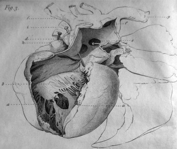
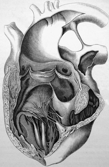
Peacock was born in York, England, in 1812. He attended University College Medical School in London. He journeyed to Paris to learn of the French approach to auscultation and care. After extensive clinical training and travel, he received his medical degree from Edinburgh University. His special interests included morbid anatomy, and he helped found the Pathological Society of London in 1846. He ultimately became editor of the Transactions of the Pathological Society of London. Over decades, Peacock published many cases of congenital heart disease in the Transactions, frequently accompanying the text with illustrations like the one displaying an obstructed subpulmonary outflow tract from the right ventricle co-existing with ventricular septal defect and aortic overriding (Fig. 3). As we have discussed above, this is the entity we now describe as tetralogy of Fallot, and as we have shown, the entity was also illustrated by Farré ahead of the description given by Fallot. Peacock died in 1882, at the age of 70.Reference Porter12, Reference Flaxman13
In this figure, Peacock illustrates the narrowed subpulmonary infiundibulum from a heart that, nowadays, we would describe as having tetralogy of Fallot. His description indicated that the heart also exhibited aortic override and a ventricular septal defect, although these features are not shown in this figure.

His treatise, entitled “On Malformations of the Human Heart with Original Cases”,Reference Peacock14 appearing initially in 1858, is the major mid 19th century book devoted specifically to congenital cardiac abnormalities. In 1866, he expanded the book in its second edition.
Peacock proposed another system for classification:
• Congenital Misplacements of the Heart, including right-sided heart and ectopic heart
• Deficiency of the Pericardium
• Malformations of the Heart. The category was subdivided into malformations dependent on arrest of development, malformations preventing the changes that should ensue after birth, and malformations that manifest later in life, this including mild abnormalities of the arterial and atrioventricular valves with progressive pathology into adulthood
• Malformations in primary development of the vessels, including transposition, interrupted aortic arch, and anomalous systemic and pulmonary venous connections.
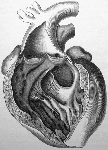
Peacock analyzed many more examples of congenitally malformed hearts than did Farré, but similar to Farré, he correlated clinical with autopsy findings. As already shown (Fig. 3), he described the phenotypic anatomy we now recognise as tetralogy of Fallot, as had Farré (Fig. 2). The article by Fallot, of course, would not be published for another 30 years, and the eponym would not become popular for another 40 years after that.Reference Evans15 Peacock produced detailed illustrative plates, and supplemented them with clinical information. Significantly, in one plate (Fig. 4), he shows an incomplete right ventricle giving rise to a transposed aorta. The second panel of this figure shows a dominant left ventricle receiving both atrioventricular valves. Peacock described the small chamber as a rudimentary right ventricle! Had we continued to follow his example, we could have avoided many tortured debates on the perceived singularity of the ventricular mass in the presence of double inlet left ventricle.
In the upper part of this figure, Peacock shows an incomplete right ventricle, supplied through a restrictive ventricular septal defect, giving rise to the aorta. In the lower part, he shows 2 atrioventricular valves entering the dominant left ventricle. In his description, he shows that he appreciated that the second chamber in the setting of double inlet left ventricle was a rudimentary right ventricle!

In a deceased 19 year-old female with cardiac findings again consistent with tetralogy of Fallot, Peacock wrote, “When ten months old she began to suffer from violent fits of crying and excitement, in which she became black in the face; and these fits sometimes terminated in convulsions…. Her mother stated that when she was pregnant with this child, and four or five months before her confinement, she was greatly alarmed by her husband, who was insane, standing over her for two hours with a loaded pistol…. When she was admitted into St. Thomas’s in July [at age nineteen] the nails were much arched and dark-coloured and the extremities of the fingers were somewhat bulbous…. [Peacock continued] The convulsive attacks, of which cyanotic persons are subject, are often relieved by the application of a few leeches to the temples or behind the ears….” His narrative clearly described cyanotic spells and classic clubbing, although the method of treatment advocated by Peacock was short lived.
Peacock was also wrong about cyanosis, concluding it occurred via venous congestion rather than venous-arterial admixture. He supported his argument by citing Morgagni and Rokitansky. He also presented, nonetheless, the correct view held by Farré and Paget, namely that cyanosis resulted from admixture of venous and arterial blood. He also developed adept auscultatory skills. He was among the first to report the inconsistency between findings from cardiac murmurs and the severity of abnormalities. He wrote, “For the production of a murmur two elements are necessary; there must be source of obstruction to the circulation, and the blood must be propelled with power through such impediment. A loud murmur may be produced by a very slight amount of obstruction, and a very great degree of obstruction may be attended by little or no murmur.” In his texts on cardiac malformations, and in 2 further treatises on valvar disease, he also reinforced the concept that valvar stenosis could cause ventricular hypertrophy.Reference Peacock16 Although not illustrating a heart with common atrioventricular junction, his description of such a heart is truly amazing. He wrote, describing the heart of an 11 year-old female, “The interesting points in the case were, first, a deficiency of the base of the inter-auricular septum, with a perfect closure of the foramen ovale. The deficient space allowed of free communication between the two auricles, so that there could only be said to be one auriculo-ventricular aperture. 2ndly. A distinctly tricuspid form of the left auriculo-ventricular valve; and 3rdly, a deficiency at the base of the septum of the ventricles, nearly closed by an extension of the anterior fold of the left auriculo-ventricular valve”. Peacock is describing the entity we now recognise as the “ostium primum defect”, or atrioventricular septal defect with common atrioventricular junction but with shunting confined at atrial level. Had we followed his example of describing the trifoliate nature of the left atrioventricular valve, we would have avoided many subsequent discussions concerning the potential presence of a cleft in an otherwise normal mitral valve.
Karl, Freiherr von Rokitansky
The Baron von Rokitansky was born in 1804, in Hradec Králové, Bohemia, currently the Czech Republic. He received his medical degree in Vienna in 1828. Early in his career, he recognized pathological examinations could aid clinical medicine. He eventually rose to become Professor of Pathology at the Vienna Medical School. In 1846, he authored his landmark 3-volume text, “Handbook of Pathological Anatomy”. He performed his first autopsy in 1827 and by retirement, 48 years later, he had performed tens of thousands. Many medical historians regard Rokitansky as the finest anatomical pathologist of the 19th century, and the founder of the specialty. He died in 1878, in Vienna.Reference Karl Freiherr17–Reference Bureš, Karel and Karl Freiherr19
Rokitansky espoused the ideas of comparative anatomy and embryology propounded by Meckel. In his magnificent atlas appearing in 1875, entitled “Die Defecte der Scheidewände des Herzens”,Reference Rokitansky20 he detailed his embryological and pathological observations of ventricular septal defects, atrial septal defects, and transposition. He differentiated the primary interatrial defect as opposed to defects within the oval fossa, albeit that, unlike Peacock, he failed to recognise the trifoliate nature of the left atrioventricular valve, promulgating the concept of a cleft in the anterior leaflet of the mitral valve (Fig. 5). From observations in chick and human embryos, he postulated an embryologic mechanism for transposition, and gave an exquisite account, along with the first illustration (Fig. 6), of the entity we now describe as congenitally corrected transposition. His text contained numerous case histories but minimal discussion of signs and symptoms.
This illustration from the atlas of Rokitansky shows an atrioventricular septal defect with separate valvar orifices for the right and left ventricles, this picture showing the left atrioventricular valve. Unlike Peacock, however, Rokitansky promoted the concept that the valve was a mitral valve with a cleft in its anterior leaflet, a misconception which was subsequently followed for at least one century.

The first illustration of congenitally corrected transposition, as shown here, was provided by Rokitansky in his atlas. He shows the entity in short axis fashion, illustrating the mitral valve entering the right-sided morphologically left ventricle, which gives rise to the pulmonary trunk, and the tricuspid valve entering the morphologically right ventricle, which gives rise to the anterior and left-sided aorta.

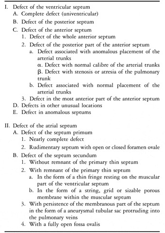
Like Farré and Peacock, he provided illustrations of the tetralogy of Fallot in advance of the publication of Fallot himself, intriguingly with his illustration (Fig. 7) showing the co-existence of the outflow morphology of tetralogy with the ventriculo-arterial connection of double outlet right ventricle. He also showed regular transposition in the setting of deficient ventricular septation (Fig. 8). His comprehensive classification system, which we have translated for the first time, as far as we are aware, from the German original, is shown in Table 1.
In this picture, Rokitanksy shows the phenotypic morphology of tetralogy of Fallot. Intriguingly, the aorta is shown as arising exclusively from the right ventricle in the setting of fibrous continuity between the leaflets of the aortic and mitral valves. Had we followed the example of Rokitansky, and subsequently Fallot himself, who described his lesion when the aorta arose predominantly from the right ventricle, we would have avoided ongoing controversies concerning the co-existence of tetralogy of Fallot and double outlet connection from the morphologically right ventricle.

In this figure, Rokitansky shows the morphologically right ventricle supporting a transposed aorta in the setting of deficient ventricular septation.

The suggested classification of the Baron von Rokitansky for congenitally malformed hearts, as translated from the German original version.

John M. Keating and William A. Edwards
John Keating was born in 1852, and William Edwards in 1860. In 1887, Keating was Obstetrician and Lecturer on the Diseases of Women and Children at the Philadelphia Hospital, and Edwards was Instructor of Clinical Medicine at the University of Pennsylvania, where at the time the chief was William Osler.Reference Keating and Edwards21
In 1888, Edwards left Philadelphia for reasons of health, and moved to San Diego, California. In 1890, Edwards married Frances Louise Taft, only sister of the President of the United States, William Howard Taft.22 In San Diego, Edwards opened a small private hospital that counselled patients on hygiene, diet, and exercise.Reference Miller23 He co-authored the book, “Two Health-Seekers in Southern California”,Reference Edwards and Harraden24 which appeared in 1897, about the advantages for health of the Californian climate. Frances and he moved to Los Angeles for a short time, but returned to San Diego, where he died in 1933 at the age of 73.
In 1891, Keating edited the multivolume “Cyclopedia of Diseases of Children”.Reference Keating25 For a time, he was also the Medical Director of the Pennsylvania Mutual Life Insurance Company. He was a founding member of the American Pediatric Society, and served as its sixth president. He, like William Edwards, had poor health, and spent most of his last few years travelling for better weather to Colorado Springs, Colorado. There he also edited the journal “Climatologist”. As was Edwards, he was a proponent of the ameliorating effects of good climate, mineral springs, and preventive medicine. Keating, nonetheless, died young at 42, in 1893.26, 27
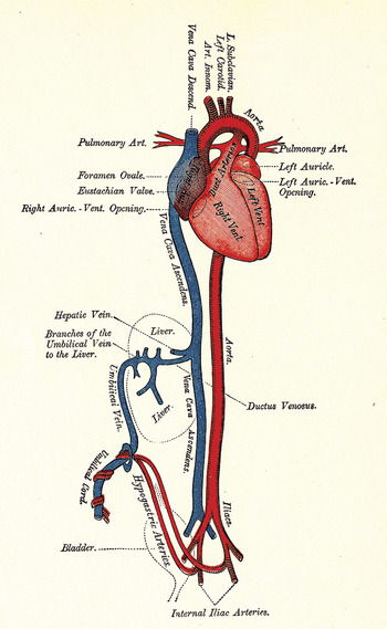
While Keating and Edwards were both in Philadelphia, they co-authored a series of articles about paediatric cardiac disease published by Archives of Pediatrics throughout 1887 in monthly instalments. They assembled the articles into “Diseases of the Heart and Circulation in Infancy and Adolescence”,Reference Keating and Edwards21 published by Blakiston Press in 1888, and subsequently by F. A. Davis in 1889. The edition of 1889 also had an appendix, “Clinical studies on the pulse in childhood,” not found in the earlier edition. They included a diagram of the fetal circulation (Fig. 9) opposite the title page, albeit less than perfect in its display of the physiological effects of the circulatory patterns.
The diagram of the fetal circulation provided by Keating and Edwards. As can be seen, the concept is less than physiologically perfect if the blue colour is intended to show the course of the richly oxygenated blood returning to the heart through the umbilical veins. The diagram also fails to show the oval foramen.

The book, with 228 pages, was comprehensive. They began, “The many excellent text-books on children’s diseases have failed to give a satisfactory account of the diseases of the heart, and, indeed as far as we know, the work now presented by us is the only systematic attempt that has been made to collect in book-form the abundant material which is scattered throughout medical literature in the form of journal articles, clinical lectures, theses, and reports to societies.”
The first section of their book covered the fetal circulation, physical examination, and congenital cardiac disease. They wrote in a modern style, and their points remain pertinent today. They discussed a recent report by J. Collins Warren concerning microscopy of the arterial duct: “His investigations led him to conclude that the ductus arteriosus at the time of birth in certain important respects differs in structure from the aorta and the pulmonary artery…. The media is thicker…it is thrown into irregular folds…. The media consists chiefly of longitudinal layers of muscular fiber, a few in circular bundles….” They noted difficulties in classifying defects, “Attempts have been made for the last thirty years to classify the circulatory anomalies and malformations into a convenient working form, so that cases may be arranged under this or that heading, with, however meager success.” They discussed cyanosis at length, concluding it was caused by both venous-arterial admixture and venous stasis. The rest of the text is divided into 9 chapters. Chapters 2 through 8 covered mostly acquired cardiac disease secondary to rheumatic fever and infectious agents. Chapter 9 and 10 discussed the secondary effects on the heart from anaemia, non-cardiac tumours, and exophthalmic goitre.
George Alfred Carpenter
George Carpenter was born in London in 1859. He died in 1910, albeit that biographical information is limited. By 1894, he was Senior Physician to out-patients at the Evelina Hospital for Sick Children. The Evelina Hospital was founded in 1869, by Baron Ferdinand de Rothschild, and was named for his wife, who died in childbirth along with their stillborn premature infant. The hospital became part of Guy’s Hospital in 1948. It has now been reconstituted as a splendid new building on the St Thomas’s site of the combined centre for paediatric cardiology for St Thomas’s and Guy’s Hospitals.28
While working at the Evelina Hospital, Carpenter published, in 1894, “Congenital affections of the heart”.Reference Carpenter29 He began, “At my outpatient clinic at Evelina Hospital for Children. I have so frequently been asked by students for demonstrations on congenital affections of the heart, and complaint has so often been raised that the ordinary text books are not sufficiently precise and explanatory of the subject that I have for some time past been in the habit of giving them demonstrations on these affections. These demonstrations I now publish in book form without any intentions or desire of rivalling the classic works by Rokitansky, Kussmaul, Peacock, and others, but in as much as they have proved of service to my classes, they may, I trust, be equally useful to other students and practitioners.”
His book was short, and only briefly discussed embryology and congenital defects, using terminology of the time, such as pulmonary stenosis, pulmonary atresia, double chambered right ventricle, patent ductus, interrupted aortic arch, coarctation, transposition, truncus, aortopulmonary window, ventricular and atrial septal defects, aortic stenosis and atresia, atrioventricular valvar abnormalities, functionally single ventricles, vascular rings, pulmonary venous return abnormalities, and right-sided heart. He discussed symptoms, findings at the cardiac examination, prognosis, and limited medical treatments. George Carpenter was an active contributor to the medical literature, writing articles on cardiology, gastrointestinal, and orthopaedic abnormalities. In 1901, he was the first to describe acrocephalopolysyndactyly, now known as Carpenter’s syndrome.Reference Carpenter30
André Charles Moussous
Biographical information is scant, but Moussous was born in 1857, and died in 1926, at the age of 69. He obtained his medical degree in 1885, from the University of Bordeaux. Following his degree, the University of Bordeaux appointed him in 1890 to a teaching position in paediatrics, and in 1898 he rose to become full Professor.Reference André31
The French dermatologist, Georges Thibierge, reviewed the book published in 1895 by Moussous, and entitled “Maladies congénitales du Coeur”.Reference Moussous32 Translated from the French, Thibierge wrote, “The symptoms of the diverse malformations are studied in detail with respect to the most recent researches.”Reference Thibierge33 Moussous began with an in-depth review of cardiac embryology. He followed with descriptions of the pathologic anatomy of both intra and extracardiac defects, including ventricular and atrial septal defects, abnormalities of the great arteries, valvar stenoses, patency of the arterial duct, and malformations of the cardiac cavities, such as functionally single ventricles, coupled with associated defects. He used broad groupings for the cardiac abnormalities, dividing them into those involving arrests in the normal developmental process, those with incomplete septation, embryologic aberrations, and conditions resulting from persistence of fetal pathways. With respect to further schemes beyond his broad designations, he concluded that the system suggested by Rokitansky was the most scientific. Regarding aetiology, Moussous considered that the evidence pointed to arrested development rather than fetal endocarditis as causative. He concluded that the evidence suggested endocarditis occurred secondary to the defect, rather than being an aetiologic agent.
His case reports exemplified clinicopathologic correlations. He discussed cyanosis at length, concluding that it likely resulted from both venous-arterial admixture coupled with venous stasis. He concluded his work with discussion of the finer points of auscultation, and a review of complications and prognosis. In his section on treatment, he emphasized diet, hygiene, prevention of infections, and the occasional use of bromides and digoxin.
Hermann Vierordt
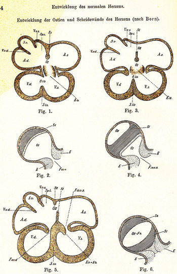
Hermann Vierordt was born in 1853 and died in 1943. He was the son of the renowned physiologist, Karl Vierordt, who lived from 1818 through 1884, and developed the haemotachometer for measuring the velocity of the flow of blood, along with an early forerunner of the modern sphygmomanometer. Both father and son were professors at the University of Tübingen.Reference Karl von34, Reference Hermann35 The work by Hermann with regard to the congenitally malformed heart was preceded by two other works on cardiovascular topics. In 1884, he produced “Kurzer Abriss der Perkussion und Auskultation”, a brief outline of percussion and auscultation, and in 1885 “Die Messung der Intensität der Herztoöne”, the measurement of the intensity of heart sounds.Reference Vierordt36, Reference Vierordt37 He later became a medical historian. Amongst other works, he published an article “Vesalius in Tübingen”.Reference Vierordt38 His book published in 1898, entitled “Die angeborenen Herzkrankheiten”Reference Vierordt39 was a systematic review of embryology, including for example the descriptions of development of the atrial septum provided by Born (Fig. 10), and an extensive review of the literature, along with a discussion of symptoms.
The concept of development of the atrial septum, adapted from Born, as used by Vierordt. Note that he correctly illustrates the mesenchymal cap on the leading edge of the primary atrial septum, but fails to recognise that the so-called “septum secundum” is a fold produced between the attachments of the superior caval vein to the right atrium and the right pulmonary veins to the left atrium. This feature of the atrial morphology is a late development. Vierordt also fails to illustrate the importance of the vestibular spine, or “spina vestibuli”, well described by His in the mid-nineteenth century.

A translation from German of a contemporary reviewer, Dr A. Ripperger, noted, “The author divides the sizable material of congenital heart defects into 14 main chapters from an anatomic-developmental perspective which unfortunately can not be further detailed at this point. Only so much may be mentioned that the author in recognition of its importance provides appropriate detail of the pathological anatomy, causes, and statistics of congenital heart defects, while the symptomatology, diagnosis, and prognosis are also adequately acknowledged.”Reference Ripperger40 The reviewer had limited space for his comments. But the impression from his words indicated his solid opinion about this work, and that it made a significant contribution to the study of congenital cardiac malformations.
Charles William Chapman
Charles Chapman was born in 1846, but we have been unable to locate a reference for the year of his death. He was a member of the Clinical Society of London, which began in 1868, and merged with the Royal Society of Medicine in 1907.Reference Rolleston41
At the time Chapman wrote his book “Heart Disease in Childhood and Youth”, Reference Chapman and Wilks42 in 1901, he was 54 and on the staff of the National Hospital for Disease of the Heart in London, which later became the National Heart Hospital. He spelled out his goals in his introduction: “The following pages are written with the object of presenting to the reader a brief outline of the more usual varieties of heart disease as they occur in young persons…. Attention is directed to the hygienic management of young heart patients, and questions as to education, sports, etc., in such cases are discussed. Finally a number of illustrative cases are quoted, followed in each instance by remarks indicating salient points in diagnosis, prognosis, and treatment.”
Chapman briefly covered rheumatic heart disease, and some cases of congenital cardiac malformations. The book is short, but a reviewer noted, “This little work, the outcome of its author’s extended experience, gives much more definitive statements and more favorable conclusions that any text book known to us.”43 The book appeared in a second edition in 1903. Chapman continued his interest in cardiac disease, publishing several case reports, and some years later, in 1927 “Heart and its Diseases: a Handbook for Students and Practitioners”.Reference Chapman44
Manuel A. Santas
Manuel Santas was born about 1872,45 and he received his medical degree from the University of Buenos Aires in 1898. Before receiving his degree, he worked with one of the leading Argentinean pathologists, Roberto Wernicke, who was a member of the faculty at the University of Buenos Aires. Following medical school, Santas spent another 2 years with Wernicke before travelling to Córdoba, Argentina, where he served for 5 years at San Roque Hospital on the paediatric service of Professor Aráoz Alfaro.Reference Santas46 The death of Manuel Santas, probably in late 1937, was noted in an obituary appearing in the British Medical Journal. The only other biographical information we could locate was from the preface of his book.47
In 1905, he wrote “Estudio Semiológico Anomalías Congénitas del Corazón”.Reference Santas46 In the introduction, translated from Spanish, Santas wrote, “It is not possible to study the semiology of a diseased organ without the previous knowledge of the anatomic lesions. To be able to relate the clinical signs to a structural abnormality it is necessary, before the beginning of the semiologic studies of the congenital anomalies of the heart, first to understand its pathologic anatomy. And because the structural anomalies are closely related to the development of the heart, is also necessary to study embryology.”
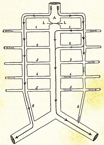
Santas discussed embryology, pathological anatomy, the possible aetiologies, including fetal endocarditis, and the teratologic theories of Rokitansky. He discussed concepts accounting for cyanosis, including venous stasis, inadequate oxygenation, and venous-arterial admixture. He concluded, in agreement with Moussous, that the cause of cyanosis was multifactorial. Another of his clinicopathologic correlations was that of coarctation of the aorta associated with palpable distal pulses. Santas reproduced an illustration (Fig. 11) from an article on coarctation by Bonnett that diagrammatically demonstrated the development of collateral circulation.Reference Bonnet48 A noteworthy inclusion was the term “tetralogía de Fallot”. With his colleague, Horatio Piñero, he used the eponym more than two decades before Maude Abbott began to popularize “tetralogy of Fallot” in her writings.Reference Evans15 Santas concluded his book with 12 cases, mainly culled from his experience on the paediatric service at San Roque Hospital. He presented histories, signs, symptoms, physical and auscultatory findings, clinical diagnoses, and pathological reviews, since all the patients died.
Santas has reproduced the diagrammatic representation of the collateral circulation found in the setting of coarctation as produced by Bonnet.Reference Bonnet48

Maude Elizabeth Abbott
Abbott was born in 1869. Following an illustrious career, which brought world fame, she died in September of 1940. She attended McGill University for undergraduate work, but as a woman, the medical school at McGill denied her admittance. Instead, she attended Bishop Medical School in Montreal. Following graduation, she sailed to Europe to study internal medicine and pathology, chiefly in Vienna. In 1896, she returned to Montreal and opened a practice in internal medicine. Charles F. Martin, one of her medical schoolteachers, sensed her dissatisfaction with private practice, and suggested she review case histories of patients hospitalised without cardiac disease, yet diagnosed with cardiac murmurs. This work led to her first article, “On so-called functional murmurs.”Reference MacDermot49–Reference Abbott53
In 1898, she was appointed at McGill as curator of the specimens held in the museum of pathology. In December of 1898, she made her first trip to the United States of America, where she visited the Army Medical Museum in Washington, D.C., and studied the collection of pathological specimens held at Johns Hopkins University in Baltimore. William Osler, a graduate of McGill, was then Chief of Medicine at Johns Hopkins Hospital. During her visit to Baltimore, Osler inspired Abbott to pursue a career in pathology.Reference MacDermot49, Reference Abbott50
Upon her return to Montreal, Abbott set upon the task of cataloguing the unorganized pathological museum. In the process, she happened upon a mislabelled specimen. The heart had previously been described by Andrew Holmes, a founder of McGill University. The specimen, having double inlet left ventricle, concordant ventriculo-arterial connections, and normally related great arterial trunks, has since become known as the Holmes heart. This specimen sparked her lasting passion for congenital cardiac disease.Reference Holmes54–Reference Jacobs and Anderson56 Knowing of her growing experience in the study of cardiac abnormalities, Osler requested that she write the chapter devoted to congenital cardiac malformations for the multivolume-textbook “Modern Medicine”Reference Abbott57 he was editing in 1907. She was enamoured with William Osler, and wished her contribution meet with his favour. Her efforts took two years. When completed, Abbott had gathered together 412 examples of congenitally malformed hearts, both from the museum at McGill, and from the literature.
The chapter, extending to 102 pages, consisted of reviews of embryology, aetiology, clinical findings, and prognosis. In her subsequent writings, Abbott classified malformations into 3 groups, namely valvar stenoses or coarctation without septal defects, so-called cyanose tardive, or late cyanosis, and those coming from patients cyanotic from birth. In her initial chapter, however, she emphasised the difficulties existing in establishing a system for classification, stating “Many attempts have been made to reduce cardiac anomalies to s scientific classification, but none have been entirely successful….no one classification will be found adequate in its practical application, and it would almost seem that a grouping ‘on mixed principles’ is the only one under which all the cases can be satisfactorily placed.”
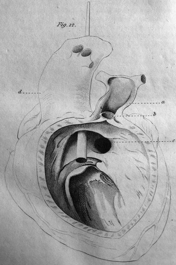
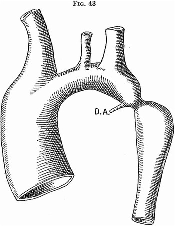
There were few illustrations in her initial chapter. For one (Fig. 12), she reproduced an illustration from the same article published by Bonnet on coarctation in 1903 that Manuel Santas had referenced in his book. Bonnet had coined the terms “infantile” and “adult” for description of the anatomic patterns of coarctation that she included in her discussion. Abbott was the first to draw attention to an association between coarctation and aortic rupture, both of the ascending and descending components of the aorta, finding this complication in 12 of the 198 cases she reviewed.
The representation of aortic coarctation used by Maude Abbott in her chapter prepared for Osler is again using the initial concept of Bonnet.Reference Bonnet48

Having read her chapter, Osler wrote to Maude expressing his compliments – ”I knew you would write a good article but I did not expect one of such extraordinary merit. It is by far and away the best thing ever written on the subject in English – possibly in any language. I cannot begin to tell you how much I appreciate the care and trouble you have taken, but I know you will find it to have been worthwhile. For years, it will be the standard work on the subject.” In a postscript, Osler added, “I have but one regret, that Rokitansky and Peacock are not alive to see it.”Reference MacDermot49
Johann Georg Mönckeberg
Johann Mönckeberg was born in 1877, and received his doctorate in 1900, from the University of Bonn. In 1912, he was appointed professor of pathology at the Academy for Medicine in Düsseldorf, subsequently being appointed in 1916 to the Chair of Pathology in Strassburg. When the French occupied Strassburg during the first Great War, Mönckeberg was expelled from the university. In 1923, an episode of influenza led to chronic renal disease, and he died in 1925. His main contributions were to cardiovascular pathology. Mönckeberg’s arteriosclerosis, also called medial calcific sclerosis, was named for him. Some 59 of his 86 publications concerned cardiac and vascular pathology.Reference Johann Georg58
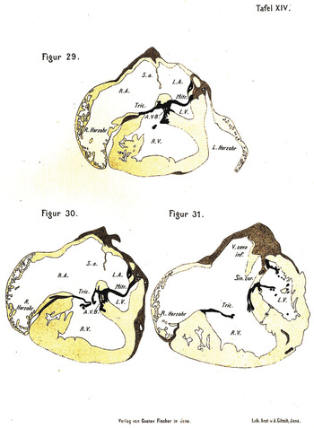
While in Düsseldorf, he published, in 1912, his atlas entitled “Herzmissbildungen; ein Atlas angeborener Herzfehler in Querschnitten mit besonderer Berücksichtigung des Atrioventrikularsystems”, which translates to “Heart defects; an atlas of congenital cardiac abnormalities in cross-sections with special consideration of the atrioventricular systems”.Reference Mönckeberg59 He did not provide clinical vignettes, so this work differs from the others reviewed. But his plates, showing cross-sectional illustrations of congenital cardiac abnormalities, were marvellous. Today, the illustrations correlate well with cross-sectional imaging techniques. In one series, he included a patient with hypoplasia of the left heart (Fig. 13). This atlas deserves to be republished.
Illustrations provided by Mönckeberg of cross sections of examples of the lesion we now describe as hypoplastic left heart syndrome.

In our review, we have not included another atlas appearing late in the 19th century. This atlas, produced by Emile Théremin in 1895, was called “Etudes sur les affections congénitales du Coeur”.Reference Théremin60 Théremin produced simple diagrammatic renderings. His text, although extensive, was primarily limited to measurements of the chambers, vessels, and valves.
George Alexander Sutherland
George Sutherland was born in 1861, and died in 1939. Biographical details are scarce. He was President of the Section for Diseases of Children of the British Medical Association before 1907. He published “Treatment of Disease in Children” Reference Sutherland61 in 1907, and issued a second edition in 1913. In 1914, he was the Senior Physician to the Hampstead and Northwest London Hospital, and Physician to the Paddington Green Children’s Hospital in London. Paddington Green Children’s Hospital opened in 1883, and closed in the 1980s.
His text of 1914, called “The Heart in Early Life”,Reference Sutherland62 was a practical early 20th century text. In contrast to previous works, Sutherland did not focus on congenital malformations. He divided the book into three sections. The first, entitled “Cardiac Disturbances (Functional),” was devoted to sinus arrhythmia, benign extrasystoles, normal variations of heart rate, and innocent murmurs. The second section was entitled “The Borderline between Functional Disturbance and Organic Disease of the Heart”. In this part, he provided detailed descriptions of paroxysmal tachycardia. The third section, called “Organic Heart Disease,” was devoted to pathologic arrhythmias, valvar abnormalities, symptoms, prognosis, and treatment. His focus for this third section was chiefly rheumatic heart disease.
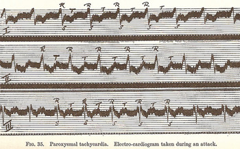
Even today, his discussion of paroxysmal tachycardia is fascinating. He began, “It may be said that the occurrence of paroxysmal tachycardia is evidence of pre-existing disease. But if it is so, then it is a form of disease of which we have no knowledge.” He presented the history of a 9 year-old male with “heart hurry” that began at the age of 7. He described the first episode as relayed to him by a fellow physician, “The boy looked well…. The radial pulse uncountable…. The condition remained unchanged for six days when the nurse…felt the heart give a ‘flop’…. Soon afterward the doctor found the heart beating at 40 per minute, with an occasional irregularity.” Sutherland saw the child in consultation two years later with a history of paroxysmal tachycardia usually resolving spontaneously or following vomiting. When Sutherland carried out his examination, the child had been in tachycardia for 21 days, during which a previous physician had treated him with “belladonna, bromides, cannabis indica, chloral, and opium” without effect. Sutherland then started digitalis in “moderate” doses, and on the 33rd day, the heart rate dropped to 80 beats per minute. He discussed this case for another 23 pages. He included an electrocardiogram (Fig. 14) performed by Thomas Lewis, a friend of Willem Einthoven, who developed the electrocardiographic recorder, and was later knighted. Sutherland wrote, “In this tracing, the auricular contraction P is not easily identified…” After a lengthy discussion Sutherland wrote, “The object aimed at in this case by means of digitalis was to diminish conductivity and so check the passage of rapid and abnormal auricular impulses through the heart.” He continued, “I do not know whether this line of treatment can be successfully adopted in other cases of the same nature.” But following more discussion of possible mechanisms, he concluded, “I am quite content to leave it [the discussion] at that for the present, but should certainly adopt the same treatment in any other persistent case of paroxysmal tachycardia.”
The electrocardiogram prepared by Thomas Lewis from a young patient with paroxysmal tachycardia, and reproduced in the book of Sutherland. This is the first example, as far as we are aware, of an illustration of a paediatric arrhythmia.

Pursuing other general cardiology books published before 1915, we could not locate another discussion of paroxysmal tachycardia with such detail. Also, to the best of our knowledge, this was the first text devoted to paediatric cardiac disease, and among the first textbooks of any type that included electrocardiographic tracings.
Discussion
Our review does not represent a comprehensive analysis of the literature of the 19th to early 20th century concerning cardiac malformations. Rather, we elected primarily to study a collection of wide-ranging works on paediatric cardiac disease. Thus, we did not include many texts, such as doctoral theses like “Über Angeborene Herzfehler” by Peiser Max,63 and the reports of single diseases similar to those by Fallot, Ebstein, and Eisenmenger.Reference Fallot3, Reference Ebstein64, Reference Eisenmenger65 Others have reviewed these eponymous works.
Written works mirror the passions of the authors. Some texts took years to complete. A few are even referenced today. Common themes of the past reflect the present. In the early 19th century, Johann F. Meckel began the systematic study of malformations.Reference Clark11 He noted the relationship between possible arrests in development and cardiac defects, showing that some defects appeared similar to normal cardiac structures as seen in lower animals. In the 20th century, Helen Taussig studied cardiac defects in birds, and championed evolutionary developmental biology.Reference Opitz and Clark66, Reference Taussig67 A book published in 2000, and devoted to cardiac malformations in mammals, by Michaëlsson and Ho, presented homologous defects between nonhuman mammals and humans.Reference Michaëlsson and Ho68 A recent review by Moorman and Christoffels, nonetheless, entitled “Cardiac Chamber Formation: Development, Genes, and Evolution,” demonstrated that the issues surrounding normal and abnormal cardiac development are yet to be resolved.Reference Moorman and Christoffels69
The pursuit of clearer understandings of clinicopathologic correlations remains important. Such correlations help clinicians select medical therapy, interventional cardiac catheterization, or a surgical procedure. With respect to cyanosis, it is understandable that physicians had difficulty grasping its cause before the instrumented detection of abnormal oxygenation of the blood. Many lesions resulting in admixture of systemic and venous streams, though desaturated, did not produce cyanosis. Recently, early detection of cyanosis as a means of screening for complex cardiac lesions has been proposed, but universal oximetry remains controversial.Reference Chang, Rodriquez and Klitzner70
Difficulties continue in establishing a universal system for the classification of congenital cardiac malformations. In the 7th edition of textbook of Moss and Adams which appeared in 2008, Bill Edwards wrote, “Is the development of a unified system of nomenclature a realistic and worthwhile goal? If so, who should decide on the acceptable terminology? And what price will be paid to gain unity at the expense of diversity? Although such a system would limit the confusing number of synonyms that now exist, it also could limit our perspective as certain terms (such as those with an embryologic basis) are purged from our rich and diverse heritage.”Reference Moss and Allen71
The quest for effective treatments also persists. We are prone to deem many current methods as curative. But future paediatric cardiologists using fetal interventions or gene therapy may read our old texts and conclude our methods were primitive, similar to our impression of the use of leeches during the 19th century for treatment of cyanosis.
The first ligation of the persistently patent arterial duct, and construction of the Blalock-Taussig shunt, heralded the birth of paediatric cardiology.Reference Gross and Hubbard72, Reference Blalock and Taussig73 The innovators of these procedures stood on the foundation of clinicopathologic investigations that had amassed for hundreds of years. Yet even today, paediatric cardiologists struggle to understand the aetiology, to define the nomenclature, and to perfect the surgical treatment for conditions as complex as the “Single Heart,” consisting of a solitary ventricle and common arterial trunk, which Farré presented to his readers as long ago as 1814.
Acknowledgements
We thank Paul R. Lurie, Emeritus Professor of Pediatrics, University of Southern California, for editorial assistance, and Luise Ahlers, MD, for translation. We are also indebted to the staff of the New York Academy of Medicine Library.