INTRODUCTION
Leishmania (Viannia) braziliensis is the most prevalent species associated with American tegumentary leishmaniasis (ATL). ATL is a public health problem with approximately 25 000 cases reported annually in Brazil (SVS-MS, 2011). Nevertheless, there is no vaccine for ATL and only a limited number of drugs are available for treating patients. Most of our knowledge on the immunopathogenesis of L. braziliensis infection comes from studies in patients and in asymptomatic individuals (Reithinger et al. Reference Reithinger, Dujardin, Louzir, Pirmez, Alexander and Brooker2007; Carvalho et al. Reference Carvalho, Passos, Schriefer and Carvalho2012). The lack of an adequate experimental model for L. braziliensis is a limiting factor for the development of biological and pharmacological health inputs to ATL.
Although relevant for cutaneous leishmaniasis (CL) studies, murine models (Balb/c and C57Bl/6) are naturally resistant to L. braziliensis (DeKrey et al. Reference DeKrey, Lima and Titus1998; Rocha et al. Reference Rocha, Schleicher, Mattner, Alber and Bogdan2007). When infected by L. braziliensis, animals develop small non-ulcerated lesions that show a progression to spontaneous healing (DeKrey et al. Reference DeKrey, Lima and Titus1998; Rocha et al. Reference Rocha, Schleicher, Mattner, Alber and Bogdan2007). There are quite a few reports employing a Balb/c model in vaccination studies that successfully obtained chronic ulcerated lesions after infection by L. braziliensis promastigotes (Salay et al. Reference Salay, Dorta, Santos, Mortara, Brodskyn, Oliveira, Barbiéri and Rodrigues2007). Other experimental models such as non-human primates (Souza-Lemos et al. Reference Souza-Lemos, de-Campos, Teva, Côrte-Real, Fonseca, Porrozzi and Grimaldi2008) and dogs (Pirmez et al. Reference Pirmez, Marzochi and Coutinho1988) require complex logistics for their maintenance under experimental conditions.
The hamster is highly susceptible to dermotropic Leishmania infection and has been largely used as a model for visceral leishmaniasis (Goto and Lindoso, Reference Goto and Lindoso2004; Dea-Ayuela et al. Reference Dea-Ayuela, Rama-Iñiguez, Alunda and Bolás-Fernandez2007). The animal develops skin lesions when infected by one of the Viannia or Leishmania species including L. (V.) braziliensis (Brazil, Reference Brazil1976; Wilson et al. Reference Wilson, Dieckmann and Childs1979; Morais-Teixeira et al. Reference Morais-Teixeira, Carvalho, Costa, Duarte, Mendonça, Boechat and Rabello2008), L. (L.) amazonensis (Figueiredo et al. Reference Figueiredo, Costa e Silva and Brazil1999), L. (V.) guyanensis, L. (V.) panamensis (Rey et al. Reference Rey, Travi, Valencia and Saravia1990; Osorio et al. Reference Osorio, Melby, Pirmez, Chandrasekar, Guarín and Travi2003), L. (V.) lainsoni (Corrêa et al. Reference Corrêa, Brazil and Soares2007) and L. (V.) peruviana (Gamboa et al. Reference Gamboa, Torres, De Doncker, Zimic, Arevalo and Dujardin2008). Indeed, the skin lesions developed by these animals are very similar to the CL ulcers observed in humans (Hommel et al. Reference Hommel, Jaffe, Travi and Milon1995). This turns the golden hamster into a promising model for the study of ATL.
A number of experimental protocols using Leishmania-infected hamsters have been described. These protocols differ on several parameters, including the Leishmania strains (Wilson et al. Reference Wilson, Dieckmann and Childs1979; Rey et al. Reference Rey, Travi, Valencia and Saravia1990; Kahl et al. Reference Kahl, Byram, David, Comerford and Von Lichtenberg1991), number of inoculated parasites (Wilson et al. Reference Wilson, Dieckmann and Childs1979; Martinez et al. Reference Martinez, Travi, Valencia and Saravia1991), route and site of inoculation (Wilson et al. Reference Wilson, Dieckmann and Childs1979; Osorio et al. Reference Osorio, Melby, Pirmez, Chandrasekar, Guarín and Travi2003), animal gender or age (Wilson et al. Reference Wilson, Dieckmann and Childs1979; Travi et al. Reference Travi, Osorio, Melby, Chandrasekar, Arteaga and Saravia2002) and biological characteristics of inoculated parasites such as the number of in vitro passages, growth phase, developmental stages or rate of metacyclic forms (Rey et al. Reference Rey, Travi, Valencia and Saravia1990; Gamboa et al. Reference Gamboa, Torres, De Doncker, Zimic, Arevalo and Dujardin2008). Most of the studies using dermotropic strains involved L. guyanensis, but the outcome of L. braziliensis infection was poorly studied. To date, no study has evaluated the reproducibility of the clinical aspects of the infection in the hamster model for CL caused by L. braziliensis.
Here we studied the natural history of L. braziliensis infection in a hamster model by using a single parasite isolate in independent experiments under similar experimental conditions. Our findings indicate that the hamster is an appropriate model for L. braziliensis infection studies.
MATERIALS AND METHODS
Animals and ethics statement
Outbred golden hamsters (Mesocricetus auratus), adult females (6–8 weeks old), weighing 80–90 g, from the animal facilities at Fundação Oswaldo Cruz, were used. Thirty-four infected animals and 13 uninfected animals were analysed. This study was specifically approved by the Ethics Committee on Animal Use (CEUA) of Fundação Oswaldo Cruz – FIOCRUZ, by the number of protocol P-0281/06.
Parasites for infection and immunological studies
Leishmania braziliensis promastigotes (MCAN/BR/98/R619) in stationary growth phase until the third in vitro passages in supplemented Schneider's Drosophila medium were used (Sigma Chemical Co., St Louis, MO, USA). Promastigotes were washed in phosphate-buffered saline, 0·15 m, pH 7·2 (PBS) and 1 × 10⁶ parasites were inoculated intradermally in the dorsal hind paw of hamsters. Disrupted antigens of L. braziliensis (MHOM/BR/75/2903) promastigotes (Lb-Ag) were obtained for immunological studies.
Clinical course of Leishmania braziliensis infection
To determine the natural history of L. braziliensis infection in hamsters, 8 independent experiments were performed during a period of 2 years. The lesion increment was monitored weekly from day 7 up to approximately 110 days post-infection. This was done by measuring the paw dorsum-ventral thickness with a digital thickness gauge (Mitutoyo America Corporation, São Paulo, Brazil) with the thickness expressed in millimetres. The lesion increment was determined as the difference of measurements between the infected and the non-infected paw of the same animal. The discrepancies in lesion increment were determined by the variance coefficient [VC = (standard deviation × mean)/100)]. For this VC analysis, 5 experiments with 5 or 6 animals per group were used. Once a week animals were checked for skin macroscopic changes and for cutaneous metastasis.
Quantification of anti-Leishmania antibodies
The anti-Leishmania IgG levels were determined in plasma samples by ELISA assay as described elsewhere (Gomes-Silva et al. Reference Gomes-Silva, Souza, Afonso-Cardoso, Lívia Resende Andrade, Dietze, Lemos, Belli, Favoreto Júnior and Ferreira2008). Plasma samples were diluted 1∶200 and horseradish peroxidase-labelled goat anti-hamster IgG was used as detector system (Santa Cruz Biotechnology, Santa Cruz, CA, USA). The results were expressed as optical density (OD).
Lymphocyte proliferative responses to Leishmania antigens
Peripheral blood mononuclear cells were collected for lymphocyte proliferative response analysis in response to Leishmania antigens as described elsewhere (Da-Cruz et al. Reference Da-Cruz, Bittar, Mattos, Oliveira-Neto, Nogueira, Pinho-Ribeiro, Azeredo-Coutinho and Coutinho2002). The cells (3 × 10⁵/well) were plated in triplicate and cultured in vitro with concanavalin A (1 μg/well) (Sigma, USA), Lb-Ag (10 μg/well), or in the absence of any stimulus as the negative control. The cell cultures were maintained for 72 h at 37 °C in a humidified atmosphere of 5% CO2 in air. Then 16 h before harvesting, 1 Ci (3H)thymidine (Amersham International, UK) was added to each well, and the radioactivity uptake was measured in a scintillation counter (1600CA, Packard Instrument Company, Downers Grove, IL, USA). Results were expressed as stimulation indices (SI= average counts per minute [cpm] of stimulated triplicates with Lb-Ag/ average cpm of negative control) where values equal to or higher than 2·5 were considered as positive.
Macroscopic analysis of lymphoid organs
The spleens of 30 hamsters were excised and weighed in a precision balance. Spleens were visualized macroscopically and registered by digital photographs. Dissemination of parasites to spleens and other anatomic structures beyond the inoculation site was evaluated by macroscopic inspection (anatomical alterations) and confirmed by visualization of Leishmania through histopathological analysis of lymphoid organs. The draining popliteal lymph nodes of 10 animals and the spleens of 14 animals were subjected to microscopic evaluation.
Histopathological analysis
Fragments from the skin (n = 24) and the draining lymph nodes (popliteal, n = 10) of the infected paw, as well as from spleen (n = 14) and liver (n = 14) were fixed in 10% buffered formalin and processed for paraffin embedding. Sections of 2–4 μm thickness were stained with haematoxylin-eosin and then observed by light microscopy (Nikon Eclipse E600 Microscope, Tokyo, Japan). The images were captured in CoolSNAP-ProcCF and edited by Image-Pro Plus program (Media Sybernetics, GA, USA). The results were expressed as a semi-quantitative analysis in which the main histopathological features were scored according to the number of animals in which the features were observed vs the number of animals analysed (given in parentheses) and scored according to the intensity of occurrence of the feature, varying from (−) absence; (±) slight presence of the histopathological feature; (+) moderate presence of the histopathological feature; to (++) full occurrence of the histopathological feature.
Statistical analysis
The data were analysed by Mann–Whitney test and Spearman's rank-correlation with the GraphPad Prism software version 4.00 for Windows (GraphPad Software, San Diego, CA, USA). The results were expressed as the mean ± standard deviation and median. Significant differences were considered when P < 0·05.
RESULTS
Variances for cutaneous lesion increment post-Leishmania braziliensis infection
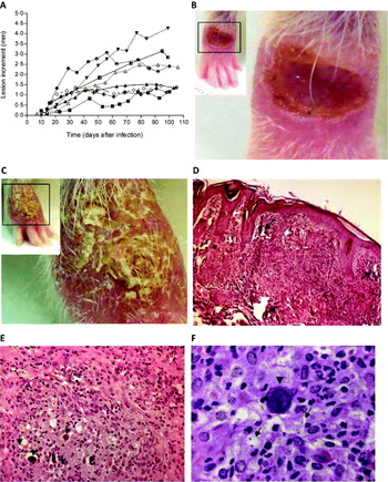
All L. braziliensis-infected hamsters developed cutaneous lesions during the observational period (∼110 days). The observation started at 2 weeks after infection, when characteristic inflammatory signs (erythema and oedema) were observed. After that there was a significant lesion increment. The first mean measure was 0·30 mm ± 0·30 mm (median = 0·22 mm, n = 34); and the final mean measure was 2·12 mm ± 1·16 mm (median = 1·9 mm, n = 34) (P < 0·001) (Fig. 1A). No spontaneous healing was observed in any animal.
Evaluation of the clinical course, macroscopic and histopathological aspects of skin lesions from golden hamsters infected with Leishmania (Viannia) braziliensis. (A) Lesion increment development in each of 8 independent experiments. Each line represents 1 experiment and the points indicate the average of lesion increments in each group. (B and C) Macroscopic aspect of the infected paw at approximately 110 days after Leishmania infection. Slides of representative lesions localized at the site of parasite inoculation (dorsal face of back right paw) taken at the end of the observational period (110 days). (D) Mixed inflammatory infiltrate reaching all over the dermis; pointed arrows indicate areas with calcification necrosis (skin, 20×). (E) Macrophages in epithelioid arrangement; arrowheads indicate Schaumann's bodies; pointed arrows indicate vacuolated macrophages with Leishmania inside (skin, 40×). (F) Granuloma; pointed arrows indicate cytoplasmatic vacuoles containing amastigotes inside; arrowheads indicate Schaumann's bodies (skin, 100×).

A cutaneous ulcerated lesion was the most frequent clinical presentation (Fig. 1B and C). Elevated erythematous borders, granular aspect, with a necrotic surface (Fig. 1B), or recovered by crusts (Fig. 1C) were also commonly seen.
The variable pattern of lesion increment was quantified by a variance coefficient. We observed homogeneous (VC < 15%, n = 1 experiment), intermediary (VC ⩾ 15% and VC ⩽ 30%, n = 2 experiments) or heterogeneous (VC > 30%, n = 2 experiments) patterns.
Histopathological findings of hamster cutaneous lesion
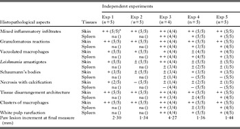
The skin histopathological findings (n = 24) are summarized in Table 1. An intense inflammatory infiltrate reaching all over the dermis (Fig. 1D), consisting predominantly of macrophages in epithelioid arrangements, and large amounts of lymphocytes, characterizing a granulomatous reaction (Fig. 1E) was visualized in all animals. Neutrophils, plasma cells and eosinophils (sometimes inside macrophage vacuoles) were also seen (data not shown). Cytoplasmic vacuoles were observed in most macrophages (22/24), whereas Leishmania were seen in all of them (Fig. 1F). Areas of necrosis with calcification (11/24) (Fig. 1D) and Schaumann's bodies (lamellar basophilic structures) (22/24) (Fig. 1E and F) were seen. A positive association between lesion increment and the frequency of amastigotes was detected (Table 1).
Histopathological features of spleen and skin lesions from Leishmania (Viannia) braziliensis infected golden hamsters

n, Number of animals per group.
a Number of animals in which the histopathological feature was observed / number of animals analysed.
(−)Absence; (±) slight presence of the histopathological feature; (+) moderate presence of the histopathological feature; (++) full occurrence of the histopathological feature.
na, Not analysed.
Parasite dissemination to other anatomical compartments
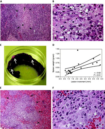
No clinical evidence of cutaneous metastasis was observed. Draining popliteal lymph nodes (n = 10 infected animals) were grey, with swollen and enlarged aspects in comparison to non-infected hamsters. They all presented disrupted architecture (10/10) with macrophages in epithelioid arrangement (Fig. 2A). Schaumann's bodies were often seen dispersed through the organ (9/10) (Fig. 2A). Macrophages exhibited vacuoles (10/10), and some of them showed moderate amounts of Leishmania (6/10), few amastigotes, or even degenerated parasites (4/10) (Fig. 2B).
Macroscopic and histopathological aspects of secondary lymphoid organs from golden hamster infected by Leishmania (Viannia) braziliensis. (A) Pointed arrows indicate macrophages in epithelioid arrangement; arrowheads indicate Schaumann's bodies (lymph node, 20×). (B) Granulomatous reaction; pointed arrows indicate vacuolated macrophages containing amastigotes (lymph node, 100×). (C) Slide of representative picture of spleen from infected animal. White arrows point to some of the several nodules found in infected spleen. (D) Correlation between spleen weight and lesion increment in paws from 20 infected animals after approximately 110 days of infection. The correlation graph shows fit line with confidence curve. r = correlation coefficient; P = significance level. (E) Pointed arrows indicate epithelioid macrophages characterizing granulomatous reaction (spleen, 20×). (F) Pointed arrows indicate Leishmania inside vacuolated macrophage in granuloma (spleen, 100×).

Spleens were severely affected and some animals presented nodules (n = 8) with a consistent aspect upon macroscopic visualization (Fig. 2C). The mean spleen weight was significantly higher (P < 0·001) in infected animals (0·47 g ± 0·32 g; median = 0·35 g; n = 20) than in non-infected ones (0·13 g ± 0·04 g; median = 0·12 g; n = 10). Indeed, a significant positive correlation between the spleen weight and lesion increment was observed in infected hamsters (r = 0·62; P < 0·01; n = 20) (Fig. 2D). The spleen histopathology (n = 14) showed mixed inflammatory infiltrate in all animals analysed, white pulp rarefaction (13/14), presence of granuloma (11/14) (Fig. 2E), vacuolated macrophages (11/14) and presence of Leishmania amastigotes (6/14) (Fig. 2F).
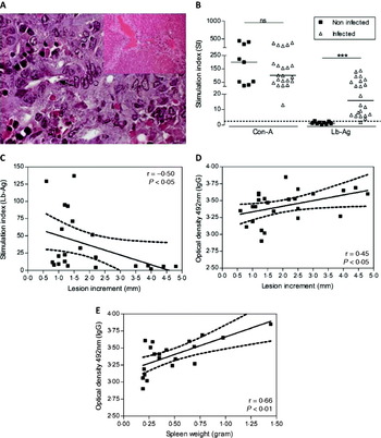
Although the macroscopic aspect of the liver seemed normal, we observed a number of histological abnormalities. The main findings were the presence of mixed inflammatory infiltrate (10/14), composed predominantly of mononuclear cells, mainly around the perivascular area (Fig. 3A). In these cases Leishmania parasites were not seen but vacuolated macrophages with acidophilic contents suggestive of amastigote forms were detected (5/14) (Fig. 3A).
Histopathological aspects of liver and immunological responses to leishmanial antigens of golden hamster infected with Leishmania (Viannia) braziliensis. (A) Mixed inflammatory infiltrate around perivascular area (window, liver, 20×); macrophages in epithelioid arrangement; pointed arrows indicate forms of amastigotes inside vacuolated macrophage (liver, 100×). (B) Lymphocyte proliferative assays using peripheral blood mononuclear cells (PBMC) obtained from uninfected and infected animals. Cells were stimulated in vitro with L. braziliensis antigens (Lb-Ag) or mitogen concanavalin A (Con-A). The results were expressed as a stimulation index (SI). *** P < 0·001. ns, not statistically significant. Each point represents 1 animal and the horizontal bar indicates the median. Dotted line indicates the cut-off SI = 2·5. (C) Correlation between lesion increment and lymphocyte proliferation intensity (stimulation index) under Leishmania antigen (Lb-Ag) stimuli. (D) Association between lesion increment in hamster paws and anti-Leishmania IgG levels (optical density values). (E) Correlation between spleen weight (in grams) and the anti-Leishmania IgG levels (optical density values). The correlation graphs (C, D and E) show fit lines with confidence curves. r = correlation coefficient; P = significance level.

Blood lymphocyte proliferation response (LPR) to leishmanial antigens may relate to the skin lesion severity
A total of 21 out of 22 infected hamsters had a positive LPR. The stimulation index (SI) to Lb-Ag was quite variable and ranged from 4·2 to 137 (mean = 33·8 ± 42·3; median = 15·9; n = 22) (Fig. 3B). SI to Lb-Ag were negatively associated with lesion increments of infected animals at the end of the monitoring period (P < 0·05; r = −0·50) (Fig. 3C). The mean SI to mitogen was similar when infected (147 ± 120; median = 100; n = 22) and non-infected hamsters (198 ± 168; median = 199; n = 9) were compared (Fig. 3B).
Levels of anti-Leishmania IgG correlated with infection severity
Anti-Leishmania IgG was detected in all infected animals. As expected, the OD values were significantly higher in infected (3·41 ± 0·22; median = 3·47; n = 29) than non-infected animals (0·58 ± 0·27; median = 0·5; n = 13; P < 0·001). The anti-Leishmania IgG levels positively correlated with lesion increments in the hamsters' paws (r = 0·45; P < 0·05; n = 25) (Fig. 3D) and with spleen weight (r = 0·66; P < 0·01; n = 20) (Fig. 3E).
DISCUSSION
The fact that the golden hamster presents susceptibility to dermotropic Leishmania (Viannia) species makes this species a better experimental model for ATL studies than the murine model, as the latter is naturally resistant to these strains (Rey et al. Reference Rey, Travi, Valencia and Saravia1990; De Oliveira et al. Reference De Oliveira, Teixeira, Gomes, Barral and Brodskyn2004). However, different Leishmania isolates or species and/or protocols have been used that do not allow prediction of the outcome of infection in this animal model as opposed to other well-established mouse/Leishmania models. Herein, all infected animals were susceptible to L. braziliensis and developed skin-ulcerated lesions. The severity of the infection in this model correlated to spleen weight, the intensity of lymphocyte proliferation to leishmanial antigens, and the titres of anti-Leishmania antibodies.
The Leishmania infection protocol used in this study was able to generate cutaneous lesions in all animals. Thus, it could be recommended for clinical, vaccine or therapeutic studies. The inflammatory signs appeared early in the course of the disease and were already observed within the first 3 weeks after infection. At 60 days post-infection all animals presented signs of disease. Shorter or longer pre-patent periods were observed by others (Wilson et al. Reference Wilson, Dieckmann and Childs1979; Kahl et al. Reference Kahl, Byra and David1990, Reference Kahl, Byram, David, Comerford and Von Lichtenberg1991). In our study, ulcerated lesion was the main macroscopic clinical presentation after 4 months of infection. This aspect, together with the chronic state of the disease, closely resembles non-healing human CL and possibly reproduces some of the immunopathological aspects of the human disease.
Although all animals developed skin lesions, the final measures of infected paws differed among intra- or inter-independent experiments. The variability coefficient analysis tended to show an intermediary variance when animals under the same experimental infection conditions were compared. This result is expected to occur in hamsters because of the outbred genetic background. However, variability is also observed even when isogenic mice are used in experimental leishmaniasis (Pereira et al. Reference Pereira, Silva, de Castilhos, Mastrantonio, Souza, Romão, Rezende, Pena, Beletti and Souza2009). Therefore, variability in lesion increment has to be taken into consideration when lesion size is a parameter to evaluate drug response or vaccine protection.
Besides the skin macroscopic aspects, another striking finding is the similarity of the skin histopathological changes when L. braziliensis-infected hamsters were compared (Laurenti et al. Reference Laurenti, Sotto, Corbett, da Matta and Duarte1990; Kahl et al. Reference Kahl, Byram, David, Comerford and Von Lichtenberg1991; Sinagra et al. Reference Sinagra, Riarte, Luna, Campanini and Segura1997) with human lesions (Magalhães et al. Reference Magalhães, Moraes, Raick, Llanos-Cuentas, Costa, Cuba and Marsden1986). As observed in CL lesions, granulomatous reactions consisting predominantly of epithelioid macrophages, lymphocytes and moderate amounts of plasma cells and scarce amounts of amastigotes were consistently seen in our experimental model. On the other hand, moderately parasitized macrophages were detected in severely clinically compromised animals. A limitation of this study was not to include the parasitic load results. The high bacterial contamination levels (probably from lesion origin) in the axenic cultures did not enable calculation of the frequency of parasites in skin-infected tissues by limited dilution assay. In the future, molecular assays such as real-time PCR to quantify Leishmania DNA products can be used.
Schaumann's bodies were a frequent finding especially in skin and lymph nodes, as observed by other authors (Kahl et al. Reference Kahl, Byra and David1990, Reference Kahl, Byram, David, Comerford and Von Lichtenberg1991; Laurenti et al. Reference Laurenti, Sotto, Corbett, da Matta and Duarte1990). This structure has been associated with a deficient phagocytic macrophage system (Laurenti et al. Reference Laurenti, Sotto, Corbett, da Matta and Duarte1990). Curiously, although Schaumann's bodies are commonly seen in infected hamsters, they are not usually described in humans or in other experimental animals with leishmaniasis (Essayag et al. Reference Essayag, Landaeta, Hartung, Magaldi, Spencer, Suárez, García and Pérez2002). In any case, the presence of Schaumann's bodies even in the absence of parasites strongly suggests Leishmania infection (Ribeiro-Romão et al. unpublished data).
In our experimental protocol, a greater cutaneous lesion increment was accompanied by systemic clinical abnormalities observed in the spleen, lymph nodes, and liver involvement (data not shown). Leishmania visceralization has been reported in hamsters infected by Viannia and Leishmania dermotropic Leishmania species (Rey et al. Reference Rey, Travi, Valencia and Saravia1990; Almeida et al. Reference Almeida, Cuba-Cuba, Moraes and Miles1996; Sinagra et al. Reference Sinagra, Riarte, Luna, Campanini and Segura1997; Soliman, Reference Soliman2006) especially when infective inoculum is high. Although skin metastases were detected in L. braziliensis infected hamters (Brazil, Reference Brazil1976; Wilson and Lollini, Reference Wilson and Lollini1980), the parasite isolate used herein did not induce metastases, agreeing with previous reports (Travi et al. Reference Travi, Rey-Ladino and Saravia1988).
The LPR assay has become an alternative for immunological studies in hamster models because of the low availability of anti-hamster monoclonal antibodies against cytokines or surface molecules. Although spleen and lymph node compartments have been frequently used as the source of mononuclear cells for LPR assays (Osorio et al. Reference Osorio, Melby, Pirmez, Chandrasekar, Guarín and Travi2003; Dea-Ayuela et al. Reference Dea-Ayuela, Rama-Iñiguez, Alunda and Bolás-Fernandez2007), we have chosen blood cells. Our rationale was to rule out the possibility that Leishmania antigens coming from these lymphoid organs may cause in vitro cell stimulation, prejudicing stimulation index calculation. As far as we know, blood cells for analysing the immune response to leishmanial antigens have not been previously used in the hamster model.
The intensity of LPR under leishmanial stimuli inversely correlated with disease severity. Similarly, snout infection by L. panamensis is accompanied by a low lymphoproliferation intensity (Osorio et al. Reference Osorio, Melby, Pirmez, Chandrasekar, Guarín and Travi2003). However, animals with small lesion increments also presented low LPR results whereas CL patients presenting low LPR stimulation indices are at high risk for cutaneous lesions relapses (Mendonça et al. Reference Mendonça, Coutinho, Amendoeira, Marzochi and Pirmez1986). This suggests that any deficiency in the effector cellular immune response could impair the parasite clearance.
The detection of anti-Leishmania antibodies has been used as a promising biomarker to assess the clinical course of leishmaniasis (Gomes-Silva et al. Reference Gomes-Silva, Pereira-Carvalho, Fagundes-Silva, Oliveira-Neto and Da-Cruz2009). Here, we showed an association between IgG anti-Leishmania levels and disease severity, similar to the results previously shown in L. panamensis infection (Osorio et al. Reference Osorio, Melby, Pirmez, Chandrasekar, Guarín and Travi2003). In our study, the anti-Leishmania antibody titres directly correlated with lesion increment and also with spleen weight.
Recently a molecular assay based on quantification of RNA transcripts has been used for the detection of cytokines, chemokines and cell-surface markers in hamster models (Espitia et al. Reference Espitia, Zhao, Saldarriaga, Osorio, Harrison, Cappello, Travi and Melby2010) as an important strategy for evaluating the immune response. This model may also allow associations between clinical outcome and more reliable immunological findings.
Here we showed that the clinical and immunological reactions can vary among independent experiments even when a single isolate of L. braziliensis is used to infect golden hamsters. Also, skin lesion increments, splenomegaly and the proliferative capacity of lymphocytes reactive to Leishmania and IgG anti-Leishmania levels correlated with disease severity. Our results indicate that the golden hamster is an appropriate model for immunopathogenesis studies, and support its use in clinical, vaccine and chemotherapy experimental protocols of cutaneous leishmaniasis caused by L. braziliensis.
ACKNOWLEDGEMENTS
We are grateful to Dr C.O. Mendes-Aguiar, Mr R.S. Nogueira and Dr E.F. Pinto for their help with the experimental procedures and for their intellectual contribution to our study. We are also grateful to Dr M.A. Souza for a critical revision of our manuscript and to Ms R. Pellegrino for secretarial assistance.
FINANCIAL SUPPORT
This work was funded by IOC/FIOCRUZ and by FAPERJ (Jovem Cientista do Nosso Estado E-26/103·111/2008). A.G.-S. and R.P.R.-R. are PhD students sponsored by CNPq and CAPES, respectively. A.M.D.-C. is a CNPq and FAPERJ fellowship researcher.