Patent ductus arteriosus is one of the most common congenital cardiac problems in children, and recently closure of patent ductus arteriosus with coil or amplatzer devices, has become an accepted alternative to the surgical ligation.Reference Moore, George and Kirkpatrick1–Reference Santoro, Gaio, Carrozza, Palladino, Russo and Calabro5 In addition, the precise measurement of patent ductus arteriosus by a non-invasive method has now become clinically more important than in the past, when surgical ligation was the only corrective procedure available for ductal closure.Reference Wong, Shim, Khoury and Meyer6, Reference Nakajima, Ogawa, Sano, Matsushita, Kayatani and Yabbuchi7
Several studies have revealed the risk factors for the failure in patent ductus arteriosus closure by coil.Reference Turner, Forbes, Epstein and Wincent8–Reference Hijazi and Geggle11 Basically, the occlusion of the patent ductus arteriosus has been performed on the basis of data obtained by catheterisation, but echocardiography is the most commonly used diagnostic tool for the assessment of the characteristics of patent ductus arteriosus and its size, either before medical or surgical intervention is carried out. The non-invasive estimation of the dimensions of patent ductus arteriosus is desirable to avoid unsuccessful catheter intervention.
The combination of two-dimensional echocardiography and colour Doppler echocardiography in children can establish the presence of a patent ductus arteriosus with a high degree of reliability – sensitivity 96% and specificity 100% – and even evaluate its morphology in many cases.Reference Wong, Shim, Khoury and Meyer6, Reference Shcneider, Hildebrandt, Schweigl and Wehner12, Reference Azhar, Abd El-Azim and Habib13 Estimation of patent ductus arteriosus size by two-dimensional echocardiography is usually difficult because of its small size and difficulty in the detection of its borders.Reference Azhar, Abd El-Azim and Habib13 In this study, the minimum and maximum size of patent ductus arteriosus were estimated by colour Doppler echocardiography and were compared with the data obtained by angiography.
Materials and methods
Patients
From January, 2008 to January, 2009, the aortic and pulmonary diameters of the patent ductus arteriosus and its length were measured in 40 children referred to the affiliated hospitals of Shiraz University of Medical Sciences, Shiraz, Iran (Nemazee and Faghihi Hospitals) consecutively. Children with other associated congenital cardiac anomalies were excluded from the study.
Study protocol
To minimise the errors, colour Doppler echocardiography examinations were performed on all the patients by one paediatric cardiologist. Before catheterisation, each patient underwent a complete echocardiographic study with a General Electric-Vivid 3 echocardiographic machine (General Electic-Vivid 3 Vingmed, Horten, Norway), using a 3-megahertz probe, in parasternal short-axis and suprasternal views. The sector angle was between 45 and 60 degrees to permit high-quality frames. The patent ductus arteriosus flow was imaged using colour Doppler echocardiography and the measurements were made at the narrowest and widest parts of the turbulent colour stream flow; gain was set at 50, the baseline was set at zero, the velocity scale was 4000 hertz, the frequency was 3 megahertz, and low velocity rejection was 3 centimetres per second in all the measurements performed three times in each view and the mean value was used for comparison.
Angiographic estimations of the minimum and maximum diameters of patent ductus arteriosus were obtained with an ascending aortogram, using 1 millilitre per kilogram non-ionic contrast through an appropriate pigtail catheter in the straight lateral camera position. The angiographic measurements were made off-line, calibrated to the catheter size (General Electric Advantex, Milwaukee, Wisconsin, United States of America). Catheterisation was performed under general anaesthesia and post-device closure aortography was performed for the assessment of the residual shunt. The children were discharged on the following morning after the procedure.
Statistical analysis
Data are presented as range of values, median or mean values, and standard deviation for the purpose of comparison with other studies. The degree of interrelation between echocardiographic and angiographic measurements was calculated using the Pearson correlation, visualised as a linear regression. The difference between the methods was also calculated and checked for significance by using a two-tailed paired Student’s t-test. Graphical visualisation of the differences was performed in Bland–Altman diagrams.Reference Bland and Altman14 A p-value of less than 0.05 was considered as statistically significant. SPSS version 16 statistical software was used for all the statistical analyses.
Results
Forty children were included in the study. The median age was 1 year with a range of 4 months and 18 years. The mean weight was 9.58 plus or minus 5.36 kilograms and sex distribution was 16 males and 24 females.
Visualisation of the patent ductus arteriosus was achieved from short-axis and suprasternal-axis views in all children with colour Doppler echocardiography. The minimal diameter of patent ductus arteriosus was 2.78 plus or minus 0.62 millimetre with a range from 1 to 4 millimetres in echocardiography and was 2.23 plus or minus 0.72 millimetre with a range from 1.3 to 5 millimetres – in angiography, which were significantly different (p less than 0.0001). In addition, there was a significant difference between the maximal diameter of the patent ductus arteriosus in echocardiography and angiography; 5.96 plus or minus 1.53 millimetres with a range from 3 to 9 millimetres and 7.02 plus or minus 1.71 millimetres with a range from 3.6 to 11 millimetres, respectively (p equal to 0.0027). The patent ductus arteriosus gradient was 64.80 plus or minus 11.98 millimetres of mercury with a range from 50 to 84 millimetres of mercury.
The ratio of the diameter of the pulmonary end of patent ductus arteriosus in echocardiography to angiography was 1.33 plus or minus 0.37 and the aortic end was 0.92 plus or minus 0.32. The mean difference in the pulmonary and aortic end of patent ductus arteriosus was 0.63 plus or minus 0.59 and −0.90 plus or minus 1.63, respectively. The measured pulmonary side diameter in echocardiography was 33.6% plus or minus 37.62% larger than angiography and the aortic side was 7.8% plus or minus 32.21% smaller. Considering the angiographic data as the standard, nine (5.4%) children had colour Doppler studies that overestimated the minimum diameter of the patent ductus arteriosus by 1.0 millimetre.
Linear regression showed the following equations related to the minimum and maximum size of the patent ductus arteriosus in echocardiography and angiography:

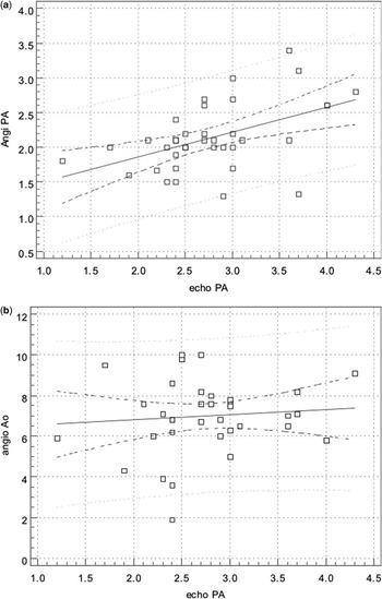
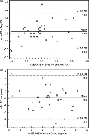
Figures 1 and 2 show the linear regression curves and the Bland–Altman diagrams that compare the aortic and pulmonary side diameters of the patent ductus arteriosus in angiography and echocardiography.
Regression diagrams (a and b) for comparison between echocardiographic and angiographic diameter of aortic and pulmonary side of patent ductus arteriosus. The solid line characterises the linear regression line; the two inner dashed lines visualise the 95% confidence interval. PA = diameter of pulmonary side of patent ductus arteriosus in echocardiography; echo AO = diameter of aortic side of patent ductus arteriosus in echocardiography; Angio PA = diameter of the pulmonary side of patent ductus arteriosus in angiography; Angio Ao = diameter of aortic side of patent ductus arteriosus in angiography.

Bland–Altman diagrams (a and b) for comparison between difference of echocardiographic and angiographic diameter of aortic and pulmonary sides of patent ductus arteriosus. The dotted horizontal line indicates agreement with a difference of 0.9 and 0.64 millimetres; the dotted lines indicate a clinically relevant difference of plus or minus 2.3 and 1.81 millimetres, respectively. PA = diameter of the pulmonary side of patent ductus arteriosus in echocardiography; echo AO = diameter of the aortic side of patent ductus arteriosus in echocardiography; Angio PA = diameter of the pulmonary side of patent ductus arteriosus in angiography; Angio Ao = diameter of the aortic side of patent ductus arteriosus in angiography.
There was not any statistically significant difference between the measurements at the suprasternal view and parasternal view for the estimation of the patent ductus arteriosus’ size (p more than 0.05).
Discussion
This study reveals that colour Doppler echocardiography overestimates the diameter of the ductus arteriosus and has a statistical correlation with angiographic data. All patients had a patent ductus arteriosus with a left to right shunt; this selection may have increased the results of visualisation in echocardiography and the accuracy of the results.Reference Shcneider, Hildebrandt, Schweigl and Wehner12 Exclusion of congenital defects that influence patent ductus arteriosus morphology, for example, pulmonary stenosis, also interferes with accurate measurement in echocardiography, because altered morphology makes the visualisation of the patent ductus arteriosus difficult. The patients were not selected based on the criteria of “weight” or “clinical signs”. Patients with a small patent ductus arteriosus and those with a large patent ductus arteriosus were included in this study.
Data on the echocardiographic visualisation of a patent ductus arteriosus and its measuring accuracy are very diverse.Reference Wong, Shim, Khoury and Meyer6, Reference Shcneider, Hildebrandt, Schweigl and Wehner12, Reference Sahn and Allen15 Transthoracic colour Doppler echocardiography can almost always visualise a patent ductus arteriosus in humans. No significant correlation with angiographic measurement has been proven in any study to date.Reference Wong, Shim, Khoury and Meyer6, Reference Sahn and Allen15 However, it has been reported that two-dimensional echocardiography can obtain a comprehensive assessment of a patent ductus arteriosus and some measurements can be used to assess the suitability for device closure before angiography.Reference Ramaciotti, Lemler, Moake and Zellers16
There are some possible explanations for the difference between the results of the studies. First, we used fixed preset colour Doppler echocardiography values in all measurements, but previous studies changed the colour baseline to lower values.Reference Wong, Shim, Khoury and Meyer6, Reference Sahn and Allen15 Second, because of the limited lateral resolution of echocardiography and the relatively small diameter of the ductus in some of the studies, it is difficult to correctly visualise and measure such a small-sized diameter. Third, a low range of measured values automatically results in a poor correlation between the two measurement methods.Reference Wong, Shim, Khoury and Meyer6, Reference Azhar, Abd El-Azim and Habib13, Reference Hiraishi, Horiguchi and Fujino17 The distance between the two examinations in terms of time may also be a reason for the differing results. Accordingly, an interval of up to 4 months was chosen between the two examinations in another study.Reference Wong, Shim, Khoury and Meyer6 During this period of time, the minimum patent ductus arteriosus diameter can increase as a result of dilatation or can decrease as a result of partial constriction. There was also a difference in the ultrasonographic technology used.Reference Azhar, Abd El-Azim and Habib13 Colour Doppler echocardiography overestimates the true diameter of a patent ductus arteriosus, because the colour field extends beyond the anatomical structures – “bleeding of the colour flow”.Reference Wong, Shim, Khoury and Meyer6 This effect is particularly strong when colour amplification is high and the Nyquist limit is low.
The true minimal diameter is particularly overestimated at low brightness settings because of the difficulty of endocardiac delimitation.Reference Sahn and Allen15 However, the investigators postulate that more modern ultrasonic devices with better lateral resolution can minimise this problem.Reference Sahn and Allen15 A further explanation may be that the minimal patent ductus arteriosus diameter cannot be visualised with exactly the same correct lateral projection in echocardiography as in angiography.Reference Wong, Shim, Khoury and Meyer6, Reference Azhar, Abd El-Azim and Habib13, Reference Ramaciotti, Lemler, Moake and Zellers16
A similar overestimation with colour Doppler echocardiography has also been reported in humans – greater than1 millimetre in 33% and greater than 2 millimetres in 19% of the patients.Reference Wong, Shim, Khoury and Meyer6 One human study only succeeded in visualising the patent ductus arteriosus by means of two-dimensional echocardiography in 30% of the patients and was not able to prove a significant correlation with angiography.Reference Wong, Shim, Khoury and Meyer6 Two other studies measured the patent ductus arteriosus in 100% of the patients with two-dimensional echocardiography; this value correlated significantly (r = 50.9711 and 50.9410) with angiographic data, and there was a slight overestimation when the patent ductus arteriosus was somewhat narrower. The data of this study for the visualisation of the patent ductus arteriosus in two-dimensional echocardiography and colour Doppler echocardiography with left parasternal views (100%) were comparable to those of the last two studies mentioned above.Reference Sahn and Allen15, Reference Ramaciotti, Lemler, Moake and Zellers16
The use of high-quality ultrasonic devices and transducers combined with examiner’s experience can increase the accuracy of echocardiographic patent ductus arteriosus size estimation. According to other studies, there is a significant correlation between the size of patent ductus arteriosus in two-dimensional echocardiography and angiography, but the small diameter of the patent ductus arteriosus makes defining of the borders difficult.Reference Wong, Shim, Khoury and Meyer6, Reference Nakajima, Ogawa, Sano, Matsushita, Kayatani and Yabbuchi7, Reference Ramaciotti, Lemler, Moake and Zellers16 This study suggests that colour Doppler echocardiography can always detect the patent ductus arteriosus easily, but it overestimates the diameters of the patent ductus arteriosus and there is a relatively fine correlation between the diameters of the patent ductus arteriosus in colour Doppler echocardiography and angiography.
Conclusion
Considering the present findings, we may conclude that it is almost always possible to measure the dimensions of the patent ductus arteriosus with colour Doppler echocardiography. However, echocardiographic measurement overestimates the angiographic measurement of the pulmonary side of the patent ductus arteriosus and underestimates its aortic side, which should be considered for the measurement of the size of the patent ductus arteriosus.
Acknowledgements
The authors thank Mrs F. Shokrpour at the Center for Development of Clinical Research of Nemazee Hospital. The authors also thank Professor G. H. Amirhakimi and H. Khajehei for their help with copy editing.