INTRODUCTION
Although Brazil has one of the highest biodiversities in the world, the knowledge about its benthic invertebrates is still unsatisfactory (Couto et al., Reference Couto, Silveira and Rocha2003; Amaral & Jablonski, Reference Amaral and Jablonski2005). In recent decades some projects have been developed to document the Brazilian biodiversity (e.g. REVIZEE, BIOTA/FAPESP, and Rede de Monitoramento Ambiental Marinho–PETROBRAS); still, the vast coastline (± 8000 km) and low number of experts in this field hinder the progress of this work.
Presently, studies on the taxonomy of echinoderms in Brazil have focused on specific classes, especially Ophiuroidea (Borges & Amaral, Reference Borges and Amaral2007; Gondim et al., Reference Gondim, Lacouth, Alonso and Manso2008; Lima et al., Reference Lima, Correia, Sovierzoski and Manso2011; Manso et al., Reference Manso, Oliveira and Ximenez2011). Holothuroidea, however, has been neglected and appears in less than 5% of the literature, most of which resulted from great expeditions (Théel, Reference Theél1886; Deichmann, Reference Deichmann1930; Tommasi, Reference Tommasi1969). To date, this class represents less than 15% of the Brazilian echinoderm fauna (Tommasi, Reference Tommasi1999), probably mainly due to the few number of experts in holothuroids in the country. For instance, the last new holothuroid species was described by Freire & Grohmann (Reference Freire and Grohmann1989) (Leptosynapta brasiliensis) and few contributions have been made since then (e.g. Tiago & Ditadi, Reference Tiago, Ditadi and Baker2001; Moura et al., Reference Moura, Campos, Curbelo-Fernandez, Cavalcanti, Harris, Boettger, Walker and Lesser2010). Furthermore, most of the genera need revision, especially with the use of new techniques (e.g. scanning electron microscopy images and/or DNA sequencing), which can help overcome the difficulties imposed by the high variability resulting from the development of the specimens and their ossicles.
In this paper, we extend the distribution range of Thyone pawsoni Tommasi, Reference Tommasi1972 and Holothuria (Theelothuria) princeps Selenka, Reference Selenka1867 to shallow waters of the north-east coast of Brazil—the first occurrence of these species in the South Atlantic Ocean.
MATERIALS AND METHODS
Specimens of H. (T.) princeps and T. pawsoni were collected using a Van Veen grab, in 2004 and 2010, respectively. They were fixed in 4% formalin, preserved in 70% ethanol, and deposited in the Museu de Zoologia at the Universidade Federal da Bahia (UFBA).
The methods used to study the specimens followed those of Rowe & Doty (Reference Rowe and Doty1977) and Samyn et al. (Reference Samyn, VandenSpiegel and Massin2006). The ossicles were removed from different tissues (introvert of T. pawsoni, tentacles, body wall, tube feet and anus) using household bleach, washed in five changes of distilled water and then in five changes of absolute ethanol. A number of samples were mounted on slides with Entellan Merck® for permanent storage, and their ossicles were then examined and measured using the software Image-Pro Express (v. 6.0) linked to an Olympus CX31-RTSF optical microscope. Other samples were dried and mounted on metal stubs with double-sided tape, coated with gold and observed with a JEOL JSM-6390LV scanning electron microscope. Photographs of specimens were taken using a Sony DSC W300 digital camera.
Abbreviations used: MCZ, Museum of Comparative Zoology at Harvard University; UFBA, Universidade Federal da Bahia; USNM, National Museum of Natural History, Washington, DC.
RESULTS
SYSTEMATICS
Order ASPIDOCHIROTIDA Grube, Reference Grube1840
Family HOLOTHURIIDAE Ludwig, Reference Ludwig1894
Genus Holothuria Linnaeus, Reference Linnaeus1767
Holothuria (Theelothuria) princeps Selenka, Reference Selenka1867
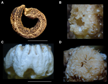
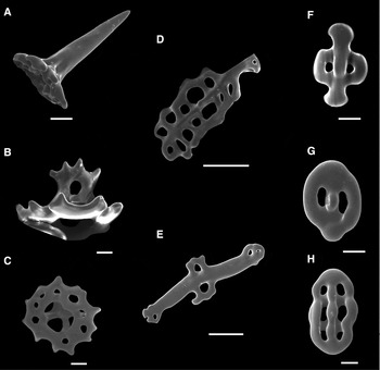
(Figures 1 & 2)
Holothuria (Theelothuria) princeps Selenka, Reference Selenka1867 (UFBA–645): (A) whole animal; (B) detail of peltate tentacles; (C) calcareous ring; (D) detail of anal papillae. Scale bars: A, 10 cm; B, 1 cm; C, 10 mm; D, 1 cm.

Holothuria (Theelothuria) princeps Selenka, Reference Selenka1867 (UFBA–645): scanning electron microscopy images (A) tack-like table; (B) table in lateral view; (C) disc of table; (D, E) supporting rods; (F, G, H) buttons at different stages of development. Scale bars: A, 50 µm; B, C, 10 µm; D, E, 50 µm; F–H, 10 µm.

Holothuria princeps Selenka, Reference Selenka1867: 332, pl. 18, figures 67–69.
Holothuria (Holothuria) princeps—Panning, Reference Panning1935: 101, figure 94.
Holothuria (Theelothuria) princeps—Rowe, Reference Rowe1969: 157, figure 19; Hendler et al., Reference Hendler, Miller, Pawson and Kier1995: 296, figures 167, 185G–J; Cutress, Reference Cutress1996: 74, figures 17–23A; Pawson et al., 2010: 39, figure 32.
MATERIAL EXAMINED
Holothuria (Theelothuria) princeps—Camamu, Barra Grande, BA, Brazil (13°56′S 38°59′W), 1 m, 15 July 2004, 2 specimens 25 and 28 cm long (UFBA–645).
DIAGNOSIS
Large form, up to 30 cm. Body wall thick, ambulacral feet numerous and scattered throughout the body. Dorsally they form conical dark papillae. 20 small paltate tentacles. Radial and interradial pieces of calcareous ring are fused together; radials with short and forked posterior processes. Cuverian organs absent; cloaca very long and wide. Body wall ossicles are small and delicate buttons and tables. Tables formed by a small disc with dentate margin possessing 12 prolongations, and a spire ending in 6–8 teeth. Tube feet ossicles are large supporting rods with central and peripheral perforations, and large and conical tables (tack-like). Colour in ethanol is dark brown.
DISTRIBUTION
North Carolina to Brazil (up to Bahia State); depth 0–73 m (Pawson et al., 2010; present paper).
BIOLOGICAL NOTES
The specimens were collected partially burrowed in sandy mud sediment from the intertidal zone. According to Wells & Wells (Reference Wells and Wells1961), the commensal crab Pinnaxodes floridensis Wells & Wells, Reference Wells and Wells1961 may be found in the cloaca and respiratory tree of H. (T.) princeps. No commensals were found in the specimens recorded herein.
SYNTYPE
At the MCZ Catologue No. 685.
TYPE LOCALITY
Florida.
REMARKS
Coloration of preserved specimens is a mosaic of brown and yellow, with some black spots mainly ventrally. Anus surrounded by long papillae (±20). Ossicles are small tables (±50 µm) with a low spire ending in 6–8 teeth; large tack-like tables with undulated margin and four pillars, which unite to form a long, conical and thick spire with one central and large hole (±150 µm long); irregular knobbed buttons (±60 µm) and rods (±200 µm).
Holothuria (T.) princeps is also the first record of the subgenus Theelothuria and the sixth record of the genus Holothuria in the south-western Atlantic Ocean. It differs from Holothuria (Cystipus) pseudofossor Deichmann, Reference Deichmann1930, Holothuria (Halodeima) grisea Selenka, Reference Selenka1867, Holothuria (Semperothuria) surinamensis Ludwig, Reference Ludwig1875, Holothuria (Thymiosycia) arenicola Semper, Reference Semper1868 and Holothuria (Vaneyothuria) lentiginosa Marenzeller von, Reference Marenzeller1892 by the presence of tack-like tables and knobbed buttons.
SYSTEMATICS
Order DENDROCHIROTIDA Grube, Reference Grube1840
Family PHYLLOPHORIDAE Östergren, Reference Östergren1907
Genus Thyone Oken, Reference Oken1815
Thyone pawsoni Tommasi, Reference Tommasi1972
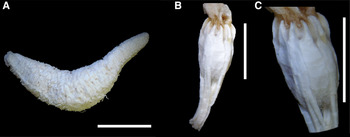
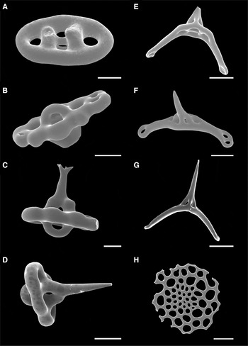
(Figures 3 & 4)
Thyone pawsoni Tommasi, Reference Tommasi1972: 19, figures 12–15.
Thyone pawsoni Tommasi, Reference Tommasi1972 (UFBA–1485): (A) whole animal; (B) calcareous ring; (C) detail of radial and interradial plates of calcareous ring without its projections. Scale bars: A, 2 mm; B, C, 10 mm.

Thyone pawsoni Tommasi, Reference Tommasi1972 (UFBA–1485): scanning electron microscopy images (A, B, C, D) tables from body wall at different stages of development; (E, F, G) supporting tables; (H) end plate from tube feet. Scale bar: A–H, 20 µm.

Thyone pawsoni—Pawson & Miller, Reference Pawson and Miller1981: 397, figures 2D–E; Miller & Pawson, Reference Miller and Pawson1984: 42, figures 34–35; Pawson et al., Reference Pawson, Pawson and King2010: 30, figure 22.
MATERIAL EXAMINED
Thyone pawsoni—Guarajuba, Camaçari, BA, Brazil (12°45′–12°48′S 38°05′–38°08′W): 26 m, 20 July 2005, 2 specimens 1.5 and 2.0 cm long (UFBA–1485); 31 m, 20 August 2009, 2 specimens 2.0 and 2.5 cm long (UFBA–1482); 31 m, 20 February 2010, 2 specimens 2.5 and 3 cm long (UFBA–1481); 31 m, 10 July 2010, 1 specimen 3.5 cm long (UFBA–1484). Off Georgia, United States (31°19′N 81°13′W), 8 m, 4 February 1980, 1 specimen 2 cm long (USNM E 19512).
DIAGNOSIS
Body cylindrical, but tapering abruptly posteriorly to form a short tail. Tube feet scattered throughout the body. 10 dendritic tentacles, ventral pair smaller. Calcareous ring with long and divided forked processes. Ossicles are tables and supporting tables. Tables with an elliptical disc with four perforations, and an elongated spire ending in one tooth. Supporting tables with a narrow and curved base with four central perforations and one in each extremity; and a spire with two pillars ending in three teeth.
DISTRIBUTION
South Carolina to the Gulf of Venezuela and Brazil (up to Bahia State); depth: 6–51 m (Pawson et al., Reference Pawson, Pawson and King2010; present paper).
BIOLOGICAL NOTES
Burrows in silt-covered quartz sand and crushed shell sediment (Pawson et al., Reference Pawson, Pawson and King2010). Present specimens were found burrowed in gravel and sand.
HOLOTYPE
Lost. The shallow water echinoderm collection from Professor L.R. Tommasi was donated to the Museum of Zoology of the University of São Paulo. This collection, however, stayed many years without proper care until it could be moved, and much material (and collecting information) was lost.
TYPE LOCALITY
Gulf of Venezuela.
REMARKS
The specimens reported herein have two types of body wall tables: one bears a high tapering spire and the other ends in three teeth (and one of these may be subdivided). Although this is in accordance with Miller & Pawson (Reference Miller and Pawson1984), Tommasi (Reference Tommasi1972) mentioned only the presence of tables with one tooth. This type may represent the high tapering spire table or it could be an error of observation, since in the figures presented by Tommasi (Reference Tommasi1972) the table has more than one tooth. Furthermore, the development of teeth at the tip of tables does not seem to be related to the size of the table since small tables with teeth and large ones without them were observed. As noted by Pawson et al. (Reference Pawson, Pawson and King2010), body wall tables with more than four perforations were found, in smaller amounts.
Although Tommasi (Reference Tommasi1972), Miller & Pawson (Reference Miller and Pawson1984) and Pawson et al. (Reference Pawson, Pawson and King2010) mentioned the presence of three teeth on the tip of the supporting tables, the specimens reported herein (including the specimens from Georgia) have supporting tables ending mainly in a tapering spire with no tooth, regardless of their size. Supporting tables ending in three teeth were rarely found, although present. This variation, however, may not be of generic importance. Unfortunately, the holotype is missing and the figures presented by Tommasi (Reference Tommasi1972) are not in good quality for comparison.
Regarding the calcareous ring, the radials and interradials of different specimens are broken in pieces of different sizes and at different places; in some specimens the radial is almost compact, recalling a Sclerodactylinae. The formation and evolution of the calcareous ring deserves further studies, especially because it is an important structure used to distinguish phyllophorids from sclerodactylids.
Besides T. pawsoni, two other Thyone species occur on the Brazilian Coast: Thyone pseudofusus Deichmann, Reference Deichmann1930 and Thyone montoucheti Tommasi, Reference Tommasi1970. However, T. montoucheti may have been misclassified since its body wall tables have four pillars (Tommasi, Reference Tommasi1970), while the tables of Thyone species have only two. Moreover, the calcareous ring of this species does not have posterior prolongations, which, accordingly, would classify T. montoucheti as a cucumariid, instead. Considering the cucumariids, however, T. montoucheti cannot simply be assigned to any of its subfamilies because of the following characteristics: ten tentacles, ventral-most two are reduced (versus 15–25 in Thyonidiinae) and presence of table and supporting tables in the body wall (versus absence in Cucumariinae and Colochirinae). In addition, the presence of tube feet scattered throughout the body and of four Polian vesicles would also exclude many genera. The holotype of T. montoucheti is also missing and, therefore, cannot be analysed to verify possible inconsistencies in the original description.
ACKNOWLEDGEMENTS
We are grateful to Gustav Paulay and Francisco A. Solis Marin for providing constructive suggestions that significantly improved the quality of this manuscript; David Pawson (Smithsonian Institution, Washington, DC) for confirming the identification of Thyone pawsoni and providing a specimen on loan; Mariano Martinez (Museo Argentino de Ciencias Naturales–Bernardino Rivadavia) for providing useful information; Marlene Peso (UFBA) for donating the specimens to MZUFBA; and the Instituto Gonçalo Moniz (FIOCRUZ/BA) for scanning electron microscopy images. L.M. received financial support from CAPES, C.S. from FAPESB, and both received financial support from PROAP-PPG Diversidade Animal.