Patient and method
A 19-year-old male with 22q11 deletion syndrome, pulmonary atresia, ventricular septal defect, and multiple aortopulmonary collaterals had a right-sided modified Blalock–Taussig shunt at 15 months, followed by a Rastelli operation using a 19-mm pulmonary homograft (right ventricle to pulmonary artery) conduit and unifocalisation of aortopulmonary collaterals aged 3.5 years. Balloon dilation of the right pulmonary artery and embolisation of an aortopulmonary collateral were performed aged 11 years. He developed exercise intolerance from severe pulmonary regurgitation with right ventricular dilatation. MRI demonstrated free pulmonary regurgitation, indexed right ventricular end-diastolic volume of 177 ml/m2, and a proximal right ventricular outflow tract aneurysm on the anterior surface measuring 5 × 4 cm. The right ventricular outflow tract and pulmonary annular dimensions (25 × 31 mm) excluded the use of currently available percutaneous valves.
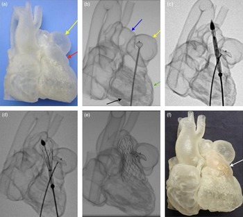
A 3D model (Fig 1a) was printed from the gated MRI images and a simulated intervention performed using a Venus P-valve (Venus MedTech, Shanghai, China) in the right ventricular outflow tract, and an Amplatzer ventricular septal defect device (Abbott Medical Inc., Minnesota, USA) to close the right ventricular outflow tract aneurysm (Fig 1b–e & Online Video 1). This confirmed that a transcatheter solution was possible (Fig 1f).
(a) A 3D model of the heart in right anterior oblique (RAO) view showing large right ventricular outflow tract aneurysm (yellow arrow) on the right ventricular outflow tract (red arrow). (b) Simulated procedure (in RAO view): the Amplatzer device in delivery catheter being advanced to the right ventricular outflow tract aneurysm. Device partially deployed forming an “onion”. Arrows: blue: main pulmonary artery; yellow: right ventricular outflow tract aneurysm; green: right ventricular body; black: left ventricular body overlapping the right ventricle. (c) Venus P-valve assembly across the right ventricular outflow tract with tip at pulmonary artery. Note: Amplatzer device fully deployed but not released. (d): Venus P-valve partially deployed. (e) Venus P-valve fully deployed; valve assembly removed and Amplatzer device released. (f) Following simulation: 3D model with Venus P-valve in situ (white arrow).

Permission was obtained from the Medicines and Healthcare products Regulatory Agency to implant an unlicensed Venus P-valve.
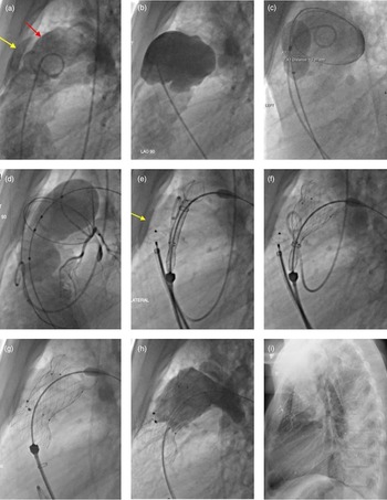
Angiography was performed in the right ventricular outflow tract (Fig 2a) and aneurysm (Fig 2b) to confirm the anatomy. The mouth of the aneurysm measured 15 mm with a 18 mm Amplatzer sizing balloon over a 0.035'' standard Amplatz wire (Abbott Medical Inc.) (Fig 2c). Coronary angiography was performed during balloon sizing of the right ventricular outflow tract (Fig 2d). A 30 × 30 mm Venus P-valve mounted on its delivery system was advanced through a 26Fr femoral venous access to the left pulmonary artery origin over a 0.035'' Lunderquist wire (Cook Medical, Bloomington, Indiana, USA) in the distal left pulmonary artery.Reference Promphan, Prachasilchai, Siripornpitak, Qureshi and Layangool 1 , Reference Garay, Pan, Zhang, Wang and Springmuller 2 A 16-mm Amplatzer muscular ventricular septal defect occluder (Abbott Medical Inc.) through a 9Fr Amplatzer 45° TorqVue sheath (Abbott Medical Inc.) was deployed in the mouth of the aneurysm but not released, while keeping the valve delivery system in place (Fig 2e). The Venus P-valve was deployed under fluoroscopic/angiographic guidance. Immediately prior to the final deployment of the valve, the ventricular septal defect device was released, followed by a release of the valve (Fig 2f–g). Pulmonary angiography confirmed no pulmonary regurgitation, and right ventricular angiography confirmed minimal through device flow into the aneurysm (Fig 2h).
Procedure shown in left lateral projection. (a) Right ventricular outflow tract (red arrow); adjacent right ventricular outflow tract aneurysm (yellow arrow); (b) right ventricular outflow tract aneurysm angiogram; (c) balloon sizing across the mouth of right ventricular outflow tract aneurysm over coiled guidewire; (d) balloon sizing across the right ventricular outflow tract with simultaneous angiogram and left main coronary angiogram; (e) valve assembly of the right ventricular outflow tract – with partially deployed distal flare. Amplatzer device deployed (yellow arrow) in the mouth of aneurysm. (f) Deployment of middle portion of the valve. Note: Amplatzer device still not released. (g) Complete deployment of the valve after the release of Amplatzer device. (h) Pulmonary angiogram demonstrating well-positioned and well-functioning Venus P-valve with Amplatzer device in position across the right ventricular outflow tract aneurysm. (i) Lateral chest X-ray at discharge showing Venus P-valve and device in position.

Fresh blood appeared in the endotracheal tube towards the end of the procedure due to the perforation of a distal branch by the guidewire. This was sealed with a 6- and a 9-mm Amplatzer Vascular Plug II (Abbott Medical Inc.). There was no further haemoptysis, and the patient was discharged 3 days later on dual anti-platelet therapy.
The patient reported improved effort tolerance 4 weeks after the procedure. Echocardiography confirmed good right ventricular function and excellent function of the Venus P-valve with a Doppler velocity of 2.3 m/s and no pulmonary regurgitation. The ventricular septal defect device was in place with no leak into the right ventricular outflow tract aneurysm. An MRI scan at 6 months confirmed good valve function, layers of thrombus in the aneurysm, and a tiny residual flow into the mouth of the aneurysm.
Discussion
Since the first reported percutaneous pulmonary valve implantationReference Bonhoeffer, Boudjemline and Saliba 3 two decades ago, there are currently only two commercially available percutaneous valves. The Melody valve (Medtronic Inc, MN, USA) is suitable for outflow tract dimensions of up to 22 mm; and the Sapien Valve (Edwards Lifesciences Corp, CA, USA) for outflow tracts of up to 29 mm. The recent introduction of the Venus P-Valve, currently undergoing a CE mark study, has further pushed the boundaries of percutaneously treating large outflow tracts of up to 32–33 mm diameter, with initial reports showing good results.Reference Promphan, Prachasilchai, Siripornpitak, Qureshi and Layangool 1 , Reference Garay, Pan, Zhang, Wang and Springmuller 2 A new Harmony transcatheter pulmonary valve (Medtronic Inc) is also undergoing clinical evaluation in the United States of America; however, this is still designed for relatively smaller outflow tractsReference Schoonbeek, Takebayashi and Aoki 4 and is currently only available in one size.
There is a single previous report of percutaneous device closure of an outflow tract aneurysm followed by implanting a balloon-expandable percutaneous valve in a small outflow tract.Reference Lurz, Taylor and Bonhoeffer 5 We now report simultaneous right ventricular outflow tract aneurysm closure aided by the implantation of a large self-expanding percutaneous pulmonary valve. We were not confident that implanting a percutaneous valve alone would also seal the aneurysm. Once the valve was deployed, approaching the aneurysm would be very difficult if there was a residual entry point because of its position. Therefore, we chose to close the aneurysm with a separate device during valve implantation.
Although there are previous reports of using 3D printed models in determining the suitability of percutaneous pulmonary valve implantation, we used a 3D model to simulate our complex procedure. While the 3D model material has a different compliance to biological tissue and ultimate positioning of the devices cannot be completely predicted, a simulation on the model encouraged us to proceed, knowing that the aneurysm device would be held firmly in place by the valve. The traditional approach to these complex lesions would have been open heart surgery; however, simulation allowed us to plan an effective procedure avoiding surgery.
Conclusion
This procedure demonstrated how 3D simulation can be a helpful adjunct in planning and performing complex percutaneous procedures.
Author ORCIDs
Salim G. M. Jivanji 0000-0003-1160-7265
Supplementary Material
To view supplementary material for this article, please visit https://doi.org/10.1017/S1047951119000106
Acknowledgement
The authors would like to thank Dr Marie Nieves Velasco Forte, Mr Nicholas Byrne, and Dr Israel Valverde for their inputs in the case.
Financial Support
This research received no specific grant from any funding agency, commercial, or not-for-profit sectors.
Conflict of Interest
None.