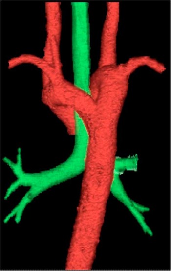
The following report describes the detection of an asymptomatic double aortic arch within a patient with Tetralogy of Fallot within a tertiary referral centre.
Case report
An 8-year-old North Indian girl presented with cyanosis, exertional dyspnoea, and palpitation only. Following clinical examination of the child – 122 centimetres, 20 kilograms, 96 beats per minute, pulse oximeter oxygen saturation 73% on air – a harsh 3/6 ejection systolic murmur was detected in the third right intercostal space, with an electrocardiogram reporting right-axis deviation and early R-wave transition. The X-ray displayed a typical coeur en sabot heart, pulmonary bay accentuation, and oligaemic lung fields.
Investigation
The echocardiography confirmed the diagnosis of Tetralogy of Fallot consisting of a 12.2-millimetre bidirectional perimembranous ventricular septal defect, infundibular pulmonary artery stenosis, with a gradient of 84 millimetres of mercury, and both right atrial and ventricular dilation with a McGoon ratio of 1.3. Computed tomography was requested to further analyse the pulmonary artery stenosis; however, a coincidental finding of a double aortic arch was detected following the echocardiography, which confirmed Tetralogy of Fallot. The vascular ring presented asymptomatically and was undetected through echocardiographic investigation. Both aortic arches displayed relatively equal patency. No cytogenetic analysis was conducted.
Intervention
The patient underwent a double-stage repair through a left thoracotomy for the ligation of the posterior aortic arch, followed by a modified Blalock–Taussig shunt through the insertion of a 5-millimetre polytetrafluoroethylene tube, anastamosing the left subclavian artery to the left pulmonary artery. Postoperatively, the patient was haemodynamically stable and recovery was unremarkable.
Three dimensional computed tomography image of double aortic arch. Original source from Narayana Hrudayalaya Department of Radiology.

Discussion
Asymptomatic double aortic arches combined with Tetralogy of Fallot in an 8-year-old are incredibly rare. Aortic arch anomalies comprise 1 per cent of operable congenital cardiac disease. This case highlights the limitations of echocardiography in the detection of extra-cardiac anomalies.