Introduction: CT techniques
X-ray computed tomography (CT) is a medical imaging method employing tomography created by computer processing. CT produces a volume of data which can be manipulated through a process known as “windowing”, in order to demonstrate various bodily structures based on their ability to block the X-ray beam. Although historically the generated images were in the axial or transverse plane, orthogonal to the long axis of the body, modern scanners allow this volume of data to be reformatted in various planes or even as volumetric representations of structures.
In CT, X-ray slice data are generated using an X-ray source that rotates around the object; X-ray sensors are positioned on the opposite side of the circle from the X-ray source. Many data scans are progressively taken as the object is gradually passed through the gantry. Newer machines with faster computer systems and newer software strategies can process not only individual cross-sections but continuously changing cross-sections as the gantry, with the object to be imaged, is slowly and smoothly slid through the X-ray circle. These are called helical or spiral CT machines.
Once the scan data have been acquired, they must be processed using a form of tomographic reconstruction which produces a series of cross-sectional images. The most common technique in general use is filtered back projection, which is straightforward to implement and can be computed rapidly. Pixels in an image obtained by CT scanning are displayed in terms of relative radiodensity. The pixel itself is displayed according to the mean attenuation of the tissue(s) on a scale from +3071 (most attenuating) to –1024 (least attenuating) on the Hounsfield scale. The pixel is a two-dimensional unit based on the matrix size and the field of view. Water has an attenuation of 0 Hounsfield units (HU) while air is –1000 HU; cancellous bone is typically +400 HU while cranial bone can reach 2000 HU or more (os temporale) and can cause artifacts.
Use of CT in dementia
The principal aim of this paper is to summarize the role of the CT scan in the routine work-up in dementia and, second, to provide some information about the role of the CT scan in the field of dementia research.
Most current guidelines for dementia require that a CT scan is done when the patient is first seen (Musicco et al., Reference Musicco, Sorbi, Bonavita and Caltagirone2004; Scottish Intercollegiate Guidelines Network, 2006; Feldman et al., Reference Feldman2008; Hort et al., Reference Hort2010). The CT scan is quite an old fashioned technique and should be considered as a first-level examination in the work-up of patients with dementia or mild cognitive impairment; however, it can still have an important role in some circumstances or centers because it is less expensive, more available, and requires less expertise than magnetic resonance imaging (MRI).
When introducing the role of neuroimaging in dementia, the first important and general concept to grasp is that dementia is a clinical diagnosis. The neuroradiologist cannot make the diagnosis of dementia; this is the role of clinician because the correlation between neuroimaging and clinical aspects is variable. In this context, neuroimaging can only support the diagnosis and will be helpful in determining the etiology of dementia. In a very few cases, neuroimaging (CT or MRI) per se can be diagnostic in dementia. However, a close collaboration between the clinician and the radiologist is advisable as far as agreement on technical aspects – such as the choice of the best imaging techniques and sequences for each individual patient – is concerned.
Role in detection of treatable causes of cognitive decline and secondary dementias
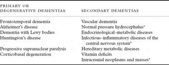
The dementia subtypes can be divided using an etiological classification: the primary or degenerative forms of dementia, and the so-called secondary types of dementia of which vascular dementia is probably the most common (Table 1).
Classification of dementia

*Causes of dementia that can be diagnosed by CT and treated specifically.
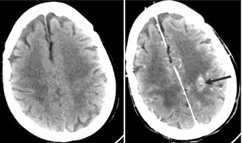
CT may have a particular role in detecting some less common causes of secondary dementia. Despite the fact that CT can immediately detect causes of cognitive decline in only a minority of cases, ranging from 1.9% to 10.4% (Gifford et al., Reference Gifford, Holloway and Vickrey2000), some of these conditions are potentially treatable, and therefore the CT scan should be done systematically in patients with dementia. Typical examples are intracranial neoplasms and other space-occupying lesions. This is one of the main reasons for requesting a CT scan at initial evaluation of a patient with cognitive decline or dementia. In fact, some brain tumors may present with cognitive decline that subsequently progresses. In this context, a contrast medium might be useful in revealing lesions not seen on first scanning, but its role is limited to a specific few cases and therefore is not recommended for routine assessment (Figure 1).
Contrast medium revealing lesion (arrow) not detected on direct scanning.

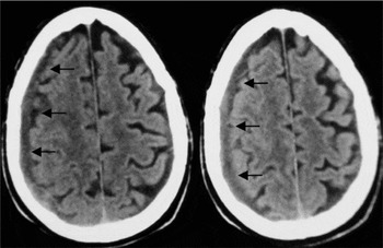
Another typical example of an intracranial lesion that can cause cognitive deterioration and can be easily detected by CT is subacute or chronic subdural hematoma (Schebesch et al., Reference Schebesch, Woertgen, Rothoerl, Ullrich and Brawanski2008). In some instances, subdural subacute hematoma can be bilateral (Figure 2). It is important to detect such a lesion because it can be surgically treated and the cognitive deterioration may be reversible.
CT scan of a chronic subdural hematoma (arrows).

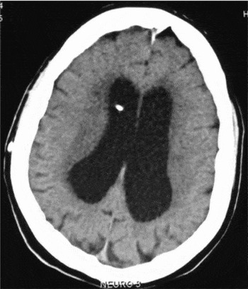
Normal pressure hydrocephalus – i.e. a progressive enlargement of the lateral ventricles not due to obstruction of cerebrospinal fluid flow – is a form of cognitive decline that can be seen in the elderly (Figure 3). It is easily recognized on a CT scan and some cases may be reversible with surgical treatment (Hebb and Cusimano, Reference Hebb and Cusimano2001).
CT scan of normal pressure hydrocephalus. Note the enlargement of the lateral ventricles. The white spot is the tip of the ventricular–peritoneal shunt tubing.

Role of CT in degenerative dementia
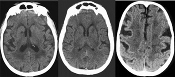
Based on routine examination, the role of the CT scan in degenerative dementias, mainly AD, is to exclude other evident causes of cognitive decline, to appreciate cortical atrophy and to outline the possible coexistence of vascular disease. Some patients present with a mixture of degenerative and vascular lesions, sometimes referred to as mixed dementia (Figure 4).
CT scan of mixed dementia with coexistence of significant cortical atrophy and vascular changes (white matter changes and lacunar infarcts).

Medial temporal lobe atrophy is nowadays considered one of the neuroimaging markers of AD (Dubois et al., Reference Dubois2007), but it is difficult to appreciate on a routine CT scan. However, if appropriate orientation of the CT scanner is applied, images are formed that allow the evaluation of this region; for this purpose, according to the Jobst procedure, a section plane 20° caudal to the orbitomeatal line in 2-mm contiguous slices through the posterior fossa is needed (Jobst et al., Reference Jobst1992; Frisoni et al., Reference Frisoni2002). However, this procedure is not as precise as the grading of the medial temporal lobe on MRI. This approach has been proposed as a tool for differentiating patients who are depressed from those with Alzheimer's disease (O'Brien et al., Reference O'Brien2000).
Role of CT in vascular dementia
According to major criteria for vascular dementia, neuroimaging may play a prominent role (Chui et al., Reference Chui, Victoroff, Margolin, Jagust, Shankle and Katzman1992; Román et al., Reference Román1993; Erkinjuntti et al., Reference Erkinjuntti2000). The role of CT in vascular dementia is not only to detect the presence of vascular changes, but also to quantify the severity of these vascular changes as they relate to the clinical picture. The second point is important because in many instances it is not the presence of vascular lesions, but rather their severity that makes a difference in terms of etiological diagnosis. For example, the observation of white matter changes on neuroimaging is not enough to state that a patient is affected by vascular dementia, given that these changes are extremely frequent and seen also in normal subjects (Pantoni and Garcia, Reference Pantoni and Garcia1995). Instead, it is important to quantify the severity of the lesions and their relationship with the presence or absence of other changes, e.g. the neurodegenerative type.
Vascular dementia is a heterogeneous group of diseases with different etiologies and mechanisms. The main subtypes are: (1) subcortical or small vessel disease dementia; (2) multi-infarct dementia; and (3) strategic infarct dementia.
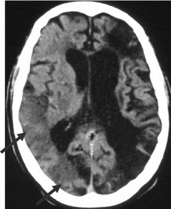
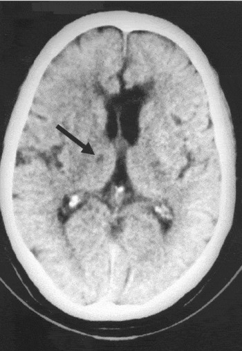
On CT, small vessel vascular dementia is characterized by the presence of lacunes and white matter changes; in multi-infarct dementia, multiple infarcts are located in cortical and/or subcortical areas (Figure 5). In our opinion, the term multi-infarct dementia is best suited to patients with multiple non-lacunar infarcts caused by large vessel disease or cardioembolism, while those with multiple lacunar infarcts are best considered with the group of small vessel disease. In strategic infarct dementia, the single lesion can be either cortical or lacunar. Typical locations of strategic lacunar infarcts are the thalamus (Figure 6), internal capsule, particularly the genu, and head of the caudate nucleus. Strategic non-lacunar infarcts are located in the angular gyrus and the posterior cerebral artery territory involving the medial part of the temporal lobe.
CT scan of multi-infarct dementia showing the presence of three large infarcts, two in the chronic stage on the left hemisphere and one subacute (arrows) in the right hemisphere, in a patient with internal bilateral carotid stenosis.

CT scan of strategic lacunar infarct (arrow) located in the right thalamus.

Hemorrhagic dementia is a rather neglected issue. Intra-parenchymal blood is easily detected by CT, but the neuroimaging picture for patients with post-hemorrhagic dementia is less clear. There are two major causes of intra-parenchymal spontaneous hemorrhage: hypertensive small vessel disease and amyloid angiopathy. In the first case, the typical location is in subcortical regions (thalamus, basal ganglia, internal capsule, pons, cerebellum); hemorrhages from cerebral amyloid angiopathy are usually lobar and involve the cortex. In the chronic stages, CT will show only the residual brain lesions and sometimes it can be difficult to differentiate these from the damage resulting from an ischemic injury. If the history is not clear or informative, only specific MRI sequences (gradient-echo) are able to show hemosiderin deposits and can be of help.
In some patients, a combination of degenerative and vascular lesions is present, and this is evidenced by neuroimaging; both lesion types can contribute to the dementia picture even if their relative contribution can be difficult to appreciate. In our opinion, in comparison with MRI, CT tends to underestimate the concomitant presence of degenerative processes when vascular lesions are obvious.
CT in dementia research
The role of CT scanning in dementia research has become less prominent over the years as MRI and other techniques have become more relevant. However, examples exist of how CT can be used for research purposes (Guo et al., Reference Guo2006; Reference Guo, Skoog, Idrizbegovic, Pantoni, Simoni and Rosenhall2008; Reference Guo2009; Simoni et al. Reference Simoni2008; Olesen et al., Reference Olesen2010).
Some years ago, Frisoni et al. (Reference Frisoni2003) presented a consensus paper outlining guidelines for the use of CT and MRI in the field of dementia research. Two levels of CT use were reported. The first level is based on routine acquisition of a CT scan to allow the visual rating of white matter changes, e.g. by means of a scale developed by consensus (Wahlund et al., Reference Wahlund2001). For this, a short period of training (1–2 weeks) is required. The second level requires the acquisition of a specific orientation and slicing of a CT scan to allow evaluation of the width of the medial temporal lobe horn. Training for this visual assessment takes 1–3 days.
The specific assessment of medial temporal lobe atrophy is relevant because the radial width of the temporal horn represents a sensitive measure in AD. As mentioned above, some data can be obtained with CT. Frisoni et al. (Reference Frisoni2002) were able to select a cut-off of 5.3 mm for this measure to differentiate patients with AD with good sensitivity and specificity. This method has been used also in studies on post-stroke dementia to assess the role of pre-existing, likely degenerative, cognitive decline and the relative contribution to the dementia picture.
More recently, it has been shown that medial temporal lobe atrophy can also be evaluated with CT using a 64-detector row CT scanner and applying oblique coronal reconstructions (Wattjes et al., Reference Wattjes2009). By using CT coronal slices, Wattjes et al. found a substantial agreement with MRI in assessing medial temporal lobe atrophy.
If only routine CT scans are used, cortical atrophy can still be graded for research purposes by means of a fairly rough visual rating scale (absent, moderate/mild, severe) (De Leon et al., Reference De Leon, Ferris, George, Reisberg, Kricheff and Gershon1980). This scale is very simple to use but not very informative; however, it does allow an assessment of the location (frontal, temporal, parietal, occipital) of atrophy. This regional assessment has been operationalized and used for scientific purposes (Gustafson et al., Reference Gustafson, Lissner, Bengtsson, Björkelund and Skoog2004).
Assessment of white matter changes on CT scans
One topic that is receiving increasing attention is that of white matter changes. On CT scans, these changes, also called leukoaraiosis, are seen as hypodense, bilateral, usually symmetric areas which are typically located in the periventricular and subcortical white matter (Hachinski et al., Reference Hachinski, Potter and Merskey1987). Although white matter changes are better visualized by MRI, CT is able to detect and, to some extent, grade them.
Many different scales have been proposed for the assessment of white matter changes and these were critically reviewed some years ago (Scheltens et al., Reference Scheltens1998). The conclusion of the review was that the ideal scale does not exist and that different scales have been developed for different purposes by different groups. To overcome this problem, one scale was developed by the European Task Force on Age-Related White Matter Changes (Wahlund et al., Reference Wahlund2001). The peculiarity of this scale is that it can be applied to both CT and MRI, thus providing immediate comparison. In one study, two CT and four MRI rating scales for white matter changes were applied simultaneously to a group of paired scans and correlation values and conversion parameters were produced (Pantoni et al., Reference Pantoni, Simoni, Pracucci, Schmidt, Barkhof and Inzitari2002). The conclusions were that, despite an apparent difference across scales, many of them are concordant and results obtained with one scale can be converted to the scoring of another.
Usefulness and limitations of CT in dementia
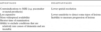
Despite clear evidence that the information provided by MRI in the field of dementia is more accurate than that provided by CT, some data still support the use of CT in this relation (Table 2). In comparison with MRI, the CT scan suffers from limited spatial definition and resolution. Furthermore, CT is limited in assessing disease progression in cognitive impairment and this aspect is becoming an important requirement, particularly in the field of AD and small vessel disease-related cognitive decline.
Advantages and disadvantages of using CT scans in dementia in comparison with MRI

However, CT is a far less expensive technique in comparison with MRI. For example, the cost of a 1.5 T MRI scanner is US$1–1.5 million. In many areas of the world, this cost is unaffordable. In addition, the construction and maintenance of MRI suites are costly. A routine CT scan of the brain is three times less expensive than a routine cerebral MRI. Moreover, it should be borne in mind that cognitive impairment and dementia are highly frequent disorders and, therefore, the number of patients to be potentially subjected to imaging is very high. In addition, this category of patients has an increased risk of comorbidities that require devices that are contraindications for MRI. In fact, most forms of medical or biostimulation implants are contraindications for MRI. These include pacemakers, vagus nerve stimulators, implantable cardioverter-defibrillators, loop recorders, insulin pumps, and cochlear implants. Another indication for the use of CT is claustrophobia; this is particularly relevant where the MRI magnet is old and requires longer examination times (up to 40 minutes for the entire procedure). This intolerability might be accentuated in people with dementia. Finally, it should be borne in mind that CT scanning may be preferable to MRI in patients with cognitive impairment who are also agitated and cannot stay still during the MR exam as required.
Conclusion
Although a CT scan is less sensitive and specific than MRI, it still has a role in the diagnostic process for patients with dementia. The main aim of prescribing a CT scan in dementia is to exclude secondary and possibly treatable causes of cognitive impairment. Moreover, CT may help in differentiating among dementia subtypes. The role in research is of course much more limited and the technique is becoming increasingly less used in this context.
Conflict of interest declaration
Leonardo Pantoni is a member of the Board of Directors and Chairman of the Publication Committee of the International Psychogeriatric Association.
Description of authors' roles
All authors contributed equally to the writing of the paper, review of the literature and preparation of the figures.