Introduction
Migration of tympanic membrane (TM) epithelia has been known for more than 100 years, and its contribution to the physiological requirements of the ear canal is generally accepted.Reference Deong, Prepageran and Raman1 This process is important to remove keratin debris and foreign bodies from the TM and external auditory canal, in order to propagate the sound wave more efficiently.
However, all previous studies of TM epithelial migration rate and pattern have focused on the normal TM, perforated TM, open mastoidectomy cavity or postmyringoplasty TM.
To the best of our knowledge, there have been no previous studies of epithelial migration in the atelectatic TM. Therefore, the question of whether epithelial migration in the atelectatic TM proceeds similarly or differently to that in the normal TM remains unanswered. This study was conducted in an attempt to answer this question.
The objectives of this study were to characterise epithelial migration in the atelectatic TM, and to compare the rate and pattern of epithelial migration in the atelectatic TM versus the normal TM.
Methods and materials
This was a prospective, non-randomised, case–control study. This study was a pilot study. Patients were selected from the otorhinolaryngology clinic of the University of Malaya Medical Centre. The study was conducted from January 2006 to January 2008. The study group comprised patients with an atelectatic pars tensa region of the TM in one ear and a normal TM in the other ear (these latter ears were used as the control group), as determined by otoscopic examination and tympanography findings.
Tympanic membrane atelectasis was graded using Sade's system, based on the severity of retraction.Reference Sade and Sade2 Thus, the study group was graded as: grade I (i.e. slight retraction of the TM over the annulus); grade II (the TM touches the long process of the incus); grade III (the TM touches the promontory); or grade IV (the TM is adherent to the promontory).
The purpose and procedure of the study were explained thoroughly to the patient, to ensure their full understanding. Following this, written consent was obtained from all patients. The study was approved by the medical ethics committee of the University of Malaya Medical Centre.
Inclusion criteria
The inclusion criteria of the study were as follows: (1) atelectatic pars tensa region of the TM, secondary to any pathology, to any grade (i.e. grades I–IV, Sade classification), with a normal contralateral TM, as determined by otoscopy and tympanography; (2) normal, dry external auditory canals bilaterally; (3) a type C tympanogram in the atelectatic TM and a type A tympanogram in the normal TM; and (4) no planned surgical intervention, and no history of allergy to methylene blue dye.
Study instruments
The following instruments were used for the study: basic demographic data record forms; diagrammatic records of epithelium migration pattern and rate measurement; an operating microscope; a micrometer; methylene blue dye; and an angled, malleable wire.
Procedures
The selected patients' ears were examined using an operating microscope. A malleable wire was used as an applicator; this wire was dipped in methylene blue and then touched onto the epithelium in the immediate vicinity of the umbo, to create a dot stain. The patients were then followed up by the investigator at weekly intervals until the dye dot had migrated to reach the annulus. The distance of migration was measured with a micrometer inserted into the eyepiece of the operating microscope. A schematic diagram with serial recording was made to document the migratory pattern for each TM.
The daily migratory rate was calculated as the distance between the umbo and the annulus, divided by the total days taken for migration.
Results
Forty patients from the otorhinolaryngology clinic were included in the study, with 40 retracted TMs and 40 normal TMs. Patients' ages ranged from 11 to 71 years, with a mean of 36.58 years. There were 21 (52.5 per cent) males and 19 (47.5 per cent) females. The ethnic distribution of the group was 45 per cent Malay, 30 per cent Chinese and 25 per cent Indian.
Tympanic membrane retraction was grade I in nine (22.5 per cent) TMs, grade II in 21 (52.5 per cent), grade III in nine (22.5 per cent) and IV in one (2.5 per cent). Tympanic membrane retraction was due either to eustachian tube dysfunction secondary to rhinosinusitis (72.5 per cent), nasopharyngeal carcinoma (12.5 per cent) or unknown causes (15 per cent).
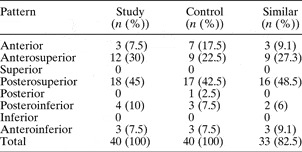
Of the atelectatic TMs, epithelial migration was posterosuperior in 18 (45 per cent), anterosuperior in 12 (30 per cent), posteroinferior in four (10 per cent), anteroinferior in three (7.5 per cent) and anterior in three (7.5 per cent) (Table I). Of the control TMs, epithelial migration was posterosuperior in 17 (42.5 per cent), anterosuperior in nine (22.5 per cent), anterior in seven (17.5 per cent), anteroinferior in three (7.5 per cent), posteroinferior in three (7.5 per cent) and posterior in one (2.5 per cent) (Table I). Thirty-three (82.5 per cent) of the 40 patients showed a similar direction of migration in their study and control TMs.
Migration pattern in study and control groups

Both study and control TMs showed a migration pattern from the umbo towards the annulus and canal wall. Sixty per cent of the retracted TMs and 35 per cent of the control TMs showed no migration during the first week after methylene blue staining. Thereafter, the rate of migration from the umbo towards the periphery was nearly constant in both the study and control TMs, assessed two to three weeks after dye application.
In the retracted TMs, the mean daily migration rate was 62.6 µm/day (range 50–78.3 µm/day). In the control TMs, the mean daily migration rate was 64.7 µm/day (range 50–80.5 µm/day) (Table II). However, this difference in mean daily migration rate was not statistically significant (p = 0.202, independent samples t-test).
Daily epithelial migration rate in study and control groups

*2.1 µm/day difference.
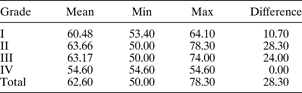
The mean daily migration rates for the different grades of TM atelectasis were: grade I, 60.48 µm/day; grade II, 63.66 µm/day; grade III, 63.17 µm/day; and grade IV, 54.60 µm/day (Table III). However, due to the small numbers in the different grading groups, it was difficult to make an accurate statistic comparison of the migration rate differences between the different grades.
Daily migration rate for different grades of TM atelectasis
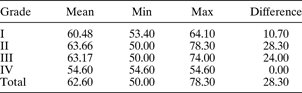
Data represent µm/day. TM = tympanic membrane; min = minimum; max = maximum
Discussion
In 1877, Burnett was the first to suggest the existence of epidermal migration in the human ear drum.Reference Alberti3 This concept was also noted by Burk in 1880 and Blake in 1882.Reference Alberti3, Reference Blake4
Stinson was the first to study epithelial migration in the TM, in 1936, after the incidental observation that a piece of reed accidentally embedded in the TM was gradually carried from the drumhead to the posterior canal wall and hence to the exterior.Reference Stinson5 Later, Stinson defined five laws of ‘epithelial escalation’. Briefly summarised, these were: (1) the epidermis migrates on its ‘original plane’ from the anterior to the posterior margin of the membrane and then along the canal wall to the exterior; (2) the epidermis over the pars flaccida fans out over the canal wall; (3) the epidermis over the processus brevis proliferates in whorls to the superior part of the canal; (4) the progress is most rapid in the posterior inferior quadrant; and (5) the epidermis does not proliferate from canal to drumhead.
Magnoni concluded in 1938 that epithelial growth was radial from the umbo, but agreed that epithelium never migrated from canal wall to drumhead. However, Magnoni's results were neglected in the English language literature.Reference Alberti3
In 1963, Litton was the first to estimate the rate of epithelial migration in the human TM, at approximately 0.05 mm/day, and also noted that an ink dot placed at the umbo disintegrated and proceeded outward in a radial fashion.Reference Litton6
Alberti repeated Litton's work in 1964, in a study of 62 human TMs, estimating an epithelial migration rate of 0.07 mm/day.Reference Alberti3 Both Litton and Alberti postulated that the rate of migration reduced steadily as the distance from the umbo increased, and also that the migration rate was proportional to the thickness of the epithelial layer. The usual pattern of migration was found to be centrifugal from the umbo, spreading out to all quadrants of the TM. There was no bar to epithelial movement between the pars tensa and the pars flaccida. The overall migration rate was equivalent to the growth rate of a finger nail.Reference Alberti3
Further studies have been carried out by Boedts and Kuijpers in 1978, O'Donoghue in 1983, Michaels and Soucek in 1989, Ong et al. in 2007, and Deong et al. in 2006; these authors have confirmed the existence of TM epithelial migration and elucidated its pathways.Reference Deong, Prepageran and Raman1, Reference Boedts and Kuijpers7–Reference Ong, Prepageran, Godbole and Raman10
The current, pilot study demonstrates that epithelial migration does occur in the atelectatic TM. The marker dye dot was clearly observed to migrate from the umbo of the TM to the annulus and ear canal wall.
A very similar migratory pattern was observed in both the study and control TMs. The most common direction of epithelial migration was posterosuperior (occurring in 45 per cent of the study TMs and 42.5 per cent of the control TMs), followed by anterosuperior migration (occurring in 30 per cent of study TMs and 22.5 per cent of control TMs). None of the study or control TMs showed any evidence of epithelial migration in the superior and inferior directions.
The migration patterns observed in this study were slightly different from those reported by Litton and Alberti. These authors reported that the ink dot disintegrated and proceeded outwards in a radial fashion, whereas, in the current study, the dye dot disintegrated from the umbo and proceeded in a particular direction and segment.Reference Alberti3, Reference Litton6 This was most probably because the dye dot had not been placed in the exact centre of the umbo.
The observed epithelial migration rate was slower near the umbo region (i.e. at the beginning of the observation period). Some TMs showed no movement of the dye dot during the first week. In almost all cases (in both study and control TMs), the epithelial migration rate increased once the dye dot had migrated further out towards the periphery.
The observed mean daily migration rates for the study and control TMs were 62.6 and 64.7 µm/day, respectively. The mean rate difference was 2.1 µm/day; however, this difference was statistically insignificant (p > 0.05). The observed mean daily migration rate was within the range documented by Litton and Alberti.Reference Alberti3, Reference Litton6
• This study aimed to determine whether epithelial migration in the atelectatic tympanic membrane (TM) (secondary to any pathology) behaves in a similar fashion to that in the normal, non-pathological TM
• Migration of dye dots was used to assess epithelial migration
• There was no difference in epithelial migration rate or pattern, comparing atelectatic and normal TMs
Clinical observation indicates that TM atelectasis and retraction pockets are unstable, dynamic phenomena. The degree of retraction of the atelectatic drum fluctuates, over weeks and months and also over hours during sleep.Reference Sade, Avraham and Brown11 In a prospective study by Luntz and Sade, 84 human, atelectatic ears were examined at different times of day.Reference Luntz and Sade12 They noticed that over one-third of adolescent and adult TMs were found to be inflated (usually even hyperinflated) following a night's sleep. It took these ‘hyperinflated’ TMs an average period of 54 minutes to return to their original, atelectatic state. In our study, most of the cases had reversible atelectasis (i.e. Sade grade I to III), and it could be argued that many of these TMs reverted back to normal during part of the day. Therefore, epithelial migration may not be significantly impeded.
This study found an increased mean daily migration rate with increased grading of TM atelectasis, especially grade IV. This difference may become statistically significant if the number of TMs of different grades is increased. Hence, further study is required.
We acknowledge that the study had a number of limitations. Firstly, it was difficult to apply the dye dot in the immediate vicinity of the umbo. Secondly, measurement was performed only at weekly intervals. Thirdly, there was a certain amount of unavoidable objective and subjective measurement error. Finally, the number of TMs in each atelectasis grade group was small, preventing accurate statistic analysis.
Conclusion
Based on the above results, we conclude that there is no statistically significant difference in epithelial migration rates and patterns, comparing atelectatic and normal TMs. However, there may be a significant difference in epithelial migration rate with increased atelectasis grading. As we only have one patient with Grade IV TM. Therefore, we are unable to analyse statistically. Further study is required.