I. INTRODUCTION
Silicon carbide (SiC) has many properties that make it a desirable candidate material for applications in nuclear energy systems. These properties include chemical resistance, high temperature mechanical stability, high thermal conductivity, and low neutron cross-section. Reference Katoh, Snead, Szlufarska and Weber1 SiC has a wide range of potential applications including the fission product barrier layer in tristructural isotropic (TRISO) fuels, Reference Ford, Hibbert and Martin2 first wall and cladding material, Reference Zinkle and Busby3 and use as matrix material for actinide burning. Reference Verrall, Vlajic and Krstic4 Improving the radiation stability of SiC would further enhance its suitability for these applications. One avenue for improving radiation resistance is refining the grain size to the nanometer length scale. Nanocrystalline (nc) materials have a high density of grain boundaries (GBs), which are known to act as sinks for point defects generated during irradiation. Increasing the sink density results in more efficient elimination of these defects, potentially leading to an increased radiation resistance. A number of studies in both metals Reference Rose, Balogh and Hahn5–Reference Birtcher and Wang9 and ceramics Reference Rose, Balogh and Hahn5,Reference Shen, Feng, Tang, Valdez, Wang and Sickafus10,Reference Zhang, Ishimaru, Varga, Oda, Hardiman, Xue, Katoh, Shannon and Weber11 have already shown the expected improvement in radiation resistance upon grain size reduction, making this approach a potential path for improving the radiation resistance of SiC.
There have been a few studies investigating the effect of grain refinement on the radiation response of SiC Reference Zhang, Ishimaru, Varga, Oda, Hardiman, Xue, Katoh, Shannon and Weber11–Reference Jiang, Jiao and Wang16 utilizing dose-to-amorphization as a measure of radiation resistance. Several of these studies found that nc-SiC had a superior radiation resistance as compared to single crystal (sc) SiC. Reference Zhang, Ishimaru, Varga, Oda, Hardiman, Xue, Katoh, Shannon and Weber11,Reference Jamison, Zheng, Shannon, Allen, Morgan and Szlufarska12 There are several possible mechanisms that could cause the observed improvement in radiation response. Based on the previously presented discussion, one could expect that the observed increase in radiation resistance is due to the increase in sink density. However, several studies have also shown a reduction Reference Jiang, Wang, Kim, Zhang and Weber13,Reference Jamison, Xu, Sridharan and Allen14,Reference Jiang, Jiao and Wang16 or no change Reference Jiang, Wang, Kim, Bae, Li, Nachimuthu, Zhu, Zhang and Weber15 in radiation resistance with grain refinement in SiC, implying that an increase in sink density cannot be the only mechanism for the observed improvement in radiation response. An increase of the sink strength in the system is another possible mechanism that could lead to an increased radiation resistance. A recent theoretical study, based on the combination of elasticity theory and molecular dynamics, has shown that in nc materials, low-angle GBs can have a larger sink strength than high-angle GBs due to the presence of GB stresses. Reference Jiang, Swaminathan, Morgan and Szlufarska17 The nc-SiC samples that exhibited an increased radiation resistance Reference Zhang, Ishimaru, Varga, Oda, Hardiman, Xue, Katoh, Shannon and Weber11,Reference Jamison, Zheng, Shannon, Allen, Morgan and Szlufarska12 had a strong texture introduced by the fabrication method, resulting in a higher concentration of low-angle GBs compared to randomly oriented grains in polycrystalline SiC. An increase in the overall sink strength in the system due to the large number of low-angle GBs may contribute to the improved radiation resistance of these nc-SiC samples. In addition to texture, these samples were characterized by a high density of stacking faults. The presence of stacking faults has been suggested as an explanation for the increased radiation resistance observed in nc-SiC as compared to sc-SiC. Reference Zhang, Ishimaru, Varga, Oda, Hardiman, Xue, Katoh, Shannon and Weber11,Reference Jamison, Zheng, Shannon, Allen, Morgan and Szlufarska12,Reference Ishimaru, Zhang, Shannon and Weber18 Stacking faults were found to improve radiation resistance by decreasing the energy barriers to Si interstitial diffusion and to the kick-out reaction (Si antisite recombining with a C interstitial) Reference Jamison, Zheng, Shannon, Allen, Morgan and Szlufarska12 so that they become active within stacking faults, even though both are inactive within the bulk in the studied temperature range. Lowering these energy barriers increases the number of point defect elimination mechanisms in the material, leading to an increased radiation resistance. Another possible mechanism for the improved resistance is a change in defect production within the initial damage cascade due to the presence of GBs. Because GBs are structurally more disordered than the bulk, displacement within the GBs may be easier, leading to an increased damage production in this region. However, it has been found that although the number of displacements in GB regions is higher than in the bulk, the average number of displacements in crystalline grains of a bicrystal sample containing a GB is the same as in a sc SiC. Reference Swaminathan, Kamenski, Morgan and Szlufarska19 Consequently, the effect of GBs on defect production is unlikely to be responsible for the observed improvement in radiation resistance of SiC with grain refinement. The increased radiation resistance of nc-SiC in these studies is likely due to a combination of increased sink density, increased sink strength, and the presence of stacking faults.
Although several studies have shown an increase in radiation resistance with grain refinement, Reference Zhang, Ishimaru, Varga, Oda, Hardiman, Xue, Katoh, Shannon and Weber11,Reference Jamison, Zheng, Shannon, Allen, Morgan and Szlufarska12 some investigations of nc-SiC found the opposite response. Reference Jiang, Wang, Kim, Zhang and Weber13,Reference Jamison, Xu, Sridharan and Allen14,Reference Jiang, Jiao and Wang16 The microstructures of SiC used in these studies are markedly different than those discussed previously. Several studies fabricated nc-SiC using pulsed-laser-deposition (PLD), resulting in SiC nanograins embedded in an amorphous SiC layer, Reference Jiang, Wang, Kim, Zhang and Weber13,Reference Jiang, Wang, Kim, Bae, Li, Nachimuthu, Zhu, Zhang and Weber15,Reference Jiang, Jiao and Wang16 and another study investigated the radiation response of two different sizes of SiC nanopowders. Reference Jamison, Xu, Sridharan and Allen14 The behavior of nc-SiC fabricated by PLD is likely dominated by growth of the large amorphous GB regions into the crystalline grain interior, accounting for the observed reduction in dose-to-amorphization with grain refinement. The studies with SiC nanopowders assumed that the reduction in radiation response was due to an interface-dominated process, though the exact mechanism was not determined. Although microstructure differences could account for the different responses to radiation damage, the irradiation species may also have an effect. Au ions, Reference Jiang, Wang, Kim, Bae, Li, Nachimuthu, Zhu, Zhang and Weber15,Reference Jiang, Jiao and Wang16 Kr2+ ions, Reference Jamison, Xu, Sridharan and Allen14 Si ions, Reference Zhang, Ishimaru, Varga, Oda, Hardiman, Xue, Katoh, Shannon and Weber11,Reference Jiang, Wang, Kim, Zhang and Weber13 and high-energy electrons Reference Jamison, Zheng, Shannon, Allen, Morgan and Szlufarska12 were all used to determine the effect of grain size on the radiation response of SiC. To determine if the irradiating species affects the radiation response of SiC, here we use Kr2+ ions to study the same nc-SiC material that had exhibited an improved radiation response under Si ion Reference Zhang, Ishimaru, Varga, Oda, Hardiman, Xue, Katoh, Shannon and Weber11 and electron irradiation. Reference Jamison, Zheng, Shannon, Allen, Morgan and Szlufarska12 We compare the response to commercial SiC samples with micrometer-scale grains (µc-SiC) over a range of temperatures to determine the grain size dependence of the radiation response in SiC under heavy-ion irradiation.
II. EXPERIMENTAL PROCEDURE
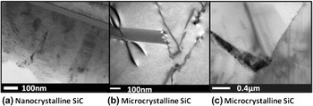
The materials used in this study included nc-SiC grown on a sc Si substrate via chemical vapor deposition (CVD) and commercially available CVD grown bulk µc-SiC. The microstructures of these samples are shown in Fig. 1. Details of the sample preparation procedure can be found in a previous paper. Reference Jamison, Zheng, Shannon, Allen, Morgan and Szlufarska12 The nc-SiC samples were characterized by a high concentration of stacking faults and the material had 90-nm-sized columnar grains extending through the thickness of the 500 nm SiC layer. The columnar structure of these grains is also representative of the structure of the SiC layer in TRISO fuel applications. Reference Petti, Buongiorno, Maki, Hobbins and Miller20 The µc-SiC samples were sourced from Rohm and Haas, and had micrometer-scale grains. To compare the radiation resistance of these materials, we measured the radiation dose required to amorphize the samples. A higher dose-to-amorphization is indicative of a higher radiation damage resistance. The experimental temperature range was kept low enough so that the only expected radiation response was amorphization of the crystal structure Reference Snead, Nozawa, Katoh, Byun, Kondo and Petti21 rather than cluster, loop, and void formation characteristic of higher temperature experiments. Therefore, any differences observed in the radiation response were caused only by differences in the initial microstructure.
TEM images of (a) nc-SiC thin film characterized by a high concentration of stacking faults, (b) µc-SiC showing the presence of dislocations and low concentration of stacking faults at the same magnification as the nc-SiC sample. (c) Lower magnification TEM image of µc-SiC microstructure showing three grains as well as intragranular dislocations.

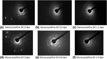
In situ Kr2+ irradiations were conducted using the IVEM-Tandem facility at Argonne National Laboratory. The dose-to-amorphization was used to compare the radiation resistance of nc-SiC and µc-SiC. The electron diffraction pattern was used to determine the degree of crystallinity of the sample. The evolution of the electron diffraction pattern is shown in Fig. 2 for both nc-SiC and µc-SiC. Before irradiation, both samples were fully crystalline and exhibited the bright spots characteristic of crystalline materials. At full amorphization, the electron diffraction pattern consisted of diffuse rings. The dose-to-amorphization was determined by gradually increasing the dose of the samples, by approximately 0.05 dpa per step. After each dose step, the electron diffraction pattern was examined to determine crystallinity. If the sample was still partially crystalline, the irradiation was continued until full amorphization was achieved. One should keep in mind that there might be a variation in thickness from sample to sample, which can affect the measured dose to amorphization. Although some samples were thick enough for Kikuchi bands to be visible, they were never thick enough for the bands to dominate the diffraction pattern. We estimate the upper bound of the sample thickness in the following way. Kikuchi bands typically start dominating the electron diffraction pattern when the sample thickness has reached one inelastic mean free path of electrons within the material. For inorganic solids, the inelastic mean free path is on the order of 100 nm. One can therefore infer that the microcrystalline SiC sample is likely less than 100 nm in thickness, as there is still a strong crystalline diffraction pattern visible in the images. To confirm that thickness plays a negligible role in our analysis, we have performed additional TRIM calculations for a range of thicknesses from 30 to 100 nm to determine how the damage conversion factor (vacancies/angstrom-ion) varies with depth. The damage conversion factor was found to be 0.750 vacancies/Angstrom-ion at 30 nm and 0.977 vacancies/Angstrom-ion at 100 nm from the surface. The damage conversion factor is then used to calculate the damage done to the sample in dpa from the measured fluence. For the data plotted in Fig. 3, all fluences were converted to dpa using the 100 nm value. The effect of a changed conversion factor will be greater for larger fluences. Therefore, for this analysis, we used the temperature of 423 K, which is the temperature with the largest fluence for which a data point for both nc-SiC and µc-SiC was collected. From these calculations we found that at 423 K, thin samples would have a dose-to-amorphization artificially increased by 0.5 dpa due to an overestimation of the damage produced. In the worst case scenario, where µc-SiC was only 30 nm and nc-SiC was 100 nm thick, µc-SiC would still have a higher dose to amorphization, as the data points are separated by more than 1 dpa. Therefore, the observed difference in the dose-to-amorphization between nc-SiC and µc-SiC is outside of the experimental error due to thickness variation.
Evolution of electron diffraction patterns during irradiation at 373 K for (a–c) nc-SiC and (d–f) µc-SiC. Samples in 2(a) and 2(d) are fully crystalline, in 2(b) and 2(e) – partially amorphous, and in 2(c) and 2(f) they are fully amorphous. A diffraction spot is visible in Fig. 2(c) and was confirmed to come from the Si substrate using dark field TEM. nc-SiC amorphized at 1.1 dpa, whereas µc-SiC did not fully amorphize until a dose of 1.3 dpa. Bright field images corresponding to these diffraction patterns are included in supplementary materials available online, figure S1.

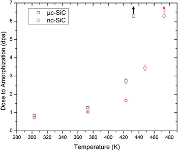
Dose-to-amorphization as a function of irradiation temperature for µc-SiC and nc-SiC under 1.0 MeV Kr2+ radiation. The symbols with arrows designate samples irradiated to the indicated dose but which have not yet amorphized. The error bars indicate the amorphization dose range, as explained in the text.

III. RESULTS AND DISCUSSION
The results of the in situ krypton irradiation of nc-SiC and µc-SiC are shown in Fig. 3. The error bars on the plot indicate the dose range in which the sample amorphized. The top of the range marks the dose where the electron diffraction pattern was indicative of fully amorphous sample, and the bottom of the range marks the dose step prior to that, where some crystallinity remained. The arrows designate samples that were irradiated to the indicated dose, but had not yet amorphized. At room temperature the ranges for nc-SiC and µc-SiC overlap, but above the room temperature µc-SiC has a higher dose-to-amorphization than nc-SiC. The increase in dose-to-amorphization indicates that nc-SiC has a lower radiation resistance compared to µc-SiC under Kr2+ ion irradiation. This experiment has shown a reduction in radiation resistance with grain refinement, in contrast to the trend observed for the same samples under electron Reference Jamison, Zheng, Shannon, Allen, Morgan and Szlufarska12 and Si ion Reference Zhang, Ishimaru, Varga, Oda, Hardiman, Xue, Katoh, Shannon and Weber11 irradiation. As the temperature increases, the dose-to-amorphization for both samples increases, as expected. Migration and the recombination rate increase with temperature, resulting in a more efficient damage recovery. Although the critical temperature (the temperature above which a material will not amorphize) was not determined in our experiments, the data indicate that nc-SiC has a higher value than µc-SiC. Inspection of Fig. 3 reveals that the critical temperature for µc-SiC is lower than that of nc-SiC. The µc-SiC sample irradiated at 430 K has not yet amorphized at 6.5 dpa, while nc-SiC has undergone amorphization at 450 K (at a dose of 3.5 dpa). From the data presented in Fig. 3, the critical temperature for nc-SiC is near 473 K, as the sample had not yet amorphized at a dose of 6.5 dpa. The difference in critical temperature between nc- and µc-SiC is a further indication that µc-SiC has a superior radiation resistance compared to nc-SiC under Kr2+ ion irradiation.
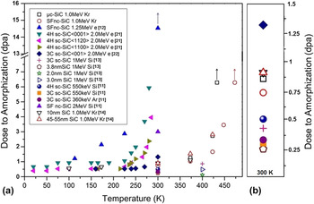
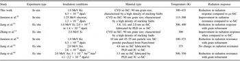
As mentioned in the introduction section, in addition to the experiments conducted in the current study, the same CVD grown nc-SiC has been investigated under electron irradiation Reference Jamison, Zheng, Shannon, Allen, Morgan and Szlufarska12 and Si irradiation. Reference Zhang, Ishimaru, Varga, Oda, Hardiman, Xue, Katoh, Shannon and Weber11 The earlier studies found an improvement in radiation resistance with grain refinement, counter to the findings of our experiments. The effect of grain refinement on the radiation response of SiC has also been investigated using nc-SiC thin films fabricated using PLD and irradiated with Si ions Reference Jiang, Wang, Kim, Zhang and Weber13 and SiC nanopowders irradiated with Kr ions. Reference Jamison, Xu, Sridharan and Allen14 These experiments found a reduction in the dose-to-amorphization with grain refinement, in agreement with the results from the current study. The results from all the discussed experiments are summarized in Fig. 4, where open symbols indicate experiments where grain refinement resulted in a reduction of radiation resistance and solid symbols indicate experiments where an increase in radiation resistance was observed. A summary of the experimental conditions for these studies can be found in Table I. In addition to the variety of sample types used to study the amorphization of SiC, a range of irradiating species (electrons, Reference Jamison, Zheng, Shannon, Allen, Morgan and Szlufarska12,Reference Inui, Mori and Fujita22,Reference Inui, Mori, Suzuki and Fujita23 heavy ion, Reference Jamison, Xu, Sridharan and Allen14 and light ion Reference Zhang, Ishimaru, Varga, Oda, Hardiman, Xue, Katoh, Shannon and Weber11,Reference Jiang, Wang, Kim, Zhang and Weber13 ), experiment type (in situ Reference Jamison, Zheng, Shannon, Allen, Morgan and Szlufarska12,Reference Jamison, Xu, Sridharan and Allen14,Reference Inui, Mori and Fujita22,Reference Inui, Mori, Suzuki and Fujita23 and ex situ Reference Zhang, Ishimaru, Varga, Oda, Hardiman, Xue, Katoh, Shannon and Weber11,Reference Jiang, Wang, Kim, Zhang and Weber13 ), and temperature ranges have been used. Heavy ions are defined here as any irradiating species more massive than the constituent elements. The following sections discuss these observed differences and how they may cause the observed reversal in the effect of grain refinement on the radiation response of SiC.
(a) A summary of several studies on the grain size dependence of the radiation response of SiC. (b) Expanded view of the data from inside the black rectangle in (a). Solid symbols indicate studies where grain refinement resulted in improved radiation resistance, whereas the open symbols indicate studies where grain refinement led to a reduction in radiation resistance of SiC. SFnc-SiC indicates samples that have a high concentration of stacking faults (these are the same samples as used in the present study). The irradiating species and energy are listed in the legend. Grain sizes Reference Jiang, Wang, Kim, Zhang and Weber13 and particle sizes Reference Jamison, Xu, Sridharan and Allen14 are also listed. Arrows label samples that were irradiated to the indicated dose, but had not yet amorphized.

Experimental details for irradiation studies of nc-SiC. We report experiment type (in situ versus ex situ), irradiation conditions (irradiating species and dose rate), irradiation temperature, and the radiation resistance relative to the comparable µc and sc SiC samples.

A. Ion type
The irradiating species used to study the dose-to-amorphization of nc-SiC included electrons, Reference Jamison, Zheng, Shannon, Allen, Morgan and Szlufarska12,Reference Inui, Mori and Fujita22,Reference Inui, Mori, Suzuki and Fujita23 Si ions, Reference Zhang, Ishimaru, Varga, Oda, Hardiman, Xue, Katoh, Shannon and Weber11,Reference Jiang, Wang, Kim, Zhang and Weber13 Kr ions, Reference Jamison, Xu, Sridharan and Allen14 and Au ions. Reference Jiang, Wang, Kim, Bae, Li, Nachimuthu, Zhu, Zhang and Weber15,Reference Jiang, Jiao and Wang16 In examining the results of these experiments a trend in which light ion experiments show an improvement in radiation resistance with grain refinement and heavy-ion irradiation studies show a decrease in radiation resistance was observed. As mentioned previously, heavy ions are defined as any ion species used for irradiation that is more massive than the constituent atoms. The exception to the observed trend is the Si-ion irradiation conducted by Jiang et al., Reference Jiang, Wang, Kim, Zhang and Weber13 where radiation resistance deteriorated with grain refinement. However, different physics may be involved with these samples, as the grains were less than 5 nm in diameter and were embedded in an amorphous matrix. The amorphization of these samples may be dominated by the growth of the amorphous matrix, rather than direct amorphization of the crystalline material. As such, this study will be left out of the following discussion.
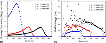
To determine how the irradiating species could affect radiation response, it is important to take into account how the damage profiles of these species differ. Under high energy electron irradiation only isolated pairs of point defects are produced, while Si and Kr ions cause radiation damage cascades. Radiation cascades result in locally high concentrations of point defects, potentially leading to direct amorphization. Figure 5 shows the damage distribution and lateral distribution of ions as a function of depth for the irradiating species used to study the same nc-SiC sample type as used in the present work. Kr ions produce a much more compact damage profile than Si ions. The damage produced per ion is significantly larger [Fig. 5(a)], and the damage is created in a smaller volume of the material [Fig. 5(b)]. Figure 5(b) is a plot of the radial range of the irradiating ion species calculated by SRIM. The radial range is the mean radial displacement range from the x axis, which is the direction of the incoming ion. Because Si ions are lighter than Kr, they are able to travel further into the material before coming to a stop after the deflection incident. The compact damage region created by 1.0 MeV Kr2+ ion irradiation may produce an amorphous pocket within the initial damage cascade, as has been observed in molecular dynamics simulations of Au-ion irradiation in SiC. Reference Weber, Gao, Devanathan, Jiang and Wang24 Si-ion irradiation produces a less dense damage profile, as shown by the lower damage level [Fig. 5(a)] and larger radial ion distribution [Fig. 5(b)], as compared to Kr ions. It is unlikely that Si-ion irradiation creates amorphous pockets directly in the radiation cascade region due to its diffuse nature. This suggests that different amorphization processes may be active in heavy ion and light ion irradiations.
(a) Damage distribution in SiC for 1 MeV Si+, 2.0 MeV Si+, and 1.0 MeV Kr2+ ions and (b) radial distribution of damage produced by the same ions. Calculations were performed using SRIM-2008 (ion distribution and quick calculation of damage, displacement energies set to 21 eV for C and 35 eV for Si Reference Devanathan and Weber25 ).

Amorphization in ceramics can proceed via a variety of mechanisms, including direct-impact, double-cascade overlap, direct impact/defect stimulated growth, and defect accumulation. Reference Weber26 In the direct impact model, an amorphous pocket is formed directly in the damage cascade and amorphization of the full sample is accomplished via the accumulation of these pockets. The double-cascade overlap mechanism requires two damage cascades to occur in the same volume before amorphization of the region can occur. In direct impact/defect stimulated amorphization, amorphous pockets are formed directly within the initial damage cascades and further growth at the crystalline/amorphous interfaces (including GBs) occurs due to diffusion of defects produced in the initial cascade, but not contained within the amorphous pocket. In the defect accumulation amorphization mechanism defects produced within the damage cascade gradually increase the overall system energy. Eventually it becomes more energetically favorable to transform into the amorphous phase rather than maintaining a highly defected crystalline structure. Amorphization in SiC has been found to match most closely with the direct-impact/defect stimulated (DI-DS) growth mechanism, Reference Weber26,Reference Bolse27 as shown with heavy-ion irradiation studies. Reference Bolse27,Reference Weber, Wang and Yu28 The DI-DS amorphization mechanism may favor materials with larger grain sizes, as GBs may be more easily amorphized than the bulk. Recent experimental data suggest that this may be the case in SiC, Reference Wang, Jamison, Shannon, Sridharan, Morgan and Szlufarska29 but the correlation between DI amorphization and the presence of GBs needs to be further investigated. Although the DI-DS amorphization mechanism is the best match for heavy-ion irradiation, light-ion irradiation may have a different amorphization mechanism. The dose-to-amorphization is dependent upon ion type, and it was found that more energy per atom must be introduced to the system under light-ion irradiation for amorphization to occur when compared to heavy-ion irradiation. Reference Wendler, Heft and Wesch30 The additional energy required to amorphize SiC under light-ion irradiation is another indication that a different amorphization mechanism may be active than under heavy-ion irradiation. The damage profile is more diffuse for light ions (such as Si) than for heavy-ion irradiation, so that the defect accumulation model may be a closer match for light-ion irradiated SiC. In this amorphization model, defect elimination becomes the dominant factor in determining radiation resistance. As nc-SiC has a higher concentration of GBs (which are sinks for point defects), nc-SiC may exhibit an increased radiation resistance compared to µc-SiC and sc-SiC. The DI-DS amorphization mechanism favors coarse-grained materials, while the damage accumulation model favors fine-grained materials. Ion type likely has a large effect on the radiation response of SiC due to a potential change in the amorphization mechanism, although further work is needed to determine if a change in mechanism occurs and how it correlates with the radiation response of SiC.
B. Experimental conditions
Studies to determine the effect of grain refinement on radiation response in SiC have used both in situ Reference Jamison, Zheng, Shannon, Allen, Morgan and Szlufarska12,Reference Jamison, Xu, Sridharan and Allen14,Reference Inui, Mori and Fujita22,Reference Inui, Mori, Suzuki and Fujita23 and ex situ Reference Zhang, Ishimaru, Varga, Oda, Hardiman, Xue, Katoh, Shannon and Weber11,Reference Jiang, Wang, Kim, Zhang and Weber13 ion irradiation, as summarized in Table I. In situ experiments could artificially increase or decrease the dose-to-amorphization as compared to ex situ results. In situ transmission electron microscopy (TEM) thin foil samples have a large surface-area to volume ratio, supplying a large sink for defects (since defects can be annihilated at surfaces). This could increase the dose-to-amorphization by providing an additional mechanism for point defect elimination. Alternatively, the large surface area could decrease the dose-to-amorphization due to interstitial starvation. This phenomenon occurs when interstitials are disproportionally eliminated from the system. When interstitials are more mobile than vacancies, as is the case in SiC in the experimental temperature range, they can more readily diffuse and annihilate at GBs or the surface. The increased elimination of interstitials from the system results in the accumulation of vacancies, leading to amorphization. Reference Swaminathan, Morgan and Szlufarska31 The large surface area in in situ experiments provides a large sink for interstitials, potentially enhancing the effects of interstitial starvation. In situ experiments would then exhibit a lower dose-to-amorphization compared to ex situ irradiation where all other parameters are identical. Depending on the active mechanisms, in situ ion irradiation experiments could lead one to conclude either enhancement or degradation in the radiation response of SiC compared to ex situ experiments. When examining Table I, no obvious trend connecting the experiment type to either improvement or reduction or radiation response can be seen. Both experiment types found contradicting results for the effect of grain size on radiation response, so experiment type cannot explain why some experiments conclude a reduction in radiation resistance with grain size and others find an improvement.
In addition to the experimental type, the flux of the irradiating species affects the dose to amorphization. A lower flux results in a higher dose-to-amorphization due to the slower damage accumulation rate. The dose rates for all experiments discussed here are listed in Table I, and are kept constant within each study. In Ref. Reference Jamison, Zheng, Shannon, Allen, Morgan and Szlufarska12, the flux for the nc-SiC study is higher than the flux used in the sc-SiC Reference Inui, Mori and Fujita22,Reference Inui, Mori, Suzuki and Fujita23 studies used for comparison. The increased damage rate would artificially decrease the dose-to-amorphization for nc-SiC. However, the nc-SiC samples were found to have an increased dose-to-amorphization compared to sc-SiC. Therefore, flux values alone cannot explain the observed differences in dose to amorphization.
C. Temperature
In Fig. 4, experiments that found an increase in radiation resistance with grain refinement are marked with solid symbols, while open symbols indicate experiments where the opposite trend was observed. There is a clear switch in behavior around 375 K, as all experiments conducted above this temperature found a decrease in radiation resistance with grain size. Experiments conducted at or below the room temperature showed that grain refinement improved radiation resistance. The only exception to these trends is the data for nanopowders. Reference Jamison, Xu, Sridharan and Allen14 However, for the powder samples, there was actually no observable difference in the dose-to-amorphization between the two particle sizes (10 nm and 45–55 nm) at or below the room temperature. For powder samples, the reduction in radiation resistance with grain refinement was only observed above the room temperature, in agreement with the hypothesis that grain refinement only results in decreased radiation resistance above the room temperature. The nc-SiC samples that exhibited an improved radiation resistance Reference Zhang, Ishimaru, Varga, Oda, Hardiman, Xue, Katoh, Shannon and Weber11,Reference Jamison, Zheng, Shannon, Allen, Morgan and Szlufarska12 had a high concentration of stacking faults, and that superior radiation resistance of nc-SiC was observed at and below the room temperature. The critical energy barrier for a chemical reaction to be active or a defect to be mobile was determined to be ∼0.71 eV Reference Jamison, Zheng, Shannon, Allen, Morgan and Szlufarska12 in this temperature regime. Stacking faults were found to lower both Si interstitial migration (from 0.8 to 0.26 eV) and antisite recombination (from 1.34 to 0.37 eV) barriers, Reference Jamison, Zheng, Shannon, Allen, Morgan and Szlufarska12 activating both mechanisms and resulting in an increased radiation resistance. At higher temperatures, Si interstitials become mobile (∼275 K) and recombination becomes active (∼400 K) even without the presence of stacking faults. Therefore, the change in the energy landscape created by the presence of stacking faults has a negligible effect on the radiation resistance. Grain size becomes the main microstructural feature that influences radiation response. Above the room temperature grain refinement reduced the dose to amorphization, Reference Jiang, Wang, Kim, Zhang and Weber13,Reference Jamison, Xu, Sridharan and Allen14 indicating that the increase of free energy due to a higher volume fraction of interfaces may drive the amorphization process. Interface-driven amorphization was previously observed in studies on nc-Si Reference Kachurin, Ruault, Gutakovsky, Kaı̈tasov, Yanovskaya, Zhuravlev and Bernas32 and nc-Cu. Reference Johannessen, Kluth, Llewellyn, Foran, Cookson and Ridgway33 The amorphization transition has been observed to initiate at GBs in nc-SiC, Reference Ishimaru, Zhang, Shannon and Weber18 suggesting that interface-driven amorphization may occur in SiC as well.
Interface-driven amorphization could occur via growth of the existing disordered GB region, or via interstitial starvation. As previously discussed, the behavior of nc-SiC with 2–3-nm-sized grains Reference Jiang, Wang, Kim, Zhang and Weber13 is likely dominated by growth of the disordered GB region into the crystalline interior due to the high GB-to-crystalline ratio. The grain size of nc-SiC used in this study is 90 nm in width, so that the GB region does not dominate the amorphization process, and interstitial starvation may play a substantial role. Interstitial starvation can occur when interstitials are more mobile than vacancies, as is the case in SiC. Interstitials are able to move more readily to the GB, or other sinks within the material, and are eliminated from the system in greater numbers than vacancies. This creates an interstitial-denuded zone near sinks, resulting in vacancy accumulation in the same region. The accumulation of vacancies can eventually lead to amorphization, Reference Swaminathan, Morgan and Szlufarska31 initiating at interfaces. Interstitial starvation is similar to production bias, Reference Was34 where interstitials cluster within the initial radiation cascade leading to a decreased number of interstitials available for recombination and to an excess of vacancies. However, the mechanisms behind interstitial starvation and production bias are different. Production bias results in excess vacancies uniformly distributed across the sample and it is not affected by grain refinement. Interstitial starvation, on the other hand, scales in effect with grain size, as absorption of interstitials at the GB leads to localized regions of excess vacancies. Rate theory models Reference Swaminathan, Morgan and Szlufarska31 predicted a cross-over temperature where grain refinement changes from improving radiation resistance to lowering it. This cross-over was determined to be due to the increasing interstitial starvation rate with temperature. A cross-over temperature was observed in Fig. 4, further supporting the hypothesis that interstitial starvation plays a role in the amorphization of SiC. The increasing gap in dose-to-amorphization between nc-SiC and µc-SiC shown in Fig. 2 also supports the interstitial starvation mechanism theory. Point defects can be eliminated from the system via recombination or annihilation at an interface (surface or GB). Due to the large number of GBs in nc-SiC, point defect annihilation at the GB dominates the radiation response. With a smaller number of GBs, the predominant mechanism for point defect elimination in µc-SiC is recombination. Both of these mechanisms become more efficient with increasing temperature. While increasing the recombination rate will increase the dose to amorphization, increasing the mobility of defects (and the rate at which interstitials are eliminated from the system) can result in a decreased dose-to-amorphization due to interstitial starvation. Therefore, µc-SiC has a rapidly increasing dose-to-amorphization with temperature, and nc-SiC exhibits a more modest increase. Although there are several indications that interstitial starvation is the cause of the interface-dominated amorphization suggested in this study, growth of the disordered GB region into the grain could also take place. To determine which mechanism is active, it will be necessary to investigate the morphology of amorphous regions near GBs and in the crystalline grains, which is beyond the scope of the current study.
IV. CONCLUSIONS
The radiation response of nc and microcrystalline SiC has been investigated by in situ observation of damage evolution during irradiation with 1.0 MeV Kr ions. Irradiations were conducted in the temperature range of 300–450 K and the dose-to-amorphization was investigated as a metric for radiation damage resistance. It was observed that under the experimental conditions and methods used in this study nc-SiC exhibited a lower radiation resistance than µc-SiC. While this observation at the outset appears to contradict some recent studies on the radiation resistance of nc-SiC, a broader survey of literature in this area indicates that factors such as the type of irradiating species used for irradiation, the microstructure, and even the method of observation (in situ versus ex situ) could lead to different results and conclusions. At lower temperatures, the presence of stacking faults within nc-SiC improves radiation resistance, while at elevated temperatures radiation resistance decreases with grain size. The temperature dependence likely comes from Si interstitial mobility and recombination becoming active above the room temperature. At lower temperatures, energy barriers are only low enough for Si interstitial migration and antisite recombination to occur within the stacking faults, resulting in an improved radiation resistance in samples that contain stacking faults. Above the room temperature, Si interstitials are mobile and recombination becomes active in the bulk. We hypothesize that in this regime interface-driven amorphization dominates the radiation response, resulting in a comparatively lower radiation resistance in nc-SiC. Our results are consistent with the phenomenon of interstitial starvation, which was proposed in earlier studies based on theory and modeling. Reference Swaminathan, Morgan and Szlufarska31 Additionally, the irradiation species may play a role in how grain refinement affects the radiation response of SiC due to a potential change in the amorphization mechanism with the irradiating species.
Supplementary Material
To view supplementary material for this article, please visit http://dx.doi.org/jmr.2013.340.
ACKNOWLEDGMENTS
The authors acknowledge the U.S. Department of Energy Basic Energy Sciences for funding this research. We thank Prof. D. Morgan from the University of Wisconsin for helpful comments on the study and Dr. Mark Kirk and the rest of the staff at IVEM-Tandem at ANL for their assistance in conducting the in situ irradiations. The electron microscopy analysis was carried out at the Electron Microscopy Center at Argonne National Laboratory, a U.S. Department of Energy Office of Science Laboratory operated under Contract No. DE-AC02-06CH11357 by UChicago Argonne, LLC.