Introduction
Stapes surgery is a very safe, standardised and successful treatment for hearing impairment in humans with otosclerosis.Reference Vincent, Sperling, Oates and Jindal1 In children with congenital stapes fixation combined with developmental defects or syndromic diseases, stapes surgery can be very challenging.Reference Bachor, Just, Wright, Pau and Karmody2, Reference Vick, Just, Terpe, Graumüller and Pau3 In the child with stapes fixation but a normal tympanic membrane and tube function, several differential diagnoses should be considered before surgery, including discontinuation of the ossicular chain and other anomalies of the middle ear,Reference Raveh, Hu, Papsin and Forte4–Reference Teunissen and Cremers6 osteogenesis imperfecta,Reference Garretsen and Cremers7 atresia of the round window niche,Reference Pappas, Pappas and Hedlin8 and isolated stapes fixation due to otosclerosisReference Cole9–Reference Robinson11 or congenital stapes fixation.Reference Dornhoffer, Helms and Höhmann12 The latter two entities are sometimes difficult to differentiate, especially when insufficient information is available with regard to family history and progression of the hearing loss. In such cases, exploratory tympanotomy should provide the diagnosis. Bachor et al. Reference Bachor, Just, Wright, Pau and Karmody2 studied children younger than six years with fixation of the posterior part of the footplate or complete footplate fixation, and found congenital stapes fixation to be more prevalent than otosclerosis. In children older than six years with progressive hearing loss, a diagnosis of juvenile otosclerosis requires a positive family history and the presence of fixation of the anterior stapediovestibular joint.
Optical coherence tomography is an imaging technology which supplies optical cross-sections of a tissue, comparable to ultrasound. Based on the interference of low coherent light, a considerably higher resolution of 5 to 20 µm is achieved without any tissue contact.Reference Tomlins and Wang13 Optical coherence tomography equipment can be integrated into the structure of an operating microscope, so that tissue structures can be visualised and their extensions measured during surgery.Reference Heermann, Hauger, Issing and Lenarz14 A new generation of optical coherence tomography devices, with technology based on the spectrally resolved detection of interference signals,Reference Häusler and Lindner15 has dramatically increased imaging speed.Reference Leitgeb, Hitzenberger and Fercher16 These spectral domain optical coherence tomography devices allow real-time B-scan imaging and rapid acquisition of three-dimensional volumes. Recently, this new technology has been fully integrated into the structure of an operating microscope,Reference Pau, Lankenau, Just and Hüttmann17 and the combined instrument has been made available for clinical use. Promising preclinical results have been obtained for various otological applications.Reference Pau, Lankenau, Just and Hüttmann17–Reference Pau, Lankenau, Just and Hüttmann19
The current experimental study aimed to assess whether optical coherence tomography could be a helpful diagnostic tool for evaluation of the oval window niche, especially the stapes footplate. In the first part of this study, on cadaveric temporal bone preparations, we aimed to identify several middle-ear structures, such as the stapes footplate and annular ligament, and thus to establish the applicability of optical coherence tomography for visualisation of the oval window niche. In the second, in vivo part of this study, we aimed to use an operating microscope with integrated optical coherence tomography technology to obtain intra-operative measurements of the oval window niche.
Materials and methods
Optical coherence tomography system, imaging and image analysis
An operating microscope (Hi Res 1000; Möller Wedel GmbH, Wedel, Germany) incorporating a modified spectral domain optical coherence tomography unit with a central wavelength of 840 nm (Thorlabs, Newton, New Jersey, USA) allowed optical coherence tomography scanning of any object in the centre of the field of vision on which the microscope was focused. Measurements were performed with a light power of 3 mW and a resolution of about 24 µm. A green pilot beam indicated the optical coherence tomography scanning field. The system was ??-certified for intra-operative documentation of tissue structures.
Temporal bone preparations
In order to demonstrate the feasibility of using optical coherence tomography for visualisation of stapes footplate structures, optical coherence tomography scans were performed on special preparations of two fresh and three formalin-fixed human temporal bones, obtained from body donors via the Anatomical Institute of Rostock University.
Optical coherence tomography measurements were carried out via the ear canal. A tympanic flap was raised in the usual way and the posterior canal wall was removed until the entire stapes was fully exposed. The stapes suprastructure was then removed (two formalin-fixed and two fresh temporal bones were used, the latter taken from the body 24 hours after death and kept deep-frozen until 3 hours before preparation).
For direct correlation between optical coherence tomography scans and histology, two temporal bones (one formalin-fixed and one fresh) were used. In the fresh temporal bone, a laser stapedotomy (CO2 laser, power 20 W, spot size 0.6 mm and pulse duration 0.3 s) was performed. After optical coherence tomography investigation, both temporal bones were fixed in 10 per cent formaldehyde, decalcified in ethylene diamine triacetic acid and processed using the standard celloidin technique. The sections were stained with haematoxylin and eosin.
Because of the large variability in the morphology of the stapes footplate, the thickness of the footplate and the ratio between bone and cartilage were not measured.
In each experiment, the scanning axis for optical coherence tomography (via the operating microscope) was both parallel and perpendicular to the facial nerve. For each temporal bone preparation, four different measurements (two parallel and two perpendicular) were taken. The scanning images were stored digitally.
Intra-operative application of optical coherence tomography
Optical coherence tomography measurements were taken in eight patients undergoing stapes surgery due to otosclerosis (n = 2), or tympanosclerosis-related stapes fixation and tympanoplasty (n = 6). After visualisation of the oval window niche during middle-ear surgery, a nominal working distance of between 224 and 280 mm was used for optical coherence tomography measurements. In all intra-operative settings, perpendicular optical coherence tomography sections were scanned through the footplate. The images obtained were stored digitally.
Results and analysis
Ex vivo optical coherence tomography measurements
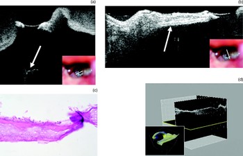
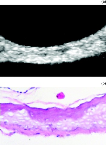
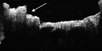
In each of the cadaveric temporal bone specimens, the footplate could be demonstrated easily on the optical coherence tomography images. The stapedotomy hole was visible in the form of a discontinuity of the footplate (Figure 1a). On some scans, the vestibule beneath the footplate could be seen (indicated by an arrow in Figure 1a). Within the footplate of four specimens, areas with homogeneous ‘echoes’ could be separated from areas where different layers were apparent (indicated by an arrow in Figure 1b). The corresponding histological sections revealed that the dense structures appearing white on the optical coherence tomography scans were bony layers, while the grey zones were consistent with cartilage (Figures 1c and 2). Three-dimensional reconstruction of the oval window niche may help the surgeon to detect irregularities within the footplate (Figure 1d).
(a) Optical coherence tomography scan showing a perpendicular section through the posterior part of the stapes footplate in the fresh temporal bone (plane indicated by white line marked on operating microscope view of middle ear; inset) (depth of measurement 2.5 mm). The stapedotomy hole is visible as a discontinuity of the footplate. Some fine structures can be identified within the cochlea (arrow indicates the vestibule). (b) Optical coherence tomography scan showing a parallel section through the anterior part of the stapes footplate in the formalin-fixed temporal bone (plane indicated by white line marked on operating microscope view of middle ear; inset) (depth of measurement 2.5 mm). Different layers are apparent within the footplate. (c) Photomicrograph showing different layers of bone and cartilage within the stapes footplate (H&E; ×40). (d) Three-dimensional reconstruction of the oval window niche from optical coherence tomography scans (inset shows computer-generated three-dimensional model).

(a) Typical optical coherence tomography scan of a normal, mobile footplate. (b) Photomicrograph showing corresponding histology (H&E; ×40).

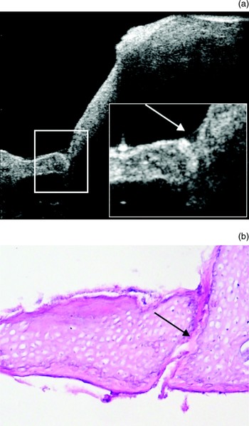
Even such delicate structures as the boundaries between the stapes footplate and the otic capsule were clearly visible (Figure 3). Due to compact ossification at the margins of the footplate and the otic capsule, seen in nearly all specimens with a non-fixed footplate, a very dense configuration could also be detected on the optical coherence tomography scans. The gap between the margins of the footplate and the otic capsule was defined as the stapediovestibular joint. A prominent facial nerve and a remnant of the posterior crus of the stapes impeded visualisation of the posterior stapediovestibular joint in two out of five specimens.
(a) Optical coherence tomography scan showing a parallel section through the posterior part of the stapes footplate in the formalin-fixed temporal bone (enlarged in the inset; arrow indicates the stapediovestibular joint) (depth of measurement 2.5 mm). The posterior margin of the footplate can be clearly differentiated from the otic capsule. (b) Photomicrograph showing the posterior stapediovestibular joint as seen in part (a); arrow indicates the stapediovestibular joint (H&E; ×40).

In vivo optical coherence tomography measurements
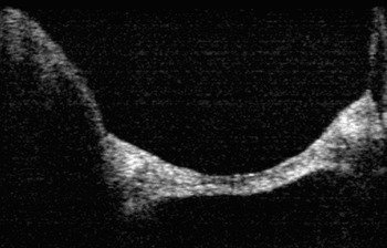
Performing the intra-operative optical coherence tomography measurements increased the operation time by just 5 to 10 minutes. Good quality optical coherence tomography images of the stapes footplate were obtained in all patients, even those operated upon under local anaesthesia (Figure 4). In all five patients with a mobile footplate, different layers could be detected within the footplate and at its margins.
In vivo optical coherence tomography scan of a non-fixed footplate, obtained during tympanoplasty (depth of measurement 1.5 mm).

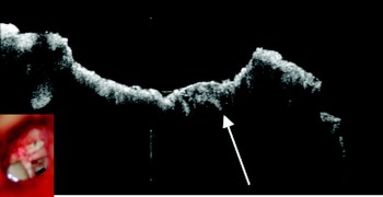
In the cases of otosclerosis (Figure 5) and tympanosclerosis (Figure 6), an inhomogeneous and irregularly thickened footplate was found (see arrows in both Figures). Figure 5 illustrates a sclerotic focus visible at the anterior stapediovestibular joint (arrow). In both cases, the border between the footplate and the otic capsule could not be visualised. A laser stapedotomy was performed for both patients. As a result, no corresponding histological images were available.
In vivo optical coherence tomography scan showing otosclerosis. Arrow indicates a sclerotic focus at the anterior stapediovestibular joint. Note the inhomogeneous footplate (depth of measurement 2.0 mm).

In vivo optical coherence tomography scan showing tympanosclerosis (inset shows intra-operative operating microscope view of middle ear). The scan represents a perpendicular section through the stapes footplate (depth of measurement 2.5 mm). The complete footplate appears inhomogeneous and thickened (arrow).

Discussion
The procedures and results of stapes surgery depend markedly upon the condition of the footplate. In some cases of stapes fixation, especially in children, it is not clear intra-operatively whether the cause is congenital stapes fixation involving the posterior stapediovestibular joint or otosclerosis. Patients with childhood onset of otosclerosis appear to have a higher risk of diffuse otosclerosis and so-called ‘biscuit’ footplate.Reference Robinson11 For these reasons, it is interesting to obtain detailed information on the oval window niche. Anatomical details of the middle and inner ears can be obtained pre-operatively by multidetector row computed tomography imaging with advanced three-dimensional reformation,Reference Chuang, Chiang, Liu and Lin20 by magnetic resonance imaging and by computer tomography with high resolution yield.Reference Veillon, Riehm, Emachescu, Haba, Roedlich and Greget21
In this paper, we report our approach to obtaining anatomical images of the oval window niche during middle-ear and stapes surgery. To the best of our knowledge, this is the first published report to describe the use of optical coherence tomography to visualise stapes footplate anatomy.
Despite the fact that corresponding histological images were obtained from only two temporal bones, the optical coherence tomography images presented here show that the intra-operative application of optical coherence tomography may offer information on the morphology of the stapes footplate (i.e. its shape and thickness) and the annular ligament. However, it is also desirable to obtain information on the footplate during revision stapes surgery or type III tympanoplasty, before any manipulation is undertaken. In the latter procedure, total ossicular replacement prosthesis related microfractures and pressure-related changes of the footplate, due to poor eustachian tube function, would be detectable. In an experimental study, it has been shown that fractures of the footplate following penetration of the prostheses into the vestibulum occur typically in the middle of the footplate at pressures of between 250 and 980 mN.Reference Beutner, Stumpf, Peuss, Zahnert and Hüttenbrink22 The varying thickness of the footplate, seen using scanning electron microscopy, correlates with the histological findings of the current study. ‘Weak spots’ were found mostly in the middle of the footplate.
Another application of intra-operative optical coherence tomography would be the visualisation of intra-operative findings in patients undergoing exploratory tympanotomy due to sudden hearing loss. Further investigations are necessary to establish the utility of optical coherence tomography in this context.
• The procedures and results of stapes surgery depend markedly upon the condition of the footplate
• Previously, no technology has been available to provide intra-operative data on the oval window
• This study indicates that optical coherence tomography technology may help the ear surgeon assess the oval window niche intra-operatively
• Further investigations are needed to clarify the clinical usefulness of these optical coherence tomography findings, including morphometric measurement of footplate anomalies
The application of optical coherence tomography technology to middle-ear surgery was first reported in 2001 by Beyer et al. Reference Beyer, Tauber, Kubasiak, Lankenau, Engelhardt and Baumgartner23 and later by Heermann et al. Reference Heermann, Hauger, Issing and Lenarz14 In our study, an operating microscope with an integrated optical coherence tomography system was used. Improvements in optical specifications and integration of the pilot beam allowed connection between the optical coherence tomography equipment and the camera port of the microscope.Reference Pau, Lankenau, Just and Hüttmann17–Reference Pau, Lankenau, Just and Hüttmann19
The technical improvements requested by Heermann et al. Reference Heermann, Hauger, Issing and Lenarz14 (i.e. pilot beam, variable focal distance and improved image quality) have been achieved. A moderate to high level measurement precision has also been achieved. The integrated pilot beam allows an exact scanning field. At the same time, the optical zoom of the operating microscope can be used over the complete range. The first clinical application of the optical coherence tomography system delivered images of good quality.
Acknowledgements
The authors would like to express their appreciation to PD Dr Friedrich Prall for his guidance and help with assessment of the histological specimens. Receipt of funding from the Innovation Fund Schleswig-Holstein (2004-12-HWT) and Möller Wedel GmbH is gratefully acknowledged.