Introduction
Miniaturization is a phylogenetic concept defined as the evolution of unusually small adult size in a lineage, reaching beyond a threshold at which dramatic changes in morphology, physiology, and ecology occur (Yeh Reference Yeh2002). It has been repeatedly documented among metazoans and studied in several vertebrate clades (Hanken Reference Hanken1984). Establishing a critical size to differentiate miniaturization from other less extreme reductions in body size is difficult and usually arbitrary because the changes between these phenomena are continuous, biological parameters scale differently with body size, and physical size is not correlated with biological size, as the latter is related with genome size (Hanken and Wake Reference Hanken and Wake1993). Larger genomes, which correlate with larger cells, are associated with slower rates of growth and development (Sessions Reference Sessions2008), and therefore, species with large genomes are biologically smaller than other taxa with the same physical size but small genomes (Hanken and Wake Reference Hanken and Wake1993).
Miniature vertebrates often have skeletons with novel morphologies and arrangements that represent the inevitable consequences of the decrease of adult size (e.g., shifts in the relative position of structures due to spatial accommodation) or adaptations that enable the size decrease (e.g., enhanced deposition of bone to compensate mechanical weakness) (Hanken Reference Hanken1993). Osteological novelties vary across size-reduced taxa; however, many lineages of miniaturized tetrapods are characterized by proportionally large nervous and sense organs and their associated skeletal elements (Rieppel 1996). These changes have been interpreted as the result of the interplay between the extrapolation of the conservative negative allometries of the brain, eyes, and otic capsules in vertebrates (Bauchot Reference Bauchot1978) and constraints imposed on nervous and sensory systems to maintain functional efficiency at a reduced head size (Hanken Reference Hanken1983). The skeletal changes concomitant with larger relative sizes of these soft-tissue structures are not restricted to the braincase; a new arrangement of the entire skull might also emerge due to the change of the spatial arrangement of its components, a phenomenon that was called “packaging effect” by Hanken (Reference Hanken1993), and the need for maintaining other skeletal functions such as feeding (Rieppel 1996). Among tetrapods, these changes in the architecture of the skull are particularly evident in extant miniaturized salamanders and frogs, groups in which the phyletic size decrease has repeatedly occurred, resulting in adult diminutive body sizes leading to convergent novel cranial arrangements (Hanken and Wake Reference Hanken and Wake1993; Yeh Reference Yeh2002).
Besides presenting morphological novelties, miniature tetrapods frequently resemble the juvenile stages of larger related species in many traits (Hanken and Wake Reference Hanken and Wake1993; Rieppel 1996), suggesting that heterochronic mechanisms leading to ontogenetic truncation (sensu Hanken Reference Hanken1993) might be involved in the decrease of the adult body size and, thus, these traits are paedomorphic. Distinguishing between morphological novelties and paedomorphic traits is relevant to understand the morphological effects of miniaturization because paedomorphic traits, in contrast to novelties, result from a conserved ancestral pathway and represent morphologies that were already expressed in the juveniles of the ancestor (Hanken Reference Hanken1993). Therefore, paedomorphic features should not be interpreted as consequences of the body-size reduction but outcomes of the ontogenetic truncation that originated the size reduction.
Because extreme size reduction may trigger substantial morphological changes that represent a pool of alternate morphological designs available for subsequent evolutionary diversification (Hanken Reference Hanken1985), miniaturization has been proposed as a key factor for the phyletic diversification above the species level and the origin of several major clades (Hanken and Wake Reference Hanken and Wake1993). Among tetrapods, miniaturization has been considered to be involved in the origin of the three major groups of living amphibians (e.g., Carroll and Holmes Reference Carroll and Holmes1980; Milner Reference Milner1988, Reference Milner1993; Carroll Reference Carroll1990; Schoch and Rubidge Reference Schoch and Rubidge2005; Fröbisch and Reisz Reference Fröbisch and Reisz2008), amniotes (e.g., Carroll Reference Carroll2001), snakes (e.g., Rieppel 1996), lizards (e.g., Carroll Reference Carroll1970), and birds (e.g., Lee et al. Reference Lee, Cau, Naish and Dyke2014). In particular, current hypotheses on the origin of the living amphibian groups (i.e., salamanders, anurans, and caecilians) involve an instance of marked body-size reduction, whether from temnospondyls (Bolt Reference Bolt1969, Reference Bolt1991; Milner Reference Milner1988, Reference Milner1993; Trueb and Cloutier Reference Trueb and Cloutier1991; Ruta et al. Reference Ruta, Coates and Quicke2003; Schoch and Milner Reference Schoch and Milner2004; Anderson Reference Anderson2007; Anderson et al. Reference Anderson, Reisz, Scott, Fröbisch and Sumida2008; Maddin and Anderson Reference Maddin and Anderson2012; Maddin et al. Reference Maddin, Jenkins and Anderson2012), lepospondyls (Laurin and Reisz Reference Laurin and Reisz1997; Vallin and Laurin Reference Vallin and Laurin2004; Marjanovic and Laurin Reference Marjanovic and Laurin2013), or both (i.e., polyphyletic origin; Carroll Reference Carroll2009). There is a growing consensus that at least frogs and salamanders originated within the smallest dissorophoid temnospondyls, the amphibamids, with many characters supporting this relationship associated with miniaturization (Milner Reference Milner1988). In this regard, the study of miniaturization in dissorophoids is particularly relevant. Moreover, it is significant to analyze whether the morphological changes possibly related with small adult size are similar to those described for extant miniaturized frogs and salamanders, especially those related to constraints on nervous and sensory systems. Comparisons with these extant groups are relevant not only because of their putative close phylogenetic relationship with dissorophoids, but also because miniaturization has been extensively studied in them and their mode of life is not associated generally with radical morphological changes, unlike those of other miniaturized fossorial/burrowing tetrapods such as caecilians and squamates.
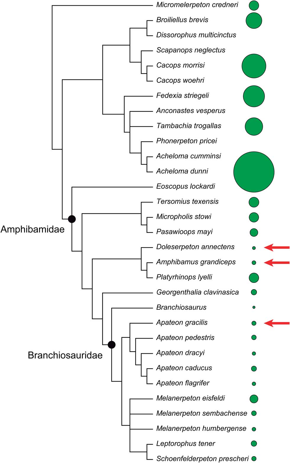
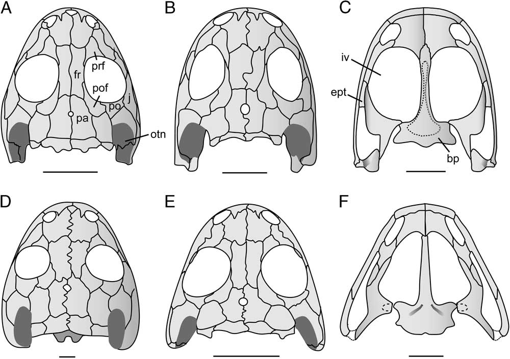
Within Dissorophoidea, the small-bodied forms are restricted to branchiosaurids and amphibamids (Fig. 1). With body sizes ranging between 5 and 15 cm, these groups also rank among the smallest temnospondyls. Branchiosaurids and amphibamids have been considered closely related (Milner Reference Milner1982; Anderson Reference Anderson2007; Anderson et al. Reference Anderson, Reisz, Scott, Fröbisch and Sumida2008; Schoch and Milner Reference Schoch and Milner2008), although recent phylogenetic analyses have recovered branchiosaurids nested within Amphibamidae (Fröbisch and Schoch Reference Fröbisch and Schoch2009). Because of historical reasons and to simplify the nomenclature, we will refer to non-branchiosaurid amphibamids simply as amphibamids, being aware that they might be paraphyletic with respect to branchiosaurids. Amphibamids are terrestrial forms (Fröbisch and Reisz Reference Fröbisch and Reisz2008) from the Carboniferous and Permian of North America and Europe, except Micropholis stowi from the Triassic of South Africa (Schoch and Milner Reference Schoch and Milner2014). The Permian Amphibamus and Doleserpeton (Fig. 2) from North America are the smallest amphibamids known; according to some recent phylogenetic analyses (e.g., Fröbisch and Schoch Reference Fröbisch and Schoch2009), they form a clade that variably includes the larger Platyrhinops and have been proposed as the closest relatives of anurans and salamanders (e.g., Maddin et al. Reference Maddin, Jenkins and Anderson2012). In turn, branchiosaurids are a clade of highly aquatic neotenic dissorophoids from Permo-Carboniferous lacustrine deposits of central Europe (Fröbisch and Schoch Reference Fröbisch and Schoch2009). Their neoteny has been recently confirmed by skeletochronology (Sanchez et al. Reference Sanchez, De Ricqlès, Schoch and Steyer2010); metamorphosed adults are only known for Apateon gracilis (Fig. 2), which resembles amphibamids (Schoch and Fröbisch Reference Schoch and Fröbisch2006) and falls within the size range of Doleserpeton and Amphibamus.
Synthetic tree of Dissorophoidea used to calculate evolutionary allometries. Circles represent the relative skull length of representative taxa. Arrows indicate the smallest metamorphic species.

Skulls of selected dissorophoids (modified from Schoch and Milner Reference Schoch and Milner2014) with relevant anatomic references. A, Doleserpeton in dorsal view. B and C, Amphibamus in dorsal and palatal views. D, Tersomius texensis, a large amphibamid, in dorsal view. E, Apateon gracilis in dorsal view. F, Apateon pedestris, a neotenic branchiosaurid, in palatal view. Scale bars, 5 mm. Abbreviations: bp, basal plate of the parasphenoid; ept, ectopterygoid; fr, frontal; iv, interpterygoid vacuity; j, jugal; otn, otic notch; pa, parietal; po, postorbital; pof, postfrontal; prf, prefrontal.

Amphibamids have been referred to as miniaturized dissorophoids (Milner Reference Milner1988; Schoch Reference Schoch1995; Fröbisch and Schoch Reference Fröbisch and Schoch2009), miniaturization being considered a critical factor in their evolution (Schoch and Rubidge Reference Schoch and Rubidge2005). Traits shared by Amphibamus and Doleserpeton, such as proportionally larger orbits, larger interpterygoid vacuities in the palate, and larger otic notches than in other dissorophoids, have been interpreted as consequences of constraints on sense organs imposed by miniaturization based on comparisons with extant miniature salamanders (Schoch and Rubidge Reference Schoch and Rubidge2005; Schoch Reference Schoch2012b). In contrast, these morphological changes with respect to the ancestral condition seem to be less pronounced in branchiosaurids (Schoch and Rubidge Reference Schoch and Rubidge2005). These statements on miniaturization in dissorophoids, however, have overlooked the phylogenetic context, occasionally confounding paedomorphosis with miniaturization, two factors that might account for the morphology of the smallest forms as discussed herein. In the present study, we used a phylogenetic framework to qualitatively and morphometrically assess morphological changes that have been considered related to size reduction in dissorophoids and discussed whether these changes are consistent with the consequences of miniaturization according to the studies in extant miniature amphibians. Morphometric analyses in temnospondyls, including dissorophoids (e.g., Witzmann et al. Reference Witzmann, Scholz and Ruta2009; Bourget and Anderson Reference Bourget and Anderson2011; Angielczyk and Ruta Reference Angielczyk and Ruta2012) have been conducted, but none of them addressed miniaturization. We focused on the skull because it is in this structure where the most conspicuous changes possibly related to miniaturization are usually observed and also because the postcranium of many dissorophoids is poorly preserved. Based on our results, we discuss the putative role of miniaturization in the origin of lissamphibians within Dissorophoidea.
Material and Methods
Phylogenetic Context
Phylogenetic effects were taken into account by using an explicit phylogenetic hypothesis to frame the qualitative comparisons among species and to apply phylogenetic comparative methods to the quantitative analyses. In the case of dissorophoids, consideration of the phylogenetic framework in the discussion of miniaturization is particularly relevant because the smallest species might form a single clade, which might lead one to confound shared morphological consequences of the size reduction with derived features not related to size. Because no comprehensive phylogeny of Dissorophoidea is currently available, we based the present study on a synthetic tree (Fig. 1) constructed by hand and combining: (1) the consensus topology recovered by Schoch (Reference Schoch2012a) in his cladistic study of Dissorophidae and Trematopidae; (2) the consensus topology recovered by Fröbisch and Schoch (Reference Fröbisch and Schoch2009) for Amphibamidae (including branchiosaurids) in their cladistic analysis based on a matrix from which characters presumably associated with a miniaturized morphology were excluded (which takes into account homoplasies related to miniaturization, as it has been observed in extant amphibians [Hanken and Wake Reference Hanken and Wake1993]) and the states of some characters were considered as ordered; (3) the taxonomic positions of Acheloma dunni and Cacops woehri, following strong qualitative statements made by previous authors (Polley and Reisz Reference Polley and Reisz2011; Fröbisch and Reisz Reference Fröbisch and Reisz2012). Also, some species were pruned from the selected topologies due to their ambiguous taxonomic positions. The resulting synthetic tree presented two polytomies that were resolved to become suitable for phylogenetic independent contrast analyses (explained in “Calculation of Evolutionary and Ontogenetic Allometries”). One of these polytomies involved Melanerpeton species, Schoenfelderpeton, and Leptorophus among branchiosaurids; the other one included Tambachia, Acheloma, and Anconastes. To eliminate the former, the polytomy was substituted for the topology recovered from the cladistic analyses of Fröbisch and Schoch (Reference Fröbisch and Schoch2009), using their total data set. Because we did not find decisive evidence supporting one of the three possible solutions of the remaining polytomy, we performed all the analyses three times, each time using one of these solutions.
Morphometric Data Collection
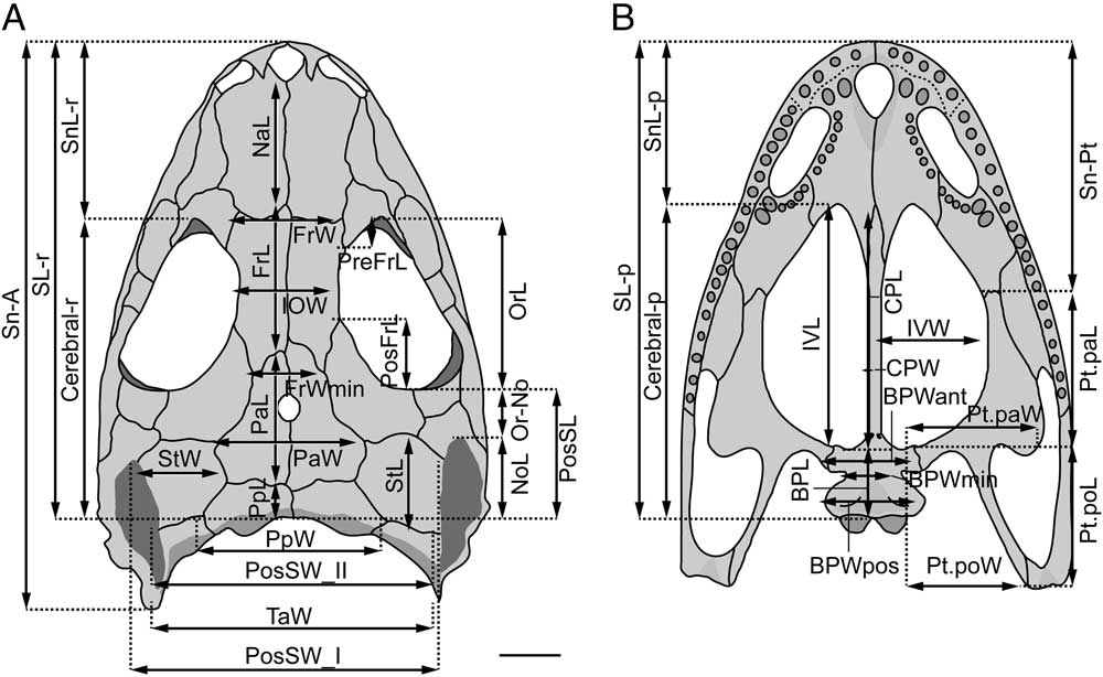
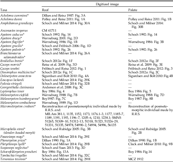
We calculated the allometric trends for several cranial variables to evaluate how morphology changes according to size in the dissorophoid clade and whether the smallest members present structures that might reflect size constraints imposed by an extreme size reduction. We focused the study on the skull roof and palate, avoiding the lateral regions, because in some species elements in these regions (e.g., jugal) are poorly preserved, which could result in erroneous or uncertain measurements. In addition, some taxa are only represented by two-dimensionally preserved material and, therefore, the lateral elements that had a vertical component in natural position are not morphometrically comparable to those of three-dimensionally preserved specimens, their reconstructions being highly subjective. We defined the morphometric variables according to the objective of evaluating structures that might be affected by size constraints and also testing previous statements on miniaturization in temnospondyls based on qualitative observations (Fig. 3). We collected 23 linear measurements of dermal bones of the skull roof and palate (Fig. 3), some of which served as proxies for the proportions of the nervous structures, because the neurocranium is not ossified in branchiosaurids and is usually poorly preserved in amphibamids. The width of the braincase was estimated by the width of the articulation between frontals and parietals (FrWmin), BWant, and BWmin. In temnospondyls, frontals, parietals, and postparietals form the roof of the neurocranium. In well-preserved specimens of Apateon pedestris, we determined that FrWmin reflects more accurately the width of the braincase than any other dorsal measurement of the dermatocranium, because at this level, the lateral flanges of the frontals are reduced to their minimum. Frontal bones bearing ridges just lateral to the braincase have also been described in the terrestrial amphibamid Tersomius (Carroll Reference Carroll1964). In palatal view, BWant matches the distance between the basipterygoid articulations and BWmin reflects the maximal possible ventral width of the basisphenoid at that level, at least in the species in which a parabasisphenoid complex was present (e.g., Acheloma dunni [Maddin et al. Reference Maddin, Reisz and Anderson2010]; Doleserpeton [Sigurdsen and Bolt Reference Sigurdsen and Bolt2010]). With respect to the size of the otic capsules, we selected BWpost as the best proxy for the width of these structures based on firsthand observations of specimens of dissorophoids with well-preserved, ossified otic capsules (e.g., Tersomius texensis) and calcareous nodules (interpreted as fillings of the endolymphatic sacs by Boy [Reference Boy1972]) in neotenic species, in which these structures remained unossified. All these measurements were taken from photographs of specimens and published reconstructions of 31 species (Table 1) that represent the total range of body sizes within the dissorophoid clade. We only included taxa whose phylogenetic placement is reasonably known and represented by specimens to which we had access by firsthand observation or through quality photographs. Only metamorphosed individuals were used, except for the species for which only neotenes are known (i.e., branchiosaurids, excepting Apateon gracilis). For each species we selected the largest individuals available to us; when we used reconstructions, we restricted the measurements to those of the structures that are well preserved in the original material. All measurements were taken using Adobe Photoshop CS5.
Reconstruction of skull of the large dissorophoid Cacops morrisi (modified from Schoch and Milner Reference Schoch and Milner2014) in (A) dorsal and (B) ventral views with the linear variables used in the allometric analyses. Scale bar, 10 mm.

List of temnospondyl taxa used in the present study with literature sources or specimens from which images were digitized. Asterisks (*) indicate taxa observed firsthand by one of the authors (C.P.-B.). Institutional abbreviations: CM, Carnegie Museum of Natural History, Pittsburgh, Pa., U.S.A.; MB, Museum für Naturkunde, Berlin, Germany; MCZ, Museum of Comparative Zoology, Harvard University, Cambridge, Mass., U.S.A.; SMNS, Staatliches Museum für Naturkunde, Stuttgart, Germany.

To recognize paedomorphic features in the adults of the smallest dissorophoids and differentiate them from those features potentially related to body-size constraints, we studied the allometries of the morphometric variables along the ontogeny of Micromelerpeton credneri and compared them with the evolutionary patterns. This also allowed us to evaluate whether mechanisms that lead to ontogenetic truncation were involved in the phylogenetic size reduction. Only variables of the skull roof were considered because the palate is seldom preserved in the available specimens. We selected this species as a model because it is medium-sized and has a basal phylogenetic placement within Dissorophoidea, possessing many putative plesiomorphic morphological and developmental features (Schoch Reference Schoch2014b). In addition, many well-preserved individuals representing different body sizes of a single “time-averaged” population are available. Although allometric studies on this species have been previously carried out by Witzmann and Pfretzschner (Reference Witzmann and Pfretzschner2003) in their very informative paper on its ontogeny, a study following the same protocol of our interspecific analysis was necessary to make comparisons between the developmental and evolutionary patterns. Furthermore, in contrast to the previous study, we restricted the sample to individuals of the time-averaged population from the lacustrine Ordenheim-Bank of Ordenheim locality (Table 1). Boy (Reference Boy1995) suggested that Micromelerpeton was facultatively neotenic and that only neotenes are probably present in the Ordenheim-Bank, in contrast to the Jeckenbach-Bank near Niederkirchen from which putative postmetamorphic individuals are known (Witzmann and Pfretzschner Reference Witzmann and Pfretzschner2003).
Calculation of Evolutionary and Ontogenetic Allometries
Evolutionary and ontogenetic allometries were first calculated using skull length as a proxy for body size. Because positive allometry was recovered for the snout (i.e., SnL-r and SnL-p variables; Fig. 3, Table 1) and this might affect the patterns of the other variables, analyses were also conducted using the variable “Cerebral-r” as proxy for body size for the skull roof and “Cerebral-p” (Fig. 3) for the palate. These proxies of size closely match the length of the braincase, which is a relatively conserved structure in temnospondyls and therefore more reliable for interspecific comparisons than the skull length, which includes the highly variable snout length.
We studied the evolutionary allometry of each variable using two phylogenetic comparative methods: phylogenetic independent contrasts (PICs; Felsenstein Reference Felsenstein1985) and phylogenetic general least squares (PGLS; Martins and Hansen Reference Martins and Hansen1997; Rohlf Reference Rohlf2001), both suitable to deal with the phylogenetic nonindependence of this kind of data (Rohlf Reference Rohlf2001; Garland et al. Reference Garland, Bennett and Rezende2005). We performed the analyses using each of the three fully resolved topologies.
Given that these methods require specifications of branch lengths and the topologies used to construct the supertree lack this information, because they are the result of cladistic analyses, we set all branch lengths to one, which corresponds to a speciational model of evolution (Garland et al. Reference Garland, Harvey and Ives1992), following previous evolutionary allometric analyses (e.g., Yeh Reference Yeh2002; Kimmel et al. Reference Kimmel, Sidlauskas and Clack2009).
We calculated the standardized PICs of the logarithms of the data using the R package ‘ape’ (Paradis et al. Reference Paradis, Claude and Strimmer2004) and checked the correct standardization, as was suggested by Garland et al. (Reference Garland, Harvey and Ives1992), with the caic.diagnostic function of the ‘caper’ package (Orme et al. Reference Orme, Freckleton, Thomas, Petzoldt, Fritz, Isaac and Pearse2013). We then performed ordinary least-squares regressions of the standardized PICs using the standardized PICs of body-size proxies (i.e., SL-r and Cerebral-r for the skull roof, and SL-p and Cerebral-p for the palate) as independent variables. We calculated the regressions through the origin, as Garland et al. (Reference Garland, Harvey and Ives1992) did.
Following the linearized allometric equation, log Y=k log X + b that describes how a dependent variable Y varies according to an independent reference of size (X), the slope (k) calculated in each of the previous regressions represents the evolutionary allometric coefficient of the regressed dependent variable (Y) (Huxley Reference Huxley1932). The 95% confidence intervals of the allometric coefficients were calculated by resampling methods to assess the allometric patterns statistically (Kowalewski and Novack-Gottshall Reference Kowalewski and Novack-Gottshall2010). Confidence intervals at 95% that include the value 1 indicate isometry, whereas allometric coefficients lower or higher than 1 represent negative or positive allomety, respectively.
PGLS regressions under a Brownian motion model of evolution were carried out for the logarithms of the morphological variables against the logarithms of body-size proxies using the R package ‘caper’ (Orme et al. Reference Orme, Freckleton, Thomas, Petzoldt, Fritz, Isaac and Pearse2013). Residuals were calculated for each PGLS analysis. Residuals of the small species were calculated for all analyses (i.e., even when they were excluded in the PGLS; see later in this section). Box plots of residuals and plots of residuals against the fitted values were generated to visualize deviations.
As demonstrated by Blomberg et al. (Reference Blomberg, Lefevre, Wells and Waterhouse2012), the slope parameter of an ordinary least-squares regression of PICs conducted through the origin is identical to that of a PGLS regression under a Brownian model. Despite the redundancy of these methods to estimate the slope parameter, both were carried out because they complement each other in further analyses: while the PICs method allows a graphical display of the regression and simpler calculations of the confidence intervals, the residuals of the PGLS regression represent each single species, their interpretation being more straightforward than interpretations of the contrast residuals.
Taking into account that neoteny might be characterized by different evolutionary allometric patterns, we performed the analyses both considering all the sampled species of dissorophoids and restricting the sampling to metamorphic dissorophoids (i.e., excluding neotenic species). We also took into account that small species might affect the general allometric trends if their skull proportions departed greatly from those of the rest of the species (which is expected under miniaturization). If this were the case and these small forms were included in the linear regressions, deviations of these species from the general trend would be masked. To address this problem, we also calculated the allometries along metamorphic species, excluding the smallest forms (i.e., Amphibamus, Doleserpeton, and Apateon gracilis). To summarize, evolutionary allometries were calculated from three data sets: (1) total data set, (2) metamorphic taxa only, and (3) metamorphic taxa excluding small species.
To study the ontogenetic allometry of each variable in Micromelerpeton credneri, ordinary least-squares regressions of the logarithm of the variables on the logarithm of the proxies for body size (i.e., SL-r and Cerebral-r) were performed, and the 95% confidence intervals of the allometric coefficients (i.e., the slopes) were calculated by resampling methods. The allometric patterns were inferred from the confidence intervals as in the evolutionary study.
Results
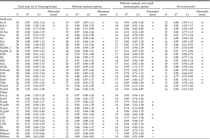
Results of the morphometric analyses are shown in Table 2. These results are the ones obtained using the skull length (SL-r and SL-p) as proxy for body size in the analyses of Sn-A, SnL-r, SnL-p, and Sn-Pt. The rest of the variables were analyzed using Cerebral-r and Cerebral-p for the skull roof and palate, respectively, as proxies for body size.
Results of the linear regressions. At the evolutionary level, where the analyses were carried out using three different topologies, only one representative result is reported for each variable. Allometric trends are only interpreted for regressions with an R 2 higher than> 0.7. All regressions with p<0.01. Abbreviations: =, isometry; +, positive allometry; –, negative allometry; N, number of specimens; CI, confidence interval; N, number of specimens.

Evolutionary Allometries
The PICs were adequately standardized using the unimetric trees, except for the few exceptions of the palate when only large metamorphic species were taken into account for the calculations. In the analyses of all the data sets, most variables showed a good fit to the linear model (i.e., R 2 >0.7). In comparison, fewer variables showed a good fit when only large metamorphosed taxa were used, probably because of the small sample size. The resulting qualitative allometric trends were rarely affected by the topology used to resolve the aforementioned polytomy. When the allometries did vary according to the topology used, the results from the majority were preferred and are reported here.
Most variables were recovered isometric in the analyses of the total data set. Positive allometry was recovered for the snout in dorsal and palatal views (i.e., SnL-r and SnL-p, respectively), PpL, and FrWmin. In contrast, orbit length and width of the basal plate of the parasphenoid (i.e., BWant, BWmin, and BWpost) showed negative allometries. These allometric patterns were recovered largely conserved when the neotenic species were excluded from the analyses. A noteworthy exception is the snout length (i.e., SnL-r and SnL-p), recovered as isometric instead of positively allometric, which is consistent with the exclusion of a large number of short-snouted forms such as branchiosaurids. With the further exclusion of the smallest metamorphic forms (i.e., Amphibamus, Apateon gracilis, and Doleserpeton), most of the allometric trends of the variables with a good fit to the linear model were recovered as before.
Box plots of the residuals of the PGLS showed that the smallest metamorphic dissorophoids were not outliers for any of the studied variables in the analyses based on the different data sets and tree topologies. In other words, small forms did not present distinctive deviations from the trends of the clade.
Comparisons between Ontogenetic and Evolutionary Allometries
When the total data set for dissorophoids was analyzed, 9 out of 18 variables of the skull roof with a good fit to the linear model showed the same allometric trend as in the ontogeny of Micromelerpeton. When the data set that includes only metamorphic taxa was used, the matching allometric trends between the evolutionary and ontogenetic levels were 8 out of 18, whereas if small species were excluded, the matches were 8 out of 16.
Discussion
Miniature vertebrates are not just dwarfed versions of closely related larger species; instead, they usually present novel morphological arrangements to maintain functional efficiency at a very reduced body size (Hanken and Wake Reference Hanken and Wake1993). As mentioned earlier, reduction of the body size in tetrapods usually correlates with novel cranial morphologies related to the increased relative size of nervous and sensory organs. Three additional features have been repeatedly documented in miniaturized tetrapods: reduced ossification, hyperossification, and increased variation (Hanken Reference Hanken1993). Both reduced ossification and hyperossification frequently occur in one individual and even in the same structural unit, such as the skull (Hanken Reference Hanken1993; Hanken and Wake Reference Hanken and Wake1993). Reduced ossification ranges from a lower degree of mineralization to the complete lack of ossification of a cartilaginous precursor (Hanken Reference Hanken1993). It has usually been interpreted as resulting from ontogenetic truncation, but secondary reduction has also been documented when ontogenetic trajectories were analyzed (Hanken Reference Hanken1993). Hyperossification in miniaturized species is often interpreted as a compensation for the mechanical weakness that might result from decreased body size and its concomitant reduced ossification in some structures (e.g., Lombard and Wake Reference Lombard and Wake1977; Wake Reference Wake1986). As noted by Hanken (Reference Hanken1993), in such cases reduced ossification and hyperossification would be functionally linked and part of the same phenomenon of skeletal growth and adaptation. Increased variation of skeletal features has also been observed in miniaturized tetrapods at the population and individual (asymmetries) levels, ranging from changes in size to complete absence of bones (Hanken and Wake Reference Hanken and Wake1993).
Establishing reduction in body size as the cause for changes in ossification, increased variation, and appearance of morphological novelty is usually difficult and not always possible, especially if one considers that the evolutionary appearance of any of the features associated with miniaturized forms does not require the reduction in size and that the adult skeleton of miniaturized forms, as in nonminiaturized ones, is the result of both phylogenetic and ontogenetic constraints and functional modifications mediated by natural selection (Hanken Reference Hanken1983, Reference Hanken1984; Trueb and Alberch Reference Trueb and Alberch1985). This implies that a rigorous analysis of the interplay between miniaturization and morphological change must consider both ontogeny and phylogeny.
The phylogenetic position of small-sized individual species or clades of such species is essential to distinguish derived features that potentially might be the outcome of miniaturization from synapomorphies whose appearance precedes the size reduction. Furthermore, an explicit phylogenetic framework allows evaluating whether the distinctive features of the small forms are extrapolations of trends already present in the lineage to a very small body size, depart from these trends, such as when a minimum functional size is reached, or represent whole new structures. Also, the phylogenetic framework might be relevant to understand differential responses to the size decrease of closely related taxa. In this regard, in her study on miniaturization in anurans, Yeh (Reference Yeh2002) suggested that the resistance of some clades, such as that of some miniature dendrobatid frogs, to typical patterns of evolution might be due to their origin from miniature ancestors.
As mentioned earlier, adult miniature tetrapods frequently resemble juvenile stages of larger related species in many traits (Hanken and Wake Reference Hanken and Wake1993). This resemblance might be due either to the retention of juvenile traits of the ancestral ontogeny (i.e., paedomorphosis) that results from mechanisms of ontogenetic truncation or to an extension of the ancestral ontogenetic trajectory wherein the juvenile features are secondarily achieved (e.g., reduced ossification by resorption). The distinction between these two developmental routes is relevant because they imply that the ancestral pathways are largely conserved or have undergone more fundamental alterations, respectively (Hanken Reference Hanken1993). Also, whereas paedomorphosis is related to the causes of the reduction of size, secondary juvenile traits might be consequences of the reduction in size.
In the case of the dissorophoid temnospondyls, it is important to consider whether the morphology of the smallest members of this group is consistent with some of these extensively documented morphological consequences of miniaturization in the skeleton: reduced ossification, hyperossification, and morphological novelty. Assessment of how the morphology changes with changes in size within Dissorophoidea both qualitatively and quantitatively (i.e., evolutionary allometry) and comparisons of adults of small species with juveniles and larvae of larger taxa led us to distinguish those features that might be the consequence of the size reduction from those related exclusively to ontogenetic truncation.
In dissorophoids, the cranial morphology of the smallest forms has been considered affected by miniaturization because of the need to accommodate the proportionally larger eyes, brain, and otic capsules (Schoch Reference Schoch2012b). However, these forms do not show major rearrangements of the skull roof and ventromedial shift of the suspensorium (i.e., verticalization) in comparison with those resulting from packaging effects in extant miniaturized salamanders and frogs (Hanken Reference Hanken1984; Yeh Reference Yeh2002). Instead, the typical temnospondyl disposition of the cranial dermal bones is conserved even at the minimum skull sizes. Furthermore, most of the morphometric variables that we evaluated were recovered isometric in dissorophoids, and the smallest metamorphic species did not depart from this trend, suggesting a largely conserved morphology within the clade, even at small sizes. The palatal configuration also seems to be conserved in dissorophoids with two noteworthy exceptions: Doleserpeton and branchiosaurids. Doleserpeton departs from other dissorophoids in lacking, or having greatly reduced, ectopterygoids (Sigurdsen and Bolt Reference Sigurdsen and Bolt2010; Fig. 2). We were unable to evaluate the evolutionary allometries of these bones quantitatively because of their poor preservation in most of the studied specimens. However, the larger, closely related Platyrhynops and other amphibamids have small ectopterygoids in comparison with those of more distantly related dissorophoids, suggesting that the reduced size of these bones in Doleserpeton might be an extrapolation of this trend. In the case of the branchiosaurids Apateon caducus and A. pedestris, the only two temnospondyls for which detailed ossification sequences of the skull are available, the ectopterygoid is the last palatal element to form (Schoch Reference Schoch1992, Reference Schoch2002); hence, reduction and even absence of ectopterygoids might be interpreted as paedomorphic. Branchiosaurids show a remarkably distinctive morphology of the palate in which the ectopterygoid and most part of the palatine are not in contact with the maxilla. Nonetheless, this arrangement is symplesiomorphic for Branchiosauridae, widespread in small and large forms; also, it has been suggested that it was part of an integrated whole adaptive for filter feeding together with the brushlike branchial denticles (Schoch and Milner Reference Schoch and Milner2008). In summary, the novel palatal morphologies of Doleserpeton and branchiosaurids do not seem to be outcomes of the decrease in size, but they might be related to ontogenetic truncation and adaptation. Therefore, as in the case of the skull roof, no major rearrangement of the palate, either related or unrelated to nervous system constraints, can be attributed unambiguously to packaging effects as in extant amphibians.
Our analyses of allometries allowed us to test whether more subtle morphological changes of the dermatocranium associated with small skull sizes reflect constraints imposed by miniaturization in eyes, brain, and otic capsules. Negative evolutionary allometric patterns were expected for these variables, indicating that the structures are proportionally larger in small forms. On the other hand, highly positive residuals for small species would indicate that a minimal functional size for these structures was reached.
Previous studies have proposed that small temnospondyls have oversized orbits, a by-product of rudimentary circumorbital bones, to accommodate the comparably huge eyes (Schoch and Rubidge Reference Schoch and Rubidge2005; Schoch Reference Schoch2012b). Accordingly, the larger interpterygoid vacuities documented in small forms, resulting from reduced palatal elements, have been associated with correspondingly larger eye muscles (Milner Reference Milner1988; Schoch and Rubidge Reference Schoch and Rubidge2005). We did recover negative allometry for the orbit length in dissorophoids, indicating proportionally larger orbits in forms with smaller skull sizes (although not necessarily because of relatively smaller pre- and postfrontal bones, whose size did not correlate to skull size). However, the length and width of the interpterygoid vacuities scaled isometrically with skull size. Because the size of the orbit does not correlate with the size of the sclerotic ring, at least in Micromelerpeton (Witzmann and Pfretzschner Reference Witzmann and Pfretzschner2003), and the latter is a better indicator of the eye size, the former variable alone is not an accurate proxy for eye size; therefore, we consider that there is not enough evidence of a correlation between proportionally larger eyes and decreasing skull size.
In our analyses of the proportions of the braincase, FrW (Fig. 3) scaled with positive allometry with body size when all the species were taken into account and isometrically when only metamorphic ones were included, while BWant and BWmin (Fig. 3) showed negative allometry for the total data set and the one excluding neotenic species, and isometry when the smallest metamorphic species were also excluded. The latter result is consistent with the conservative trend of larger brains in smaller forms among tetrapods as a consequence of size constraints; however, the smallest species evaluated for this character (i.e., Amphibamus and Doleserpeton) did not depart significantly from the general trend, indicating that no further size limitations affected their morphologies. Furthermore, a broad basal plate can be interpreted as paedomorphic because it was noted in qualitative studies of the ontogenies of Micromelerpeton (Boy Reference Boy1995) and Acheloma dunni (Maddin et al. Reference Maddin, Reisz and Anderson2010), a large dissorophoid, that this structure has a negative allometric pattern.
Regarding the size of the otic capsules, the proxy (BWpost; Fig.3) did show the expected pattern of negative allometry, indicating that otic capsules do tend to be proportionally larger in smaller species, although Doleserpeton and Amphibamus did not depart from the general trend of the clade. However, as in the case of the other variables related to the width of the basal plate (i.e., BWant and BWmin, proxies of braincase width), a proportionally high BWpost seems to be a paedomorphic feature.
Another critical organ system that has been proposed to potentially impose morphological changes to the skull under miniaturization in temnospondyls is the tympanic–stapedial system (Schoch and Rubidge Reference Schoch and Rubidge2005; Schoch Reference Schoch2012b), the presence of which in North American dissorophoids was convincingly demonstrated by Bolt and Lombard (Reference Bolt and Lombard1985). In Amphibamus and Doleserpeton, the tympanum lay partly within a large otic notch, which was described as medially expanded, leading to a constricted posterior skull (Schoch and Rubidge Reference Schoch and Rubidge2005). According to our results, the skull width at the level of the articulation between supratemporals and tabulars (i.e., PosSW_I; Fig. 3) is isometric in dissorophoids, instead of being positively allometric, as might be expected for a disproportionally medially expanded otic notch in small forms. On the other hand, no relationship was found between small size and length of the otic notch (i.e., NoL).
With regard to the degree of ossification, it is difficult to interpret this in the context of small body size in the case of amphibamids because this feature is seemingly linked to habitat: terrestrial taxa are much more ossified than aquatic branchiosaurids of similar size, as the neurocranium, otic capsules, quadrates, and vertebral centra are ossified in the former group but not in the latter. A further difficulty is that the known individuals of the terrestrial Amphibamus have been interpreted as juvenile postmetamorphic forms (Carroll Reference Carroll1964; Bolt Reference Bolt1979; Daly Reference Daly1994; Anderson et al. Reference Anderson, Reisz, Scott, Fröbisch and Sumida2008). In the case of Apateon gracilis, the lack of ossification of the neurocranium and otic capsules might be interpreted either as a juvenile condition or as owing to a more aquatic lifestyle than that of amphibamids. In contrast, Doleserpeton shows a mosaic of different degrees of ossification, a situation similar to that documented in extant miniaturized taxa (Hanken Reference Hanken1993). Whereas the dermal elements of the skull and girdles are lightly built and the external surfaces of the dermal skull bones are smoother than in any other amphibamid, including the also small Amphibamus (Sigurdsen and Bolt Reference Sigurdsen and Bolt2010), the braincase and some parts of the appendicular skeleton are highly ossified. In contrast, the braincase of Doleserpeton is more highly ossified than that of any other amphibamid, including the much larger Pasawioops (Fröbisch and Reisz Reference Fröbisch and Reisz2008). Furthermore, both the prootic and opisthotic are ossified, whereas in other larger amphibamids such as Micropholis and Tersomius, only the latter bone is present (Carroll Reference Carroll1964; Schoch and Rubidge Reference Schoch and Rubidge2005). Also, in some specimens of Doleserpeton, the distal ends of the humeri are highly ossified (Sigurdsen and Bolt Reference Sigurdsen and Bolt2010), unlike those of other known amphibamids. No such variation in the degree of ossification has been described for Amphibamus or Apateon gracilis.
Paedomorphosis, Miniaturization, and Morphological Evolution
When the ontogenetic trajectory of dissorophoids is considered, the morphology of small metamorphic dissorophoids clearly resembles that of immature stages of larger taxa, including features that have been associated with miniaturization. Given the extent of the resemblance, a truncation of the ancestral ontogenetic trajectory that results in paedomorphic features appears to be the most parsimonious explanation for this pattern (although an alternative possibility was considered earlier). In this context, paedomorphosis would not have been restricted to neotenic branchiosaurids and micromelerpetontids among dissorophoids. In contrast to previous proposals, poor dermal ornamentation, overall thin dermal bones, and some rudimentarily developed skull bones in small metamorphic dissorophoids were recovered here as paedomorphic and not as consequences of miniaturization. The former two features (Schoch and Rubidge Reference Schoch and Rubidge2005; Schoch Reference Schoch2012b) are consistent with paedomorphosis because ossification and ornamentation increase throughout the ontogeny (Boy Reference Boy1974; Schoch Reference Schoch1992) and there is no evidence of bone resorption in any known temnospondyl taxon. Rudimentarily developed bones usually associated with miniaturization are circumorbital bones (i.e., prefrontal, postfrontal, and postorbital), tabulars in the skull roof, and ectopterygoids in the palate. Because these bones form late in the ontogeny of the branchiosaurids Apateon pedestris and A. caducus (Schoch Reference Schoch1992, Reference Schoch2002), under ontogenetic truncation they are expected to be relatively smaller than other dermal bones that form earlier, such as frontals and parietals. Oversized otic capsules also have been related to miniaturization (Schoch and Rubidge Reference Schoch and Rubidge2005; Schoch Reference Schoch2012b) and, as discussed earlier, are consistent with both the general evolutionary negative allometric trend in tetrapods, which is also recovered for dissorophoids, and the ontogenetic negative allometric trend in temnospondyls. Therefore, enlarged otic capsules are not strictly an outcome of a precocious truncation of the ontogeny but are also related to the conservative retention of an ancestral pattern of tetrapods. Based on comparisons with juveniles of Acheloma, Maddin et al. (Reference Maddin, Reisz and Anderson2010) proposed in addition that an anteriorly restricted dental field on the basal plate of the parasphenoid, a basal plate wider than long, and a gap between exoccipitals and postparietals are all evidence of paedomorphosis in amphibamids. One-half of the morphometric variables of the skull roof that we evaluated show the same allometric trend at the evolutionary and ontogenetic levels, even when neotenic branchiosaurids are excluded from the analyses, a result that adds evidence of paedomorphic features in metamorphic dissorophoids.
The smallest known metamorphic dissorophoids (i.e., Doleserpeton, Amphibamus, and Apateon gracilis) resemble miniaturized extant frogs and salamanders in having an overall paedomorphic morphological condition. However, the cranial morphologies of these temnospondyls do not match the radical novel arrangements of the skull documented in miniaturized extant taxa, indicating that the impact of the size reduction was less extreme in temnospondyls. Although this might be due to a differential resistance to constraints imposed by a small body size, a more parsimonious explanation is that the decrease in size was less severe in temnopondyls than in extant amphibians. In fact, the skull length of the smallest known temnospondyls is about 20 mm, whereas those of some miniaturized extant lissamphibian taxa reach 2 mm. In addition, a recent study on the evolution of the genome size in several extant and fossil vertebrate groups (Organ et al. Reference Organ, Struble, Canoville, de Buffrénil and Laurin2016) showed that the dissorophoids, as well as the other few temnospondyls considered, had smaller genome sizes than extant frogs and salamanders, indicating that temnospondyls were not only physically but also biologically larger than miniaturized batrachians. In contrast to extant amphibians, the reduction in body size in dissorophoids might have not reached the threshold at which the extrapolation of evolutionary allometries and minimum functional sizes result in novel morphological arrangements.
From the foregoing, it seems that the truncation of the ancestral ontogeny, and not miniaturization, might have been the factor that played a major role in the morphological evolution of small dissorophoids. In this context, the heterochronic processes related to the origin of extant amphibian groups from small amphibamids proposed by Bolt (Reference Bolt1979), Roček and Rage (Reference Roček and Rage2000), Boy and Sues (Reference Boy and Sues2000), and Schoch (Reference Schoch2014a), among others, would be an extrapolation of a trend already present in dissorophoids.
Miniaturization or Heterochrony as Major Factor in the Origin of Lissamphibians?
Miniaturization has been invoked in the origin of modern amphibians (lissamphibians) (Milner Reference Milner1988; Schoch Reference Schoch1995). This was proposed as an explanation for far-reaching architectural changes in the skull and pectoral girdle, which correlate with the loss of a number of dermal bones. In fact, all three extant amphibian clades lack the interclavicle in the pectoral girdle and the postfrontal, postorbital, jugal, supratemporal, and tabular bones in the skull roof, at least as discrete bony elements. When only living species are examined, this set of absence characters gives a distinct pattern that has been used as a source of character codings in phylogenetic analyses, giving additional support to a scenario in which the putative common ancestor of lissamphibians lost all these dermal elements presumably not stepwise but in concert, thus suggesting some major evolutionary rearrangement. The concerted loss of so many bones not only prompted an attractive parsimonious hypothesis but also revealed a pattern that might be readily explained by an evolutionary hypothesis: miniaturization.
This hypothesis of miniaturization probably emerged because many lissamphibians are substantially smaller than even the tiniest temnospondyls (Amphibamidae, Branchiosauridae). Furthermore, developmental biologists have proposed an explanation for this evolutionary loss of bones. They report that the formation of bony tissue depends on the size of stem cell populations in the mesenchymal tissue layer in which these bones form (Atchley and Hall Reference Atchley and Hall1991). For instance, in the mouse, ossification centers of the mandible can only form if mitosis of stem cell condensations produces population sizes large enough to reach the state in which they may initiate bone growth. In the miniaturization scenario, size reduction might have reduced the number of mesenchymal cells beyond the condition at which they could still maintain operational cellular condensations, thus precluding formation of certain ossification centers. Bone loss may thus not have been selected specifically but instead may be a by-product of body-size reduction.
The discovery and anatomical description of the stem caecilian Eocaecilia micropodia have added substantial new evidence (Jenkins et al. Reference Jenkins, Walsh and Carroll2007). Whereas the geologically oldest and most plesiomorphic stem anuran Triadobatrachus and stem salamanders Karaurus and Kokartus already lack the full complement of dermal skull bones (Skutschas and Martin Reference Skutschas and Martin2011; Ascarrunz et al. Reference Ascarrunz, Rage, Legreneur and Laurin2016), Eocaecilia retains most of these elements (Maddin and Anderson Reference Maddin and Anderson2012). The arrangement and morphologies of these elements indicate that stem caecilians had a temnospondyl-like skull after they diverged from the common ancestor of batrachians (salamanders and frogs). This evidence points to the loss of the same set of dermal skull bones asynchronously in caecilians and batrachians, precluding a single event of miniaturization in lissamphibian evolution from temnospondyls.
An alternative scenario based on the evolution of development rather than the morphological consequences of extreme reduction in body size can be proposed. The study of the cranial ossification sequence in the branchiosaurid temnospondyl Apateon revealed remarkable similarities with the development of basal salamanders, such as the extant Siberian Ranodon (Schoch Reference Schoch2002). In Apateon the dermal skull bones develop in a sequence that closely parallels that of salamanders. Interestingly, the last bones to appear in the skull of Apateon are consistently absent in salamanders and other lissamphibians. This supports an alternative scenario to that of miniaturization, in which dermal bones were lost due to a truncation of the developmental trajectory. This hypothesis of heterochrony is consistent with other observations of instances of heterochrony and thus a likely factor in lissamphibian origins (Schoch Reference Schoch2014a).
Acknowledgments
The authors thank D. B. Wake (Museum of Vertebrate Zoology, University of California, Berkeley, California), M. A. Norell (American Museum of Natural History, New York), F. Witzmann (Museum für Naturkunde, Berlin, Germany), O. W. M. Rauhut (Bayerische Staatssammlung für Paläontologie und Geologie, Munich Germany), K. Padian (University of California Museum of Paleontology, Berkeley, California), and S. E. Pierce (Museum of Comparative Zoology, Harvard University, Cambridge, Massachusetts) for specimen access. C. P.-B. also thanks Luigi Mastrangelo for fruitful discussions. The article was greatly improved by suggestions from anonymous reviewers. This work was funded by doctoral fellowships from CONICET, DAAD together with the Ministry of Education of Argentina, and the Konrad Lorenz Institute (awarded to C.P.-B.).