Autoimmune disease, characterised by a host immune response against self-antigens, affects more than 8% of the world's population (Ref. Reference Cooper, Bynum and Somers1). As for complex diseases in general, autoimmune disease is said to be caused by a combination of genes and environmental factors. The involvement of genes in autoimmune disease is unequivocal, with hundreds of risk loci identified across the more than 80 different diseases, many shared between diseases (Refs Reference Cotsapas2, Reference Javierre, Hernando and Ballestar3, Reference Richard-Miceli and Criswell4). The involvement of environmental factors in autoimmune disease is less well established (Ref. Reference Ellis, Munro and Ponsonby5), but is becoming increasingly recognised. Most research effort to date has focused on identifying these factors in isolation. However, it is likely that at least a component of overall risk (or conversely, protection) will be determined by the interaction of the genes with the environment; that is, the degree of disease risk contributed by one factor will be influenced by the presence or absence of another (Fig. 1). Evidence for such interactions in autoimmune disease is now beginning to emerge, and is providing important mechanistic insight into disease causation.
The involvement of genetic and environmental factors in disease. Genes and the environment may influence disease risk independently, or they may interact to determine risk. There are many models for such an interaction, for example, the environment might influence the risk conferred by the genes, the genes might influence the risk conferred by the environment, or both factors might be necessary for any impact on disease risk. The environment may impact the genome via epigenetic mechanisms such as alterations to DNA methylation.

In this review, following a brief summary of current knowledge as to the genetic and environmental risk factors for autoimmune disease, we will focus on the data generated to date that support the concept of gene–environment (G–E) interaction as an important component to overall autoimmune disease risk. While such interactions are often initially identified using statistical approaches, importantly, various molecular mechanisms of interaction between these intrinsic and extrinsic factors are also being studied to support the statistical findings. Thus, our understanding of the causes of autoimmune disease is growing significantly through multifaceted approaches to research, and an ongoing focus on G–E interaction, although challenging, is highly justified.
What is autoimmune disease?
‘Autoimmunity’ has been defined as immune activation in the absence of known external drivers such as infection (Ref. Reference Davidson and Diamond6). ‘Autoimmune disease’ refers to the progression of autoimmunity to a pathogenic state. Both the adaptive and innate immune systems are thought to play a role in autoimmunity. In contrast to autoinflammatory conditions, where innate immune dysregulation is predominant, in autoimmune disease, adaptive dysregulation of the T cell and/or B cell populations also occurs (Refs Reference Doria7, Reference Hayter and Cook8). In relation to the adaptive immune system, self-tolerance is a highly regulated immune process in that self-reactive T and B cells are normally produced by the immune system but are selected against via various central and peripheral tolerance mechanisms. For example, centrally in the thymus, thymocytes are exposed to a vast array of self-peptides. Approximately 98% of thymocytes are found to be self-reactive and are destroyed. In the periphery, production of T cells from the remaining functionally competent (and non-self-reactive) thymocytes is tightly controlled by genomic factors, such as FOXP3. Comparable processes occur for the regulation of B cell production from the bone marrow (Ref. Reference Cho and Gregersen9). Most autoimmune diseases are believed to be caused by a breakdown of these immune tolerance processes via mechanisms that remain largely unclear. This results in inflammation and tissue destruction in a systemic- or organ-specific fashion (Ref. Reference Javierre, Hernando and Ballestar3). Examples of systemic autoimmune diseases include systemic lupus erythematosus (SLE), which may involve damage to skin, kidney, lungs, brain and heart (Ref. Reference Tsokos10); and rheumatoid arthritis (RA) that primarily affects the joints but can also involve inflammation of organs, such as the lungs and heart (Ref. Reference McInnes and Schett11). Examples of organ-specific autoimmune diseases include multiple sclerosis (MS) in which demyelination of the central nervous system occurs (Ref. Reference Ramagopalan12); and type 1 diabetes (T1D) that involves destruction of pancreatic beta cells (Ref. Reference Bluestone, Herold and Eisenbarth13). The classification of a disorder as autoimmune can be controversial, and Crohn's disease (CD) represents one disease where such controversy exists (Ref. Reference Behr, Divangahi and Lalande14).
Genetic risk factors for autoimmune disease
A key feature of most autoimmune diseases is familial clustering, indicating the presence of shared genes and/or shared environmental risk factors. A 2006 systematic review demonstrated familial clustering of RA, T1D, MS and autoimmune thyroid disease (Ref. Reference Somers15). It is assumed that this multi-disease clustering indicates an overlap in causal pathways. It is perhaps therefore not surprising that many identified genetic risk variants are common to more than one autoimmune disease, whereas others are specific to a single disease (Refs Reference Cotsapas2, Reference Richard-Miceli and Criswell4).
The association of genetic variation within the human leukocyte antigen (HLA) region on chromosome 6 with multiple autoimmune diseases has been well established for some time, and is often the single greatest contributory factor to genetic predisposition (Ref. Reference Cho and Gregersen9). Mechanistically, this likely reflects that HLA genes encode cell-surface antigen-presenting proteins. The HLA region is exceptionally variable at a sequence level, providing variability that is crucial in providing the capacity to present a variety of foreign, but also self-antigens, to the immune system. This may impact on individual immune tolerance in two ways – through the efficiency of presentation of self-antigens to thymocytes in order to eliminate those that show self-reactivity, and in the determination of the repertoire of antigens able to be recognised by T cells in the periphery (Ref. Reference Thorsby and Lie16). Associations of particular HLA alleles are often specific to a particular autoimmune disease (Ref. Reference Cho and Gregersen9), reflecting that the relevant self-antigen is likely to be different for each form of disease.
Outside of the MHC, the advent of the genome-wide association study (GWAS) approach has seen the number of identified loci associated with various autoimmune diseases reach the hundreds in recent years (Ref. Reference Cho and Gregersen9). Interestingly, these genes generally fall into distinct functional network groups, most prominently lymphocyte activation and intracellular signalling (e.g. PTPN22, CTLA4, PTPN2), cytokines and receptors (e.g. IL23R, IL2RA), innate immunity and microbial recognition (e.g. NOD2, IRF5) and transcription factors (e.g. STAT4). Recently several analyses have shown that the degree of sharing of genetic effects between autoimmune diseases is highly significant (Refs Reference Cotsapas2, Reference Richard-Miceli and Criswell4). For example, Cotsapas et al. examined 107 autoimmune disease loci in relation to RA, T1D, SLE, MS, celiac disease (CeD), CD and psoriasis. They presented evidence that single nucleotide polymorphisms (SNPs) at 47 loci are associated with more than one immune-mediated disease, where only one was expected by chance. One SNP at SH2B3 (involved in T cell receptor activation signalling), showed evidence of association to all seven diseases. Others associated to various disease subgroups, for example a SNP at PTPN22 associated with RA, SLE, CD and T1D; at CTLA4 with RA, CeD and T1D; and at IL23R with psoriasis, MS and CD (Ref. Reference Cotsapas2).
Despite recent progress, for each distinct autoimmune disease, much of the heritability remains unaccounted for (Ref. Reference Cho and Gregersen9). A 2009 review of the ‘missing heritability’ of various complex diseases indicated that only 15 and 20% of the heritability of SLE and CD respectively could be explained by identified risk loci (Ref. Reference Manolio17). Currently, it is still estimated that over half of the genetic predisposition to autoimmune disease is still unable to be explained (Ref. Reference Cho and Gregersen9). It may be found within multiple yet-to-be-identified common variants contributing very modestly to overall risk, in rare variants that are not adequately captured by GWAS, or in gene–gene (G–G) interaction (Refs Reference Manolio17, Reference Eichler18). Furthermore, and the subject of this review, the missing heritability is likely to reflect that genes do not operate alone but also in the context of the environment, leading to interactions between risk loci and environmental risk factors (Refs Reference Manolio17, Reference Eichler18).
Environmental risk factors for autoimmune disease
The environmental risk factors associated with autoimmune disorders are varied, as is the strength of evidence for each. A comprehensive review of this field was recently published by an expert panel assembled by the US National Institute of Environmental Health Sciences (Ref. Reference Miller19). The various exposures and the strength of the evidence in various diseases and further research requirements were outlined. Here, we briefly focus on a selection of these environmental factors as a necessary introduction to the consideration of G–E interaction in autoimmune disease.
Tobacco smoke
Tobacco smoke contains more than 6000 components, many of which are cytotoxic, carcinogenic, mutagenic and/or antigenic (Ref. Reference Arnson, Shoenfeld and Amital20). Studies of the role of tobacco smoke exposure in autoimmune disease are many and varied in terms of study quality, sample size and approach to exposure measurement. The most convincing and well-replicated association is with risk of developing RA. A recent meta-analysis of over 13 000 cases from 16 studies in which a primary purpose was to examine the relationship between smoking status and RA (Ref. Reference Sugiyama21), confirmed the relationship. The meta-analysis demonstrated that smoking is a risk factor for RA for both men (around twice the risk) and women (about 1.3 times the risk, when compared with non-smokers). It mattered little whether smoking status was classified as ever, current or past. For MS, evidence for association with smoking is also quite strong, as demonstrated by a recent meta-analysis of 14 studies (Ref. Reference Antonovsky22). For SLE, the evidence is somewhat weaker. A meta-analysis of nine studies demonstrated a significant increase in risk in those who were current smokers but there was no increase in risk among those who were former smokers (Ref. Reference Costenbader23). In relation to autoimmune thyroid diseases, smoking has also been associated with an increased risk of Grave's hyperthyroidism and particularly with Grave's ophthalmopathy, in a dose-dependent fashion. It also appears associated with a decreased risk of the hypothyroid disease Hashimoto's thyroiditis (Ref. Reference Wiersinga24). The biological mechanisms for these associations remain unknown. Other autoimmune diseases examined in relation to smoking include CD and ulcerative colitis, however; studies have been more limited and the outcomes more variable [for review, see (Ref. Reference Miller19)].
Sun exposure and vitamin D status
Sun exposure provides ultraviolet radiation (UVR) and thus assists the generation of UV-derived vitamin D. There is a growing body of evidence of association of low vitamin D and autoimmune disease risk but the specific roles of UVR and vitamin D are difficult to tease apart (Ref. Reference Hart, Gorman and Finlay-Jones25). For MS, a striking ecological feature is the association between higher latitude, thus reduced ambient UV levels, and increased prevalence [e.g. (Refs Reference Hammond26, Reference Kurtzke, Beebe and Norman27, Reference Taylor28, Reference Vukusic29)]. Markers of individual low past sun exposure (Ref. Reference van der Mei30), season of birth (Refs Reference Dobson, Giovannoni and Ramagopalan31, Reference Staples, Ponsonby and Lim32), and low maternal exposure to sun (Ref. Reference Staples, Ponsonby and Lim32), have all provided further support for a role of low sun exposure in increasing MS risk. Low vitamin D has also been associated with higher MS risk (Ref. Reference van der Mei33). In a prospective US Military cohort, serum 25-hydroxyvitamin D was measured in samples collected prior to disease onset, and risk of MS significantly decreased with increasing vitamin D (Ref. Reference Munger34). In a cohort of US nurses, vitamin D supplementation was also associated with reduced MS onset (Ref. Reference Munger35).
For some other autoimmune diseases, similar but often less well-replicated associations with both sun exposure and vitamin D, have been identified. In relation to latitude, there is published evidence of an inverse association with T1D (Refs 36, 37), CD (Ref. Reference Armitage38) and RA (Refs Reference Ramos-Remus39, Reference Vieira40). A study by Disanto et al. recently examined the relationship between month of birth and a number of autoimmune diseases including RA, CD, UC and SLE. All diseases, with the exception of CD, showed seasonality (Ref. Reference Disanto41). T1D is also known to be related to season of birth (Refs Reference Kahn42, Reference Rothwell43). Lower serum vitamin D levels, and some evidence of a positive effect of supplementation, have been reported in diseases including RA and T1D (Refs Reference Bener44, 45, Reference Hypponen46, Reference Kroger, Penttila and Alhava47, Reference Merlino48, Reference Oelzner49, Reference Stene50).
Microbial exposure
There are two hypotheses related to microbial exposure that are not mutually exclusive. The first pertains to microbial infection as a trigger for the onset of disease. The second deals with what is commonly referred to as the ‘hygiene hypothesis’, whereby microbial exposure during early life serves to ‘prime’ the developing immune system, providing increased protection against immune disorders later in life.
There are multiple lines of evidence that support an association between Epstein-Barr virus (EBV) infection and higher risk of subsequent MS (Ref. Reference Lucas51). A meta-analysis of more than 19 000 cases and 16 000 controls demonstrated an increased risk of MS in those with a past history of infectious mononucleosis (Ref. Reference Handel52). Meta-analysis of 13 studies has reported an odds ratio (OR) of 13.5 for MS in those who are EBV seropositive (Ref. Reference Ascherio and Munch53). Evidence of primary EBV infection coinciding with MS presentation is lacking (Ref. Reference Wagner54), suggesting that it is prior EBV infection that is of greatest importance to disease initiation (Ref. Reference Lucas51). There is some evidence that EBV may also increase risk of developing other autoimmune diseases, including SLE (Ref. Reference Draborg, Duus and Houen55) and RA (Ref. Reference Meron56), but the data are generally less convincing (Ref. Reference Miller19).
A number of other microbes have also been considered in relation to autoimmunity. For parvovirus, anti-B19 antibodies have been reported as more prevalent in the plasma and synovial fluid of RA cases (Ref. Reference Chen57), however others failed to confirm these findings (Refs Reference Kozireva58, Reference Peterlana59). The observed co-occurrence of periodontitis and RA (Ref. Reference Chen60) has led to the hypothesis that Porphyromonas gingivalis may be a contributory factor to RA, and some data exist to support this (Refs Reference Mikuls61, Reference Mikuls62). There have been a number of studies considering the role of the gram-negative bacterium Helicobacter pylori in autoimmune disease risk, including in RA and SLE, but relationships remain unconfirmed [for review, see (Ref. Reference Hasni, Ippolito and Illei63)]. Enterovirus infection appears to be associated with T1D onset, as shown by meta-analysis drawing together data from 26 case–control studies (Ref. Reference Yeung, Rawlinson and Craig64).
The hygiene hypothesis, as proposed by Strachan in 1989 (Ref. Reference Strachan65), suggests that the decreasing incidence of early life infection through improvements to western hygiene is contributing to the increasing incidence of immune disorders such as allergic disease. The concept has also been postulated for autoimmune diseases (Ref. Reference Okada66). Higher exposure to siblings, a proxy for a higher early life microbial load, has been associated with a reduced risk of MS (Ref. Reference Ponsonby67) and T1D (Ref. Reference Cardwell68).
Diet
There is a well-established link between dietary elements, including vitamins, minerals and trace elements, and immune competence (Ref. Reference Selmi and Tsuneyama69). This suggests that the particular nutrients may associate with risk of autoimmune diseases. It has recently been shown that dietary micronutrients that affect DNA methylation can exacerbate or suppress murine lupus through genetic–epigenetic interactions (Ref. Reference Strickland70). In addition to vitamin D as discussed above, beneficial effects of supplementation of other nutrients have been demonstrated, such as vitamin A for psoriasis, and omega-3 fatty acids for RA and possibly SLE (Ref. Reference Selmi and Tsuneyama69). Diet can also modify the intestinal microbiota, which have been postulated to influence autoimmune disease (Ref. Reference Brown71). Recently, a high salt diet has been shown to promote the development of Th17 helper T cells, cells that are mediators of autoimmune inflammation (Ref. Reference Wu72). Increased NaCl concentrations at physiological levels markedly boosted the induction of murine and human Th17 cells, driving autoimmune disease (Ref. Reference Kleinewietfeld73). The consumption of cow's milk-based infant formulae has been implicated in the development of childhood T1D (Ref. Reference Virtanen74). A more obvious role for diet in autoimmune disease is apparent for CeD, where ingestion of gluten triggers disease onset (Ref. Reference Fasano and Catassi75).
The interaction of genes and environment – how is ‘interaction’ defined?
We now turn to current understanding of the ways in which genes are impacted by environment to determine risk of disease. Before considering the literature on G–E interactions in autoimmune disease, it is necessary to consider the meaning of ‘interaction’ in both a statistical and biological sense.
The term ‘interaction’ means, in general, a reciprocal action, effect or influence. In relation to genetic and environmental risk factors for disease therefore, it can be defined as the influence of one such risk factor on another, or a lack of independence between the two risk factors in determining disease risk. If two factors are independent, the presence or absence of the second risk factor has no influence over the degree of risk that is attributed by the first and vice versa. Put together, the attributable risk is the sum of the risk attributed by the two individual factors (Ref. Reference Klareskog76). Rothman's sufficient cause ‘pie-model’ states that two risk factors can be said to be independent where there is no pathway to disease that requires the involvement of both factors. Conversely, if one pathway to disease requires the presence of both factors, those factors are said to interact (Refs Reference Klareskog76, Reference Rothman77).
As reviewed by Wang et al., the term ‘interaction’ will have different interpretations depending on the field of reference. In the biological fields the term may imply physical interaction, such as G–G interaction that involves the physical contact of far-reaching genomic loci (Ref. Reference Lanctot78). Proteins may interact, for example the physical contact of a ligand and its receptor. In the field of statistics, however, interaction is often defined as a statistically significant deviation from a specified model, although there are a number of other interpretations (Ref. Reference Wang, Elston and Zhu79). The combined effect of two factors may be synergistic or antagonistic, in that the combined effect might be greater or less than the sum (identified using an additive statistical model) or product (identified using a multiplicative statistical model) of the individual effects (Ref. Reference Wang, Elston and Zhu79). The Strengthening the Reporting of Observational Studies in Epidemiology (STROBE) statement advises reporting the separate effect of each exposure as well as the joint effect compared with the unexposed group in order to provide sufficient information to interpret interaction on both additive (combined effect deviates significantly from the sum of the individual effects) and multiplicative (combined effect deviates significantly from the product of the individual effects) scales (Refs Reference de Mutsert80, Reference Vandenbroucke81). Unfortunately, such data have been presented in only a very small proportion of past articles reporting interactions (Ref. Reference Knol82).
The identification of G–E interaction is useful for several reasons. It is becoming increasingly recognised that the inclusion of G–E interactions can increase statistical power to detect gene–disease associations in some settings (Refs Reference Dwyer83, Reference Khoury and Wacholder84, Reference Lindstrom85, Reference Williamson86, Reference Hancock87). Furthermore, for observational studies of adverse environmental factors, where randomised controlled trials are not feasible, demonstration of expected G–E interaction can provide further evidence to support a causal inference. Allowing for G–E interaction can also provide stratum-specific estimates of the environmental effect, which may be more relevant for clinical and public health applications than population-averaged effects (Ref. Reference Lindstrom85). It can also provide findings that lead to an understanding of the underlying biological interactions that are taking place at a molecular level.
Evidence for the interaction of genes and environment in determining autoimmune disease risk
Direct and indirect evidence of interaction
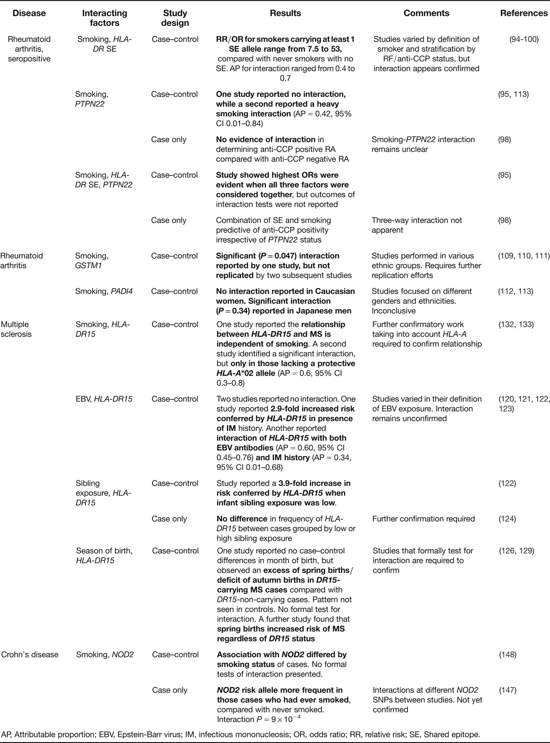
In reviewing G–E interaction in various autoimmune diseases it is clear that there are two broad forms of evidence. Direct evidence of interaction is apparent where studies have used various methods to statistically evaluate G–E interaction. That is, both the gene(s) and environmental factor(s) were directly measured in the study, and the presence of interaction was specifically considered. The strength of the direct evidence varies widely, and in many cases, the power to detect interactions was likely inadequate due to small sample sizes. Few interactions could be categorised as ‘convincing’ according to recently proposed criteria for joint G–E effects on cancer (Ref. Reference Boffetta88). Table 1 summarises the autoimmune disease data, concentrating specifically on those interactions that have been examined in more than one study.
Direct evidence of G–E interaction in autoimmune disease – selected interactions examined by two or more studies
AP, Attributable proportion; EBV, Epstein-Barr virus; IM, infectious mononucleosis; OR, odds ratio; RR, relative risk; SE, Shared epitope.
In addition to the direct evidence, for many autoimmune diseases there is more indirect evidence of G–E interaction. This may be anecdotal rather than based on a formal test for interaction; derived from bioinformatics approaches; or assumed due to an apparent role of epigenetics in disease risk. Epigenetics concerns the chemical modification of DNA, for example DNA methylation, that has the capacity to alter gene transcription and thus cell function. Epigenetic marks are malleable, potentially altering across the life course, and there is considerable evidence that they are impacted by environmental exposures (Ref. Reference Foley89). For example, smoking has been shown to alter DNA methylation at various loci across the genome (Refs Reference Breitling90, Reference Wan91), and there is evidence that many other pollutants and chemicals can induce changes to the epigenome (for review, see (Ref. Reference Feil and Fraga92)). Therefore, epigenetics provides a conduit through which the environment can influence genes, and thus affords a biological mechanism for an interplay between environment and genes in autoimmune, and other, diseases (Ref. Reference Javierre, Hernando and Ballestar3).
Disease-specific evidence
Rheumatoid Arthritis
Of all the autoimmune diseases, the direct evidence for G–E interaction in determining risk of disease is strongest for RA. The RA ‘shared epitope’ (SE) defines a group of HLA types that share a common amino acid sequence in the third hypervariable region of the HLA DRβ1 chain, encoded by the DRB1 gene (Ref. Reference Gregersen, Silver and Winchester93). The SE incorporates a number of different alleles of HLA–DRB1 that have been associated with RA in different populations. The statistical interaction of smoking with the HLA–DR SE was first reported in a Swedish population-based case–control study by Padyukov et al. in 2004 (Ref. Reference Padyukov94). It was observed that smoking was only a modest risk factor for RA in those not carrying an SE allele (SE-negative), however, in those SE-positive, the contribution of smoking to RA risk was striking. A dose-dependent effect was also seen for SE, with risk increasing with a single, or double SE allele (current smokers with no SE allele relative risk (RR) = 1.1, 95% CI 0.7–1.9; current smokers with a single SE allele RR = 2.3, 95% CI 1.4–3.7; current smokers with double SE allele RR = 5.6, 95% CI 2.9–11.1; all compared to never smokers with no SE allele) (Ref. Reference Padyukov94). This interaction has now been widely replicated (Refs Reference Kallberg95, Reference Karlson96, Reference Mahdi97, Reference Morgan98, Reference Pedersen99, Reference Too100, Reference van der Helm-van Mil101, Reference van der Woude102, Reference Willemze103).
The study by Padyukov et al. also reported another striking finding – that the association of the combination of smoking and SE with RA was only true for the subtype of RA characterised by rheumatoid factor (RF) seropositivity (Ref. Reference Padyukov94). This finding was further extended by data that showed citrullination of certain peptides renders them more able to bind to the HLA-DR SE (Ref. Reference Hill104), and knowledge that RF positivity is highly correlated with anti-cyclic citrullinated peptide (anti-CCP) antibody positivity in RA. When anti-CCP positivity was used to subclassify RA, the association of the combination of smoking and HLA-DR SE was even stronger, suggesting that the association with RF positivity was likely secondary to a primary association with anti-CCP positivity (Ref. Reference Klareskog105). Again, this has been widely confirmed (Refs Reference Mahdi97, Reference Pedersen99, Reference Too100, Reference van der Helm-van Mil101, Reference van der Woude102). It has been proposed that the likely biological mechanism behind this interaction involves the tobacco smoke-induced citrullination of self-peptides in the lungs. Citrullination involves the post-translational modification of protein that is catalysed by the enzyme peptidylarginine deiminase (PAD) (Ref. Reference Wegner106). Thus, smoking may preferentially induce an autoimmune response to citrullinated peptides in those who are HLA-DR SE carriers (Ref. Reference Klareskog107) (Fig. 2). Thus, self-peptides may be chemically modified by tobacco smoke, rendering them more open to recognition by the immune system as non-self. The exact peptide(s) that is rendered arthritogenic by citrullination is the subject of considerable research effort. It has also been noted that the presence of anti-CCP can often be detected years before the onset of any clinical signs of disease, potentially providing the opportunity for preventive intervention (Ref. Reference Klareskog76).
Proposed mechanism through which exposure to tobacco smoke, in interaction with genetic factors, may lead to the development of rheumatoid arthritis. Smoking may cause citrullination of peptides present in the lung. Antigen-presenting cells (APCs) of genetically susceptible individuals who carry the HLA-DR shared epitope (SE) may be more prone to presenting these citrullinated self-peptides to T cells, resulting in an immune response against the peptides. The mechanism through which this process then initiates joint inflammation remains unclear. However, a ‘second hit’ such as a joint trauma may induce joint peptide citrullination, to which the immune system is already primed to respond (Ref. Reference Klareskog76).
Further work, introducing a second genetic locus, PTPN22, into the interaction analysis has demonstrated that this gene too may play an important role in the interaction. PTPN22 encodes a tyrosine phosphatase known to play a role in lymphocyte activation (for review, see (Ref. Reference Rhee and Veillette108)), and contains a non-synonymous single nucleotide variant associated with autoimmune disease. Across three distinct RA case–control sample sets (Sweden, US, Netherlands), a G–G interaction was found between HLA-DR SE and PTPN22 in determining risk of anti-CCP positive RA. No interaction between PTPN22 and smoking was identified (Ref. Reference Kallberg95).
A number of other smoking-gene interaction analyses in RA risk have been described. A 2006 report demonstrated interaction between smoking and the glutathione s-transferase M1 gene (GSTM1) (Ref. Reference Criswell109). This was not replicated by later studies (Refs Reference Keenan110, Reference Mikuls111), although one such later study did detect interaction with other genes also encoding enzymes known to be involved in carcinogen detoxification and protection from oxidative stress (GSTT1, HMOX1) (Ref. Reference Keenan110). Interaction of smoking with PADI-4, encoding a PAD enzyme involved in post-translational citrullination, was reported in Japanese (Ref. Reference Kochi112), although this was in contrast to a previous study in Caucasian women where no interaction was demonstrated (Ref. Reference Costenbader113). Some evidence also exists to suggest interaction of smoking with complement-activating mannan-binding lectin (MBL, involved in innate immune defences) in Caucasians (Ref. Reference Saevarsdottir114) and with N-acetyltransferase 2 (NAT2, a carcinogen detoxifier) in African Americans (Ref. Reference Mikuls111).
Indirect evidence for G–E interaction in RA comes from a growing number of studies reporting changes to the DNA methylation profile of various tissues from RA cases compared to unaffected controls. Earlier studies demonstrated a global hypomethylation of RA synovial fibroblast DNA (Ref. Reference Karouzakis115), and changes to the methylation state of various candidate loci such as IL6 (Ref. Reference Nile116) and DR3 (Ref. Reference Takami117) in disease-relevant tissues. More recent genome-scale studies have reported numerous loci across the genome that appear differentially methylated in cases compared to controls (e.g. (Refs Reference Liu118, Reference Nakano119)). Collectively, this evidence directly implicates a role for epigenetics in the RA disease state, and indirectly suggests that the environment may be interacting with the genome at numerous loci in this disease.
Multiple sclerosis
Direct evidence of G–E interaction is also relatively strong for MS. Following from the knowledge that both HLA-DRB1*15 and EBV exposure are associated with risk of MS, the interaction of these two factors was examined. A 2008 nested case–control study of 148 female MS cases and 296 matched controls from the US Nurses’ Health Study demonstrated expected single factor associations of DR15 and measures of EBV exposure (infectious mononucleosis, anti-EBV antibody titre) with disease (Ref. Reference De Jager120). However, statistical analysis of DR15 and anti-EBV antibody titres revealed no evidence of interaction in determining MS disease risk (Ref. Reference De Jager120). The results of independent follow-up studies have been mixed. A 2009 study examining Danish and Swedish cohorts identified a significant interaction between IM and HLA-DRB1*15, with risk of MS associated with HLA-DRB1*15 being 2.9 times higher in those with history of IM (95% CI 1.3–6.5, interaction P = 0.007) (Ref. Reference Nielsen121). However, a lack of statistical interaction between anti-EBV antibodies and DR15 was reported by a 2010 Australian study (Ref. Reference van der Mei122). A 2012 Swedish case–control study (Ref. Reference Sundqvist123) looked for interaction between presence of risk allele HLA-DRB1*15, absence of protective allele HLA-A*02 and anti-EBV antibody titres. They found significant interaction, as defined by departure from additivity, between EBV antibody titres and DRB1*15 (AP = 0.6, 95% CI 0.45–0.76). This interaction persisted when data were restricted to those who were A*02 negative or A*02 positive. Similarly, interaction was found between EBV antibodies and absence of A*02 (AP = 0.39, 95% CI 0.18–0.61), which persisted only in the DRB1*15 negative restricted group. They argued that previous studies with a negative outcome had reported interaction only on the multiplicative scale, and that their detection of departure from additivity is more biologically relevant (Ref. Reference Sundqvist123). Thus detection of the interaction may be quite sensitive to study design and analytical approaches.
Studies have also examined interaction of HLA with infant sibling exposure (as a proxy for early life microbial exposure) in MS. In cases and controls recruited from Tasmania, Australia (Ref. Reference van der Mei122), an interaction between low infant sibling exposure and DR15 was detected, such that the combined effect of these two factors was 3.9 times greater on a multiplicative scale than that expected if no interaction were present (interaction P = 0.019). Therefore, infant sibling exposure may be more protective against MS in those who are HLA-DR15 positive. No interaction of anti-EBV antibodies with HLA-DR15 was detected, nor was there a statistically significant three-way interaction between the factors (Ref. Reference van der Mei122). This was followed by a report of 197 Canadian MS probands with cumulative sibling exposure <1 year, and 1112 probands with higher exposure (Ref. Reference Ramagopalan124). No difference in the frequency of DRB1*15 alleles was observed between the exposure groups, suggesting a lack of interaction. The authors point out that unlike in the Australian study, they were unable to identify a single-factor association between MS and infant sibling exposure, and suggest that the reported phenomena may be population specific (Ref. Reference Ramagopalan124).
A very recent study took a bioinformatics approach to indirectly considering G–E interaction in MS (Ref. Reference Mechelli125). The authors defined candidate ‘interactomes’ – sets of genes for which some evidence exists for interactions with various viral factors in determining disease risk. They then tested for statistical enrichment of genes within these interactomes amongst loci identified as associated with MS by GWAS. In particular, they found genes of the EBV interactome were significantly enriched within GWAS-identified loci, providing further support that G–E interaction is an important component of MS risk (Ref. Reference Mechelli125).
The interaction of genes and sun exposure/vitamin D in determining risk of MS has also been considered. Season/month of birth has been used as a proxy measure of sun exposure/vitamin D in early life, although this measure does not capture variations in individual behaviours, skin pigmentation, or dietary intake of vitamin D (Ref. Reference Staples, Ponsonby and Lim32), and is likely associated with other seasonal factors. An analysis of month of birth in relation to HLA-DRB1*15 status was carried out in a large sample of MS patients from Canada, Sweden and Norway, and in two groups of unaffected controls (proband siblings and unrelated individuals from Sweden) (Ref. Reference Ramagopalan126). A higher proportion of DRB1*15-positive MS cases were born in April (spring) compared to DRB1*15-negative cases, and conversely, a lower proportion of DRB1*15-positive cases were born in November (autumn) compared to DRB1*15-negative cases. No such pattern was observed for controls. While no formal statistical test for interaction was performed, the opposing effect suggests that the combination of season of birth and DRB1*15 is important in MS (Ref. Reference Ramagopalan126). The reason for this may lie in the recent discovery of DRB1 allele-specific functionality of an upstream vitamin D response element (VDRE). In HLA-DRB1*15-bearing haplotypes, the VDRE is functional, and vitamin D is capable of controlling the expression of the gene. On the background of other DRB1 alleles not associated with MS, the VDRE is non-functional and vitamin D does not stimulate gene expression (Ref. Reference Ramagopalan127). Therefore, exposure to vitamin D at a crucial early life time point that is captured by season of birth may be more relevant to MS risk in those carrying the HLA*DRB1*15 allele. Furthermore, a recent study characterised the functionality of the VDRE associated with various HLA-DRB1 alleles, then tested for association of MS with alleles carrying/not carrying a functional VDRE (Ref. Reference Nolan128). Increased risk of MS was associated with the functional VDRE-carrying DRB1 allele. DRB1 alleles carrying non-responsive VDREs were associated with reduced MS risk (Ref. Reference Nolan128). One proposed mechanism is that thymic deletion of autoreactive T cells may be impaired when early life vitamin D is insufficient in those carrying HLA-DRB1 alleles with responsive VDREs (Ref. Reference Ramagopalan127). Not all studies though have reported consistent findings. In over 8000 Finnish MS cases and controls, a direction of effect opposite to that previously observed was reported, in that the increase in May (spring) births was more pronounced in DR15 − MS cases than in DR15+ MS cases (Ref. Reference Saastamoinen, Auvinen and Tienari129). Similarly, no relationship between month of birth and HLA*DRB15 was found within samples utilised for a large MS GWAS (Ref. 130).
Furthermore, a study showed that a promoter variant of the vitamin D receptor gene (VDR) was not associated with overall risk of MS in Tasmanian cases compared to controls. However, the VDR variant was associated with an increased risk of MS in those with low winter sun exposure (≤2 h/day), but not in those with high exposure. The interaction was statistically significant (P = 0.012) (Ref. Reference Dickinson131). Again, this illustrates the potential to identify new genetic associations through stratification by relevant environmental exposures.
In relation to smoking, one study found that association with the HLA-DRB1*15 risk allele was independent of smoking (Ref. Reference Simon132). A later study identified an interaction between presence of HLA-DRB1*15 and absence of HLA-A*02 (protective allele) in disease risk that was observed only in smokers (Ref. Reference Hedstrom133). The biological mechanism(s) through which any HLA-smoking interaction might act in MS are unknown. Further work is required to confirm the findings and to increase mechanistic understanding.
Systemic lupus erythematosus
NAT2 has also been examined for interaction with smoking in SLE in Japanese. The combination of smoking and the genotype associated with slower detoxification significantly increased risk compared to the rapid genotype with no smoking history (AP = 0.05, 95% CI 0.12–0.88) (Ref. Reference Kiyohara134). Similarly, in Japanese women, an interaction between smoking and TNFRSF1B (a member of the tumour necrosis factor receptor family) has been reported (Ref. Reference Kiyohara135). Additionally, CYP1A1 and GSTM1 have been tested for interaction with smoking in Japanese SLE. Both these genes encode enzymes that metabolically activate carcinogenic or toxic substances to produce reactive oxygen species (ROS) (Ref. Reference Kiyohara136). ROS have been implicated in SLE pathogenesis (Ref. Reference Griffiths137). The combined ‘at risk’ CYP1A1/GSTM1 genotype was associated with a 17.5-fold increase in disease risk in ever smokers (95% CI 3.2–95.9), but only a 2.8-fold increase in risk in non-smokers (95% CI 1.7–4.6) (Ref. Reference Kiyohara136). All these findings await further independent confirmation.
Fraser et al. looked for interaction between occupational sun exposure and GSTM1 in SLE. They found a 3-fold increase in risk in those with high sun exposure who were homozygous for the GSTM1 null allele, but there was no association with sun exposure in those who carried a functional GSTM1 (Ref. Reference Fraser138).
There is also substantial evidence, gathered over many years, for a role for epigenetics in SLE (for recent review, refer to (Ref. Reference Zhang139)). In brief, early studies demonstrated decreased global DNA methylation levels in T cells of SLE patients (Ref. Reference Richardson140). Candidate gene studies have demonstrated altered SLE methylation states at various loci, including ITGAL (Ref. Reference Lu141) and PRF1 (Ref. Reference Kaplan142). Recent genome-scale efforts comparing methylation between discordant twin pairs (Ref. Reference Javierre143) and unrelated cases and controls (e.g. (Ref. Reference Jeffries144)) have also demonstrated differences in methylation patterns across the genome that are associated with SLE. Furthermore, recent work has demonstrated that, while exposure of peripheral blood mononuclear cells to ultraviolet B can induce an inhibition of DNA methylation, PBMCs from patients with active SLE are more sensitive to this process (Ref. Reference Wang145). Photosensitivity is known to be associated with SLE (Ref. Reference Wysenbeek, Block and Fries146); therefore this finding provides a potential mechanism through which UVB, as an environmental exposure, may interact with the genome in disease.
An interesting example in which differential DNA methylation could be linked to both environment and altered gene expression was published very recently in a mouse model of SLE (Ref. Reference Strickland70). Genetically lupus-resistant and lupus-susceptible mouse strains were fed diets either high or low in dietary micronutrients important for the process of DNA methylation (methyl donors), such as methionine, choline, folate and other B vitamins. The authors showed that a diet higher in methyl donors decreased autoantibody levels and kidney disease severity in lupus-susceptible mice. They also showed that in mice on a methyl donor restricted diet, DNA methylation was reduced at the gene encoding CD40L, a protein that is elevated on T cells from human and mouse females with lupus. They concluded therefore that a diet high in methyl donors may be beneficial to lupus patients via an increase in methylation at lupus-susceptibility genes, reducing their expression (Ref. Reference Strickland70).
Crohn's disease
A small number of studies of CD have examined interactions of genes with smoking in disease risk. NOD2 plays an important role in regulation of host response to intestinal bacteria, and has been robustly associated with CD. A large German case-only study recently identified interaction between smoking (current and ever) and a NOD2 CD risk allele (Ref. Reference Helbig147). This was consistent with an earlier Dutch case–control study (Ref. Reference van der Heide148). Other genes in the Dutch study apparently interacting with smoking included DLG5, IL23R and PTPN2. Association with PTPN2 was only detectable if the smoking CD group alone was compared with controls, showing that stratification by relevant environmental phenotypes is likely to have the capacity to identify new risk genes (Ref. Reference van der Heide148).
A moderate level of indirect evidence of G–E interaction in CD comes from a limited number of studies examining the CD epigenome. A genome-scale effort examining around 27 000 CpG sites identified more than 1000 differentially methylated loci in peripheral blood mononuclear cells (Ref. Reference Nimmo149). The authors also reported significant enrichment of differential methylation near genes associated with CD by GWAS studies, including NOD2. This suggests that CD risk genes altered by sequence variation may be further impacted by epigenetic variability, possibly brought about by differing environmental exposures such as smoking. Smoking is known to impact DNA methylation (Ref. Reference Zeilinger150).
Thyroid autoimmunity
Age is likely a proxy measure for cumulative exposure to numerous non-genetic factors. For the autoimmune thyroid disease Grave's disease, there are data to suggest that some genetic associations with this condition differ by age. For example, an age-dependent gene dosage effect of the risk allele of PTPN22 has been reported (Ref. Reference Skorka151), as has association of variants of HLA-DRB1 only in those diagnosed at a younger age (Ref. Reference Jurecka-Lubieniecka152). Assuming that the use of age indirectly captures one or more relevant environmental factors, these data may indirectly indicate a G–E interaction in determining disease risk. Further indirect evidence comes from work showing that the thyroglobulin gene (TG) a gene associated with thyroid autoimmune disease, is up-regulated by interferon alpha (IFNα), which in turn is stimulated by environmental factors such as viral infection (Ref. Reference Hasham and Tomer153).
Type 1 diabetes
There is some evidence of G–E interaction for T1D onset. However, this issue has been relatively understudied in T1D. In a case-only analysis, Badenhoop et al. examined the pattern of month of birth among those carrying various HLA-DQ genotypes, and observed differences in birth month distributions (Ref. Reference Badenhoop154). This may reflect an impact of environmental factors related to season, including vitamin D levels and rates of viral infections, on the degree of disease risk attributed by HLA alleles. No such variability in month of birth was noted between cases carrying non-HLA risk alleles, suggesting that the impact may be specific to HLA. Moreover, in 15 pairs of monozygotic twins discordant for T1D, over 100 loci across the genome were found to be differentially methylated in monocytes (Ref. Reference Rakyan155), providing indirect epigenetic evidence for G–E interaction in this disease.
Celiac disease
An example of indirect evidence for G–E interaction, rather than having been formally tested for using statistical methods, is that of HLA and gluten in CeD. Virtually 100% of all individuals diagnosed with CeD carry a HLA-DQ2 or DQ8 haplotype. Additionally, following deamidation by the enzyme transglutamidase 2 (Ref. Reference Sollid and Jabri156), gluten peptides are known to be the pivotal trigger of disease initiation, demonstrating that at least one causal pathway to disease must include both of these factors (Ref. Reference Fasano and Catassi75). Furthermore, a gene-dosage effect has been shown in which those homozygous for the HLA-DQ2 risk allele have a greater magnitude and breadth of gluten-specific T cell responses compared with heterozygotes (Ref. Reference Vader157).
Other diseases for which indirect evidence exists
A number of other autoimmune diseases carry indirect epigenetic evidence of G–E interaction. Aberrant DNA methylation has been associated with psoriasis, systemic sclerosis, primary biliary cirrhosis and primary Sjögren's syndrome, as reviewed elsewhere (e.g. (Ref. Reference Lu158)).
Clinical implications/applications
Ultimately, a better understanding of disease risk genes and environmental exposures, and the ways in which they interact, will enable the development of more effective approaches to disease treatment and prevention. In complex diseases, including the autoimmune diseases discussed in this review, various causal pathways are likely to exist in the population, and thus the number of contributing risk factors and their combinations may vary from person to person. An ability to determine an individual's potentially unique profile of risk factors and their interactions will likely shed light on the underlying biological mechanisms responsible for disease onset, and will therefore be a powerful tool in determining the most effective treatment strategies. Understanding the influence of environment on genetic profile is likely to enhance the field of pharmacogenomics, in which the aim is to predict the efficacy or toxicity of a drug based on genetic makeup (Ref. Reference Yu and Huang159), moving closer to more refined personalised medicine approaches. Additionally, disease-related changes to the epigenome brought about by environmental exposure may be able to be pharmaceutically corrected (Ref. Reference Kelly, De Carvalho and Jones160). In terms of prevention, understanding how genes influence risk in different environmental strata, and vice versa, will help better define groups of individuals in the population for which preventive interventions are more likely to be effective (Ref. Reference Song, Lee and Kang161).
Research progress and outstanding research questions
One of the biggest challenges facing G–E interaction research at the present time is that international, multi-study, harmonised measurement of environmental exposures is very difficult to achieve (Ref. Reference Bookman162). The explosion of genetic associations via GWAS approaches has been successful for the simple reason that genome-wide genotyping is an established technique that can be carried out anywhere in the world with high precision and at low cost. DNA is easy to collect, and genotypes do not change over time so it does not matter when (pre- or post-disease onset) DNA was collected from study participants (Ref. Reference Khoury and Wacholder84). Thus, international disease consortia that bring together thousands of case DNA samples have become commonplace. Often samples are genotyped using various methods but there are well-established methodologies to harmonise genotype data between case groups (Ref. Reference Ioannidis, Thomas and Daly163). Thus, it is relatively easy to collect GWAS sample sets of sufficient size to provide power to detect loci with modest risk effects. It is also straightforward to access independent sample sets within which to perform replication analyses, the gold standard for confirmation of associations (Ref. Reference Ioannidis, Thomas and Daly163).
In contrast, extensive environmental data are often not collected alongside cohort or case–control DNA collections (Ref. Reference Bookman162). The resources required to collect such data are often significant, and thus not prioritised in studies aimed primarily at the largest possible DNA sample size. Even if some environmental data are captured, the way in which it is captured may be (and often is) different or imprecise between studies, making the task of data harmonisation to enable the assembly of large, well-powered, discovery and replication datasets a difficult one (Ref. Reference Cornelis164). It has been concluded that the trade-off between bigger studies versus more precise and informative measurement should balance more towards the latter in G–E interaction studies of continuous traits (Ref. Reference Khoury and Wacholder84). Further work to develop more sophisticated methods of assessing G–E that account for gene allele dose responses and genotype risk scores within gene networks, rather than single gene variants, is likely to be worthwhile (Ref. Reference Lin165).
Efforts towards larger-scale G–E studies are building, with several international data collections and data harmonisation efforts now underway (Ref. Reference Aschard166). To provide further clarity in this field, it was recommended in 2012 that the causal inference for future G–E interactions could be reported using guidelines which incorporate: (i) a score for strength of evidence for the (a) environmental and (b) genetic factors; (ii) establish a prior score and decide on the pattern of interaction to be expected; (iii) score interaction strength; and (iv) evaluate the overall plausibility for interaction (Ref. Reference Boffetta88). Progress in this field is likely to uncover new and more complete information about the breadth of autoimmune disease risk factors; help to further understand the proportion of risk that has not yet been explained by genetic research, and provide new insights into biological mechanisms behind disease development.
In summary, the number of established genetic and environmental risk factors associated with autoimmune disease is growing. Some of these factors appear shared between a number of autoimmune diseases and some appear specific for single diseases. Similarly, some particular combinations of G–E interactions appear to be shared between diseases, but the biological mechanisms behind the interactions may be different. However, a lack of well-established standardised analytical approaches, coupled with the inherent difficulties of large-scale collection of validated and harmonised environmental exposure data provide significant challenges in moving the field further forward. But the importance of identifying G–E interaction in autoimmune, and other complex diseases, is becoming increasingly acknowledged.
Financial support
This work was supported by the Victorian Government's Operational Infrastructure Support Programme. J. A. E. was supported by an Australian Research Council Future Fellowship, Project ID FT120100253. A. L. P. was supported by a National Health and Medical Research Council Senior Research Fellowship, ID 1008396.
Conflicts of Interest
None.