Introduction
Nasal septal perforations are full thickness defects of the nasal septum, and may result from trauma, chemical exposure or various pathologies; they may also be idiopathic.Reference Foda1 Whilst most patients remain asymptomatic,Reference Brain, Mackay and Bull2 a significant minority suffer from debilitating symptoms including nasal crusting, nasal obstruction, epistaxis, pain and whistling caused by turbulence of airflow through the septal perforation. Symptomatic patients can be treated either conservatively (with nasal douching, emollients, humidification or septal obturators) or surgically.Reference Barraclough, Ellis and Proops3
Various surgical methods have been described for the closure of nasal septal perforation. However, reported results are variable and rarely statistically significant.Reference Goy and Husain4 Techniques described include transplantation of septal cartilage,Reference Newton, White and Lee5 advancement and suture of perforation margins,Reference Gollom6 oral mucosal flaps,Reference Tardy7 temporalis fascia grafts,Reference Fairbanks8 inferior turbinate flaps,Reference Vyuk and Versluis9 grafting with tragal cartilage and temporalis fascia,Reference Hussain and Murthy10 acellular human dermal allograft,Reference Kridel, Foda and Lunde11 and a two-staged procedure with tissue expanders.Reference Nunez-Fernadez, Vokyrka and Chrobok12 To date, surgical repair of nasal septal perforation remains a distinct challenge to otolaryngologists and facial plastic surgeons.
This study aimed to assess the success of nasal septal perforation closure using bilateral advancement superior and inferior nasal mucosal flaps and porcine collagen, in terms of perforation closure and symptom control.
Materials and methods
This was a prospective study of 28 consecutive patients. All had symptomatic nasal septal perforation, closed surgically by the same surgeon (UR) using the same technique, between November 2005 and October 2007.
Every attempt was made to identify the cause of patients' nasal septal perforation, from their clinical history, case notes and haematological investigation results (e.g. cytoplasmic anti neutrophilic cytoplasmic antibody (cANCA), Treponema pallidum haemagglutination assay (TPHA) and angiotensin converting enzyme (ACE) test). When no clear cause was identified, a biopsy of the septal perforation margin was taken.
Patients were asked to assess the severity of their symptoms using a visual analogue scale (VAS), where 0 represented no symptoms and 10 represented maximal symptoms. This was done pre-operatively and six months post-operatively, for each symptom. Closure of the perforation was objectively confirmed at each six-monthly review.
An endoscopic photograph of the nasal valve angle was taken pre-operatively and six months post-operatively. Two independent otolaryngologists were randomly allocated these photographs and asked to measure the nasal valve angle. The nasal valve angle was measured by drawing one line along the upper lateral cartilage and another along the septal cartilage, and then projecting these lines until they intersected. The angle formed by the two lines was then measured using a protractor.
Surgical technique
Every patient underwent an open rhinoplasty approach under general anaesthesia. Pre-operative antibiotics were given. Two per cent lignocaine with 1:80 000 adrenaline was injected into the septum and nasal dorsum for hydrodissection and vasoconstriction. In addition, neurosurgical patties soaked in 2 ml of 10 per cent cocaine with 1:10 000 adrenaline were kept in both nasal cavities for 15 minutes, for local vasoconstriction.
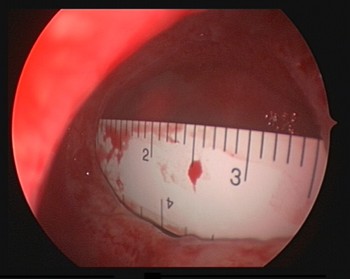
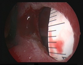
The horizontal and vertical dimensions of the nasal septal perforation were measured using a sterile, flexible ruler (Figures 1 and 2).
Measurement of horizontal dimension of nasal septum perforation.

Measurement of vertical dimension of nasal septum perforation.

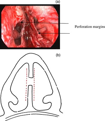
An inverted V-shaped, trans-columellar incision is made at the mid-point. The skin flap was elevated as for a standard open rhinoplasty approach. Mucoperichondrial flaps were raised without tear on both sides of the septum. This was continued around the perforation using a Freer's elevator, and the perforation margins were separated (Figure 3).
(a) Bilateral intact mucoperichondrial flaps; arrows indicate perforation margins. (b) Diagrammatic representation of (a).

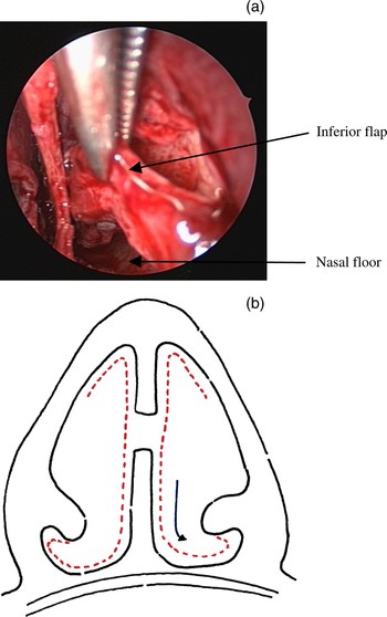
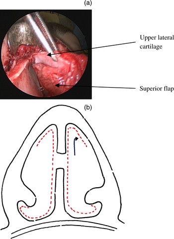
The lower part of each flap was elevated along the floor of the nose, inferior meatus and inferior surface of the inferior turbinate, leaving it attached along the border of the inferior turbinate (Figure 4). The upper lateral cartilage was separated from the dorsal border of the septum. The superior part of the septal mucoperichondrial flap was then dissected off the undersurface of the upper lateral cartilage, taking care not to damage the mucosa (Figure 5).
(a) Elevation of the inferior flap; upper arrow indicates inferior flap and lower arrow indicates nasal floor. (b) Diagrammatic representation.

(a) Elevation of the superior flap; upper arrow indicates upper lateral cartilage and lower arrow indicates superior flap. (b) Diagrammatic representation.

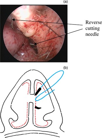
The lateral extent of this dissection should have been sufficient to provide an adequate flap for a tension-free approximation of perforation margins. If more inferior flap length was required, an incision could be made along the border of the inferior turbinate to convert this inferior flap into a bipedicle flap. However, we did not require the use of a bipedicle flap in this series. After sufficiently mobilising the flaps, the perforation margins were approximated without tension and sutured with vertical mattress sutures using 4 ‘0’ polydioxanone (PDS) with a reverse cutting needle (Figure 6).
(a) Vertical mattress sutures to approximate bilateral advancement flap; arrows indicate reverse cutting needle. (b) Diagrammatic representation.

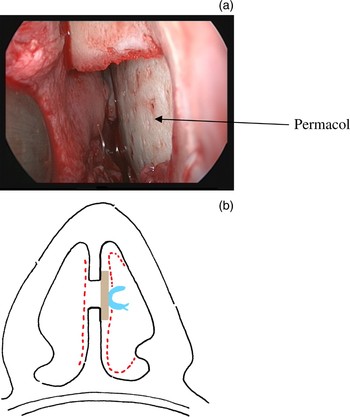
Acellular porcine collagen was positioned in the perforation between the right and left mucoperichondrial flaps and sutured to the cartilage to overlap the repaired site (Figure 7). Acellular porcine collagen (Permacol®; Tissue Science Laboratories, Castleford, UK) was preferred because it: (1) was easily available; (2) was ready to use from the packet; (3) was very similar in its architecture to human tissue; (4) was acellular and therefore non-immunogenic; (5) did not involve a donor site; (6) was resistant to collagenase enzymes (preferable if a revision was necessary); and (7) it supported the repaired perforation site.13
(a) Insertion of porcine collagen; arrow indicates Permacol. (b) Diagrammatic representation.

Soft Silastic® nasal splints were placed in each nasal passage and secured in position without pressing on the superior and inferior flaps. The trans-columellar incisions were closed with 6.0 prolene sutures and marginal incisions with 5.0 vicryl sutures. SteristripsTM and a nasal thermal splint were then applied on the dorsum. All these were removed after a week.
Patients were discharged the following day with advice to continue antibiotics for a week and to undertake regular nasal douching with saline.
Results
We enrolled 28 consecutive patients with symptomatic nasal septal perforations who presented to us between November 2005 and October 2007. No patient was excluded from the study.
Patients comprised 12 women and 16 men, of mean age 45 years (range: 21–76).
The mean perforation size was 22 mm (range: 10–35) vertically and 27 mm (range: 10–47) horizontally.
The mean follow up was 16 months (range: 6–24), with a minimum of six months' follow up.
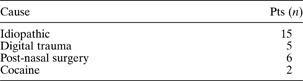
The causes of nasal septal perforation in our patients are shown in Table I.
Patients' causes of nasal septal perforation

Pts = patients
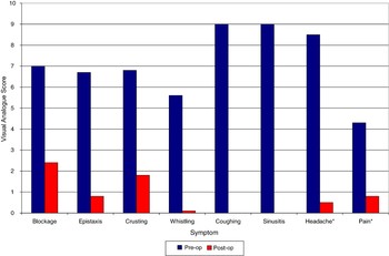
There were significant differences between the pre- and post-operative mean VAS scores for the symptoms assessed (Figure 8).
Preoperative (pre-op) and postoperative (post-op) mean visual analogue scale for the symptoms assessed.

Statistical assessment was done using the paired t-test (Table II).
Statistical analysis* of patients' pre- and post-operative VAS symptom scores

* Paired t-test. †Results should be interpreted with caution due to small number of observations (<10). Pre-op = pre-operative; post-op = post-operative; VAS = visual analogue scale; SD = standard deviation
Statistical comparison of pre- and post-operative VAS scores produced the following results: for epistaxis, p < 0.001; for crusting, p < 0.001; for whistling, p < 0.001; and for nasal obstruction, p < 0.001. A similar comparison of pain scores had a statistical significance of p < 0.002, but as we had less than 10 observations for this symptom, we are cautious about this result.
One patient (4 per cent) had a residual perforation of 1 × 2 mm. This was a reduction from an initial perforation measuring 25 × 30 mm. However, this patient did have a clinically significant improvement in symptoms.
Therefore, the success rate for nasal perforation closure in our study was 96 per cent, with a 100 per cent improvement in epistaxis, crusting and whistling and a 92 per cent improvement in nasal blockage.
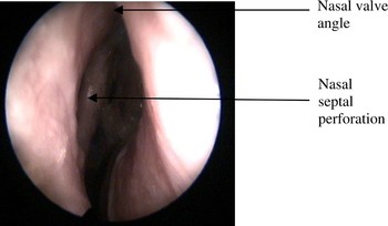
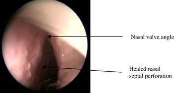
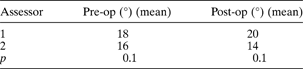
Two independent assessors compared the pre- and post-operative photographs of the nasal valve angle (Figures 9 and 10). These assessments were statistically analysed using the paired t-test (Table III). There were no statistically significant differences, comparing the two assessors' pre-operative nasal valve angle assessments, and also comparing their post-operative nasal valve angle assessments.
Pre-operative nasal valve angle. Upper arrow = nasal valve angle; lower arrow = nasal septal perforation

Post-operative nasal valve angle. Upper angle = nasal valve angle; lower arrow = healed nasal septal perforation

Nasal valve angle

Pre-op = pre-operative; post-op = post-operative
Discussion
Previous authors have described various surgical approaches for nasal septal perforation closure.
FodaReference Foda1 used an open rhinoplasty approach for closing nasal septal perforations in 20 patients, and achieved 90 per cent success. In this series, reduction rhinoplasty techniques were used to increase the availability of flaps, and bipedicled and advancement flaps were used on both sides. Symptoms resolved completely in 80 per cent of patients, but only partially in the remaining 20 per cent.
Goy and Hussain,Reference Goy and Husain4 from Dundee, critically evaluated reports on different surgical treatments for nasal septal perforation. They concluded that, although an extensive range of surgical treatments had been described, the results were rarely statistically significant as the numbers involved in the studies were small.
Newton et al. Reference Newton, White and Lee5 achieved 90 per cent success in closing nasal septal perforations using unilateral bipedicled flaps and temporalis fascia. Retrospective telephone questionnaires were conducted to assess pre- and post-operative symptom scores, using linear analogue scores. A statistically significant improvement in symptom scores was observed after septal perforation closure.
Kridel et al. Reference Kridel, Foda and Lunde11 described surgical septal perforation repair with bipedicle flaps and acellular human dermal allografts in 12 patients. They managed to achieve a 91 per cent success rate in patients in whom the acellular human allograft was placed between the flaps. Of the 12 patients, seven had closure of both sides during the operation. Four patients had closure of flaps on one side, although closure on the other side could not be achieved. Despite this, these patients' perforations healed. The remaining one patient had a residual perforation, as neither side flaps could be approximated. Symptom control was not assessed.
Pedroza et al. Reference Pedroza, Patrocinio and Arevalo14 utilised both external and internal approaches, using temporalis fascia, conchal cartilage and mastoid cortical bone, either in isolation or combination, concomitantly. Only 14 of the 68 patients studied underwent an external approach, but the authors did not specify how many of these 68 patients had a bipedicle flap or bilateral advancement flap. Their success rate was 97 per cent, the best reported thus far. All patients with complete closure of septal perforation experienced an improvement in their symptoms. However, the study did not mention the method of symptom evaluation, nor whether the change in symptoms was statistically significant.
Woolford and JonesReference Woolford and Jones15 used local mucosal flaps and composite conchal cartilage grafts to achieve septal perforation closure. They suggested that composite cartilage was preferable to fascia as a graft, as it provided greater solidity and support. They commented that the role of skin in the composite graft was less clear, as skin sometimes formed a yellowish covering that could take weeks to re-epithelialise, whilst on other occasions the graft took immediately. All but one of their patients was asymptomatic post-operatively. No mention was made of the types or severity of patients' symptoms.
In our study, we tried to minimise variation by ensuring that the same surgeon performed the same technique in all patients with symptomatic nasal septal perforation. All our patients underwent nasal septal perforation repair by the open rhinoplasty approach. This gives wide access for mobilising mucosal flaps, with minimal trauma. It also allows approximation of the perforation margins with vertical mattress sutures, particularly the posterior suture, which is the most difficult to insert.
We believe our favourable success rate for nasal septal perforation closure was due to tension-free approximation of the perforation margins, and to the use of vertical mattress sutures (which helped to keep the margins everted so that the raw surface remained in contact, ensuring healing). Previous reports on nasal septal perforation closure have not highlighted the importance of approximating the edges using vertical mattress suturing. We decided to use four mucosal advancement flaps to achieve tension-free closure of the perforation margins, as well as to preserve the vascularity of the flaps.
We did not have difficulty in closing perforations even up to 47 mm long horizontally, because of the access provided by the open approach. The maximum vertical dimension we closed was 35 mm. In no case did we have to change an advancement flap to a bipedicled flap. This helped to avoid external exposure of the superior part of the septal cartilage.
We do not believe that acellular porcine collagen played a major role in successfully closing our patients' perforations. However, using a sheet of collagen between the mucosal flaps helped to recreate the three layers of the septum. In addition to this, it helped support the repaired site while it was healing, and prevented the flaps from adhering to each other.
As we had repaired the perforation margins meticulously on both sides, there was no need for the mucosa to grow over the acellular porcine collagen, in contrast to the acellular dermis used by Kridel et al. Reference Kridel, Foda and Lunde11 The use of acellular porcine collagen for surgical closure of septal perforations has not previously been reported.
• Various surgical methods for closing nasal septal perforations have been described; however, reported results have been variable and frequently poor
• This study aimed to assess perforation closure and symptom control in patients with nasal septal perforation receiving bilateral superior and inferior nasal mucosal advancement flaps and porcine collagen
• Closure of nasal septal perforation using vertical mattress sutures to approximate bilateral mucosal advancement flaps, and using a porcine collagen sandwich, gave a 96 per cent closure rate and 100 per cent improvement in nasal symptoms
One of our patients had a residual small, posterior septal perforation, our first in this series. This patient's initial perforation measured 25 mm vertically and 30 mm horizontally. The residual perforation measured 1 mm vertically and 2 mm horizontally, and was in the posterior part of the original perforation. This may have occurred due to the initial difficulty in using a vertical mattress suture in the posterior part of the perforation.
Our patients' presenting symptoms were assessed prospectively using a VAS score. Previous studies have assessed the success of perforation closure, but only a few have assessed symptom control. Our patient's prospectively recorded symptom scores were statistically analysed, to determine whether successful septal perforation closure made any significant difference to the presenting symptoms. All our patients (including the patient with the residual perforation) reported a statistically significant post-operative improvement in their presenting symptoms.
Conclusion
In this study, closure of nasal septal perforation using vertical mattress sutures to approximate bilateral mucosal advancement flaps, using a porcine collagen sandwich, enabled a 96 per cent closure rate and 100 per cent improvement in nasal symptoms.
We believe that this method may prove a pertinent and reliable alternative in the surgical management of nasal septal perforation. However, we do not deny that the success rate is proportionally dependent on the surgeon's skills.