Constrictive pericarditis is rare in children and is the end stage of an inflammatory process involving the pericardium caused by idiopathic, infection, post-surgery, radiation injury, connective tissue disorders, or trauma. Reference Cameron, Oesterle, Baldwin and Hancock1 Protein losing enteropathy is characterised by an excessive loss of serum proteins into the gastrointestinal tract, resulting in hypoproteinaemia, oedema, and in some cases, pleural and pericardial effusions. Protein losing enteropathy as the principal manifestation of constrictive pericarditis is extremely rare in younger children. Protein losing enteropathy due to constrictive pericarditis usually occurs without any obvious signs of heart disease, and many of its clinical manifestations may actually result from severe hypoproteinaemia. Therefore, it is not uncommon that constrictive pericarditis as the cause of protein losing enteropathy may be overlooked translating into a delay in the diagnosis and definitive treatment. Here, we report a case of protein losing enteropathy caused by constrictive pericarditis in a toddler. Protein losing enteropathy completely resolved after pericardiectomy with remarkable improvement.
Case report
Two-year and 4-month-old male child was admitted with the history of abdominal distension and intermittent abdominal pain persisting for 1 month. Symptoms started with an episode of tonsillitis. On clinical examination, he had cervical lymphadenopathy, hepatosplenomegaly, and signs of ascites. Initial blood tests showed mildly elevated liver enzymes, low serum albumin (31 g/dl), low total protein (68 g/dl), and positive antibody titres (IgM as well as IgG) for Epstein Barr virus. Epstein Barr virus load was 21,875 copies per ml. Abdominal ultrasound confirmed hepatomegaly and moderate ascites. First echocardiography at our centre showed mild pericardial effusion along with fibrinous strands suggesting inflammatory pericarditis in an otherwise normal anatomical heart. Subsequently, his Epstein Barr virus load dropped with complete resolution of pericardial effusion. However, hypoalbuminaemia worsened with generalised anasarca, ascites, and pleural effusion requiring frequent albumin infusions. His stool was positive for alpha one antitrypsin confirming the diagnosis of protein losing enteropathy. He underwent extensive workup that included hepatitis panel, autoimmune antibodies, biochemical markers for storage disorders, liver, and lymph node biopsy; none of the results revealed any specific aetiology. He was again referred to paediatric cardiology for further workup to identify the cause of protein losing enteropathy.
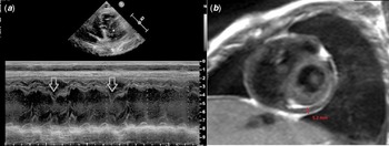
Repeat echocardiograms showed dilated atria out of the proportion to ventricular size. There was mild-to-moderate tricuspid regurgitation with normal right ventricular systolic pressure estimated from the tricuspid regurgitation jet. Exaggerated mitral and tricuspid valve flow variance (>40%) and septal bouncing (Fig 1a) were present. Mitral annular tissue Doppler Indices across the medial and lateral mitral annuls were preserved (12.6 and 13 cm/seconds, respectively). E/e′ ratio was 6.6 which is lower than expected in the setting of increased filling pressure. All these findings were consistent with dissociation of intrathoracic and intracardiac pressures, interventricular dependence, and preserved ventricular relaxation. Therefore, constrictive pericarditis was suspected. Henceforth, diagnostic cardiac catheterisation and cardiac MRI were planned.
( a ) Septal bouncing on m-mode echocardiography and ( b ) thickened pericardium seen in cardiac MRI.

Diagnostic cardiac catheterisation showed elevated left and right ventricular end diastolic pressures (21–23 mmHg). Right and left ventricular systolic pressures were 42 and 73 mmHg, respectively. Left pulmonary artery pressure was 39/24 mmHg with mean of 30 mmHg. Pulmonary vascular resistance index was 4 Wood Units × m2. Cardiac MRI confirmed significantly thickened pericardium measuring 5.2 mm in thickness (Fig 1b). In addition, cardiac MRI showed ventricular septal bouncing, excessive tricuspid annular motion, and a dilated inferior vena cava.
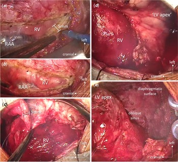
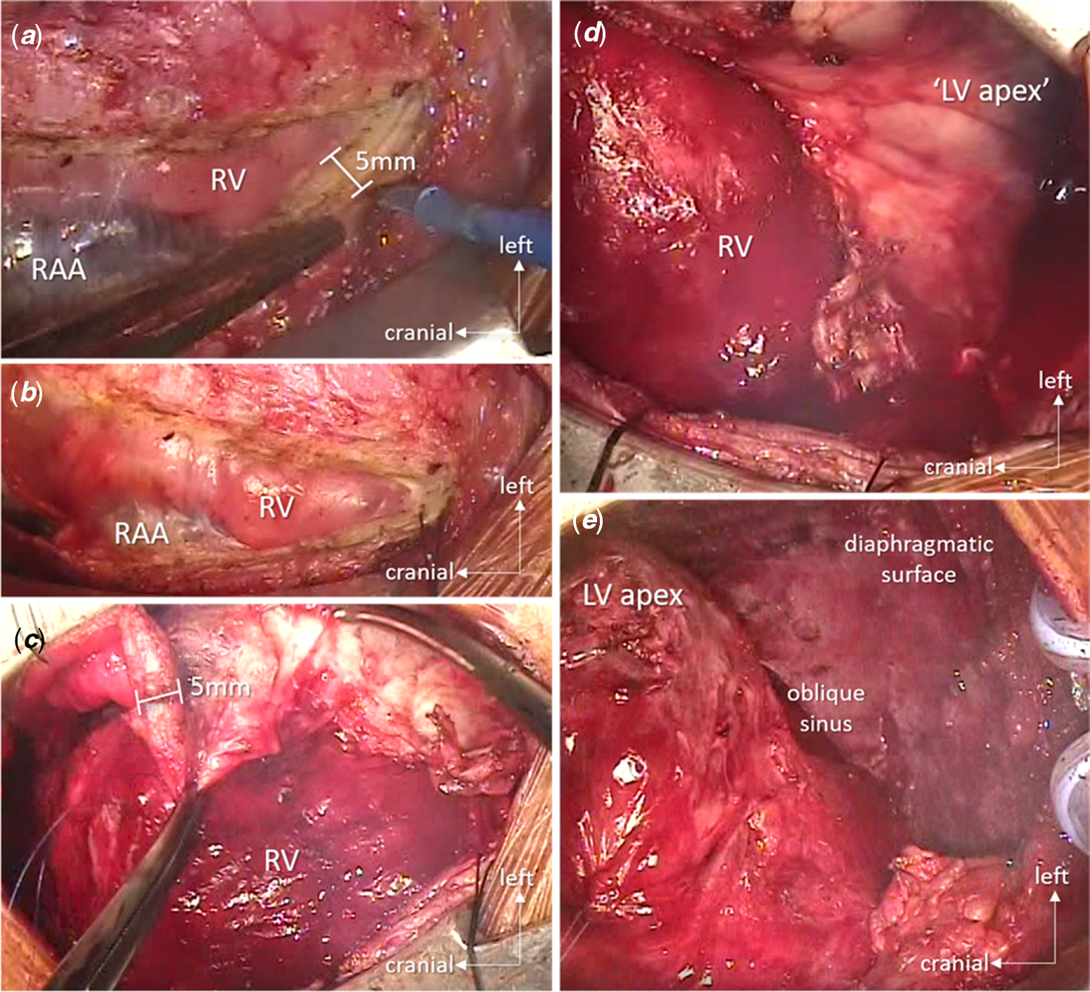
The patient subsequently underwent surgical pericardiectomy. Intra-operative findings confirmed grossly thickened pericardium (5 mm anteriorly and 7 mm around the left ventricular apex), shown in Figure 2. Parietal pericardium was removed around the anterior aspect and the apex of the ventricles; the visceral pericardium was peeled off. There were no intra-operative complications. Post-operative recovery was excellent. The patient was discharged on the ninth post-operative day. His serum protein and albumin levels normalised (78 g and 40 g/dl, respectively) by the time of discharge. At the latest follow-up (2-year post-operatively), the patient remained asymptomatic and appeared as a normally growing, healthy child. Protein losing enteropathy completely resolved; echocardiography demonstrated normal cardiac function without any evidence of constrictive pericarditis.
Intra-operative image of grossly thickened pericardium. ( a , b ) Thickened (5 mm) pericardium opened above the right ventricle (RV), the right atrial appendage (RAA) is freed; ( c ) RV anterior surface is liberated; ( d ) left ventricle (LV) apex is still encased in thickened pericardium; ( e ) LV apex is mobilised by having resected thickened/calcified pericardium around the cardiac apex and diaphragmatic surface liberating the posterior surface of the LV.
Discussion
Although rare, since its first report in 1961, Reference Davidson, Waldmann, Goodman and Gordon2 protein losing enteropathy is a known complication of constrictive pericarditis in children. To the best of our knowledge, there are less than 20 cases reported in children with protein losing enteropathy secondary to constrictive pericarditis. This is only the third report in a toddler and prior two reports Reference Simcha and Taylor3,Reference Kumpe, Jaffe, Waldmann and Weinstein4 were also in 2-year-old children. It is extremely rare to occur at such a young age as it was in this case. Despite its rarity, a high degree of suspicion is advised, because protein losing enteropathy instigated by constrictive pericarditis is completely curable with excellent outcome.
Diagnosis of constrictive pericarditis can be challenging and requires careful evaluation of clinical signs, laboratory investigations, and various data obtained from non-invasive as well as invasive modalities. A multi-disciplinary team approach involving a gastroenterologist, hepatologist, cardiologist, and cardiovascular surgeon should be used in the management of patients with constrictive pericarditis. Echocardiography and cardiac catheterisation can provide haemodynamic information to identify or exclude the diagnosis of constrictive pericarditis. CT is able to detect pericardial calcification. Cardiac MRI is an excellent modality to evaluate pericardial thickness precisely. Reference Breen5 Presence of biatrial enlargement with normal-sized ventricles usually raises suspicion of restrictive cardiomyopathy. It is important to differentiate constrictive pericarditis from restrictive cardiomyopathy Reference Geske, Anavekar, Nishimura, Oh and Gersh6 and both can present with common manifestations. While constrictive pericarditis is treatable with pericardiectomy and, thus, has an excellent outcome, restrictive cardiomyopathy has no curative treatment currently. Traditional haemodynamic criteria for constrictive pericarditis differentiating it from restrictive cardiomyopathy include a left ventricular end diastolic pressure not exceeding right ventricular end diastolic pressure by more than 5 mm Hg, a right ventricular systolic pressure not exceeding 50 mmHg, and a right ventricular end diastolic pressure above the one-third of the right ventricular systolic pressure. Vaitkus et al Reference Vaitkus and Kussmaul7 demonstrated that 91% of patients with constrictive pericarditis met all three abovementioned criteria in their study. All criteria were fulfilled in our case, too. Last but not least, around 20% of patients with constrictive pericarditis can have normal pericardial thickness Reference Talreja, Edwards and Danielson8 making diagnosis of constrictive pericarditis more challenging. In such cases, diagnosis of dissociation of intrathoracic and intracardiac pressures, interventricular dependence, and preserved ventricular relaxation can be the key to differentiate constrictive pericarditis from restrictive cardiomyopathy.
Diagnosis of constrictive pericarditis in a very young child – as presented in this case – can be quite challenging since the classic diagnostic tools may yield inadequate information. Echocardiogram on a crying toddler may not show the classical findings of constrictive pericarditis. Cardiac catheterisation in a very sick child often requires intubation and positive-pressure ventilation; thus, subtle differences in ventricular pressures and interactions that help differentiating between constrictive and restrictive physiology may be obscured. Similarly, CT scans offer a better predictive value in adults, allowing precise measurement of pericardial thickness and calcifications. However, calcifications are rare in very young children and the typical heart rates exceeding 100 beats/minute may result in blurred images. In our patient, history of inflammatory pericarditis and the echocardiographic findings raised suspicion of constrictive pericarditis. Thickened pericardium visualised by cardiac MRI confirmed the diagnosis.
Surgical removal of pericardium is the definitive treatment of constrictive pericarditis. Diuretics should be used sparingly with the goal of reducing elevated venous pressure, ascites, and oedema while awaiting surgical intervention. Removal of the thickened and inflamed pericardium can be technically challenging. Efforts should be made to remove as much pericardium as technically feasible. Excision of pericardium can be performed safely with high success and excellent long-term outcome in this debilitating disease.
In conclusion, constrictive pericarditis should be considered as a cause of protein losing enteropathy even at very young age. Diagnosis of constrictive pericarditis should be made after careful analysis of various non-invasive and invasive diagnostic tools. Once constrictive pericarditis is confirmed, pericardiectomy is the definitive treatment in children and should not be denied solely on the basis of normal pericardial thickness or severity of clinical condition.
Acknowledgement
None.
Financial support
This research received no specific grant from any funding agency, commercial, or not-for-profit sectors.
Conflicts of interest
None.
Ethical standards
Not applicable.