Introduction
Right atrial appendage aneurysms or giant right atrial appendages are uncommon occurrences. They are characterised by an abnormal dilation or outpouching of the right atrial appendage. This structural anomaly may result from congenital weakness of the atrial wall or secondary factors such as increased right atrial pressure. Reference Aryal, Hakim and Giri1 There are congenital as well as acquired cases, but, unlike left atrial appendage aneurysm, a formal definition of right atrial appendage aneurysm is currently lacking. Reference Foale, Gibson, Guyer, Gillam, King and Weyman2 Right atrial appendage aneurysms are less frequent than those of the left atrial appendage. Reference Aryal, Hakim and Giri1 This condition can affect individuals of all ages and is frequently discovered incidentally during cardiac imaging done for other purposes. These aneurysms may be asymptomatic or cause symptoms. Reference Aryal, Hakim and Giri1
So far, the first and only systematic review on right atrial appendage aneurysm dates back to 2014, encompassing just 17 cases published between 2001 and July 2013. Reference Aryal, Hakim and Giri1 The present review aims at updating the list of the analysed right atrial appendage aneurysm cases. While the earlier review captured the state of knowledge on right atrial appendage aneurysm with remarkable clarity, subsequent advances necessitate a reassessment of its conclusions. In fact, many additional cases published in recent years are likely to have expanded the understanding of the clinical spectrum, potential complications, and therapeutic options for right atrial appendage aneurysm.
Search methodology and data gathering
An independent electronic search of Medline, PubMed, and Scopus was carried out by two authors (PPB and KM) for case reports on right atrial appendage aneurysm published up until March 2025. The search utilised terms such as “right atrium,” “appendage,” “aneurysm,” “cardiac,” “congenital,” “atrial,” and “acquired,” along with the medical subject heading terms “atrial appendage” and “aneurysm.” The search was restricted to human studies.
Cases that only described right atrial aneurysm were excluded from the study. No restrictions based on time or language were applied. The references of the reviewed articles were manually searched to find additional cases, with efforts made to prevent duplication. A total of forty-four cases were identified (see Figure 1 for the PRISMA diagram of the paper selection process). Various factors, including demographic information, clinical presentations, electrocardiographic features, diagnostic methods, treatment options, and outcomes, were analysed.
Echocardiographic 4 chambers view displaying a right atrial appendage aneurysm. Acronyms: RA = Right Atrium; RV = Right Ventricle; LA = Left Atrium; LV = Left Ventricle.

Results
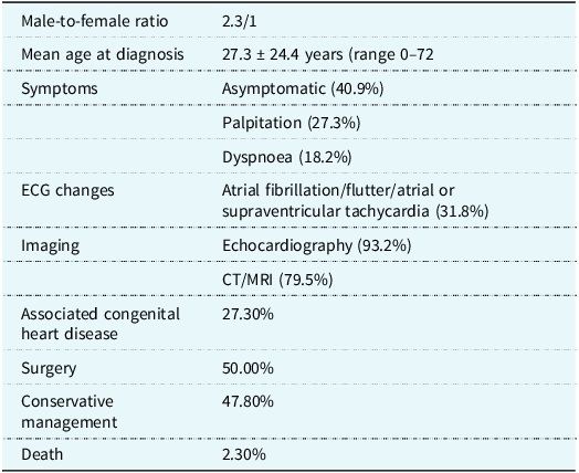
A total of 44 cases of right atrial appendage aneurysm were identified, comprising 30 males (68.2%) and 13 females (29.5%) (see Table 1). Reference Luo and Yuan3–Reference Yoon, Kim and Lee46 In one case sex was not specified. Reference Tunks, Malysz and Clark12 The median age at presentation was 27.3 ± 24.4 years, with an age range between 0 and 72 years. The highest right atrial appendage aneurysm prevalence occurred in the third decade of life. Eighteen patients (40.9%) were asymptomatic. The most frequent clinical symptom was palpitation, affecting 12 patients (27.3%), followed by dyspnoea in 8 patients (18.2%). Other symptoms were seen in 13.6% of the sample. Electrocardiography was documented for 34 patients (77.3%), of whom 14 (31.8%) had an atrial arrhythmia (atrial fibrillation or flutter, atrial tachycardia, supraventricular tachycardia). Normal sinus rhythm was detected in 19 subjects (43.2%). Chest radiography was done in 29/44 (65.9%) and revealed cardiomegaly in 20 (45.5%). Echocardiography was the most common diagnostic modality (93.2%). Right atrial appendage aneurysm diagnosis was confirmed on CT and/or MRI in 79.5%. These imaging studies were conducted to further assess and confirm the diagnosis of right atrial appendage aneurysm identified through echocardiographic examinations. Right atrial appendage aneurysm dimensions were measured in 19/44 (43.2%). The mean size of right atrial appendage aneurysm was 93 × 70 mm. In 12 patients (27.3%), an associated congenital cardiac abnormality was found, mostly in the form of an atrial septal defect/patent foramen ovale (22.7%). One patient presented with aneurysms in both the right and left atrial appendages, along with Eisenmenger’s syndrome resulting from underlying atrial and ventricular septal defects. Reference Sevimli, Gundogdu, Aksakal, Arslan, Gurlertop and Senocak40 Half of the patients (50.0%) were treated surgically with or without the Maze procedure (performed in 4.5%), whilst 47.8% were treated medically with close follow-up. The indications for surgery were not clearly mentioned in some cases. Medical treatment included antiarrhythmic drugs and anticoagulant therapy against thromboembolic risk. In a few case reports the outcome was not reported. One death (2.3%) was reported also (see Table 2).
List of the case reports which have been published in the field

Right atrial appendage aneurysm patients’ features

Discussion
Right atrial appendage aneurysm is an uncommon condition which can be either congenital or acquired. Reference Aryal, Hakim and Giri1 In our review of 44 documented cases, males were affected way more often than females. Reference Aryal, Hakim and Giri1,Reference Mercuro, Bassareo, Mariucci, Deidda, Zedda and Bonvicini47 Although right atrial appendage aneurysm has been observed across all age groups, it is most commonly diagnosed in individuals in their 30s. It has also been identified in newborns and even during prenatal development.
The congenital form of right atrial appendage aneurysm is thought to result from abnormal development of the right atrial muscular wall. Reference Ishii, Inamura and Kayatani30 The exact cause of the acquired form remains unclear, but it is suspected to be associated with prolonged elevation of right atrial pressure and increased atrial volume due to underlying conditions such as pulmonary hypertension, congenital heart defects like atrial septal defect, or heart failure. Reference Aryal, Hakim and Giri1
The coexistence of left atrial appendage aneurysm and patients with right atrial appendage aneurysm is anecdotal. Reference Sevimli, Gundogdu, Aksakal, Arslan, Gurlertop and Senocak40 Histological examination often reveals endocardial fibrosis, and in some cases, lipomatous degeneration of the aneurysm is observed without signs of inflammation. Reference Guaricci, Tarantini and Basso19,Reference Corti, Sciuchetti, Ferro, Porta, Redaelli and Paolini44
The most frequent symptom of right atrial appendage aneurysm was palpitation caused by atrial tachyarrhythmias, such as atrial fibrillation/flutter, atrial tachycardia, or supraventricular tachycardia. Some individuals with right atrial appendage aneurysm exhibited no symptoms, with the condition being discovered incidentally during cardiac imaging, autopsy, or surgeries conducted for unrelated reasons. Reference Kurata, Shishido-Hara and Hosaki45 Others presented with thromboembolic complications. Reference Tejero-Hernández, Espejo-Pérez and Suárez-de-Lezo-Cruz-Conde29 Dyspnoea, often linked to underlying cardiovascular issues such as heart failure and pulmonary hypertension, can also be a symptom. In our review, some patients had pulmonary hypertension with or without Eisenmenger’s syndrome or had left ventricular dysfunction. Reference Sevimli, Gundogdu, Aksakal, Arslan, Gurlertop and Senocak40–Reference Mizui, Mori and Kuroda42
Cardiomegaly, indicative of underlying heart disease, was observed on chest X-rays in 45.5% of patients with right atrial appendage aneurysm. Transthoracic echocardiography served as the initial imaging modality for right atrial appendage aneurysm diagnosis. In cases where transthoracic images were inconclusive, transoesophageal echocardiography with or without contrast enhancement was employed. Reference Fang, Zeng, Chen and Cui13,Reference Guaricci, Tarantini and Basso19 Advanced imaging techniques such as CT and MRI were valuable adjuncts, offering high sensitivity and specificity. These modalities were particularly useful for confirming the connection of the aneurysm to the right atrial appendage and assessing its relationship to adjacent structures. Reference Sondhi, Wardhan, Pandit and Nath16
Surgical resection is recommended for symptomatic patients and those with enlarging right atrial appendage aneurysms to reduce the risk of arrhythmias and thromboembolic events. Concomitant procedures, such as bi-atrial aneurysm resections, the Maze procedure, and CHD repair, can be performed simultaneously to address associated cardiac conditions. Reference Contreras, Courtis, Brenna, Amuchastegui and Amuchastegui18,Reference Guaricci, Tarantini and Basso19,Reference Sevimli, Gundogdu, Aksakal, Arslan, Gurlertop and Senocak40,Reference Mizui, Mori and Kuroda42 Currently, there is no information available regarding the use of device direct closure for right atrial appendage aneurysm. While device-based closures are commonly employed for left atrial appendage management to reduce stroke risk in atrial fibrillation patients, Reference Lorenzoni, Merella, Pischedda and Casu48 their application in right atrial appendage aneurysm cases is not documented yet. One patient experienced right atrial appendage aneurysm reduction in size after atrial septal defect device closure. Reference Duan, Li and Sun28 Surgical resection remains the standard treatment for symptomatic or enlarging right atrial appendage aneurysms to mitigate risks such as arrhythmias and thromboembolic events. Reference Aryal, Hakim and Giri1
Less than half of right atrial appendage aneurysm patients were managed conservatively without reported complications. A conservative approach is typically recommended for asymptomatic individuals. Reference Aryal, Hakim and Giri1 However, factors such as large right atrial appendage aneurysm size, enlarged right atrium, and associated congenital heart defects have been linked to increased risks of thromboembolic events and arrhythmias. Reference Contreras, Courtis, Brenna, Amuchastegui and Amuchastegui18,Reference Guaricci, Tarantini and Basso19,Reference Sevimli, Gundogdu, Aksakal, Arslan, Gurlertop and Senocak40,Reference Mizui, Mori and Kuroda42 Regular monitoring of asymptomatic patients using CT imaging or cardiac magnetic resonance to detect right atrial appendage aneurysm enlargement has proven useful in identifying candidates who may benefit from early surgical intervention. Reference Gulati, Gheta and Chan32
Asymptomatic right atrial appendage aneurysm patients undergoing conservative management should receive anticoagulation therapy to prevent thrombus formation. Antiarrhythmic medications are used to manage atrial tachyarrhythmias. The prognosis for these patients is generally favourable, but they require close monitoring. Reference Cianciulli, Rubinetti, Saccheri, Dethinne and Prezioso17
A large or progressively enlarging right atrial appendage aneurysm has been identified as a risk factor for thromboembolic complications and atrial arrhythmias. Although there is limited long-term data supporting routine repeat imaging, some experts suggest follow-up cardiac MRI every two years. Reference Gulati, Gheta and Chan32
Patients with evidence of aneurysm enlargement should be considered for surgical excision due to their heightened risk of arrhythmias and thromboembolic events. Those with a patent foramen ovale or atrial septal defect should be carefully monitored for systemic thromboembolism, and these defects should be closed during right atrial appendage aneurysm surgical treatment. Reference Ishii, Inamura and Kayatani30,Reference Leonard, Marras, Calcaterra and Bassareo49
Potential complications of right atrial appendage aneurysm include pulmonary embolism, rupture of the aneurysmal right atrial appendage, and sudden death. Reference Cianciulli, Rubinetti, Saccheri, Dethinne and Prezioso17
In conclusion, right atrial appendage aneurysm can lead to significant health issues. Surgical removal is advised for symptomatic individuals and those with large aneurysms. Asymptomatic patients may be managed conservatively, focusing on preventing thromboembolic events and atrial tachyarrhythmias. Regular follow-up imaging, such as echocardiography and cardiac magnetic resonance, can be considered to monitor aneurysm size and progression (see Figures 2, 3, 4).
Axial four chamber view on cardiac MRI showing a massive right atrial dilatation in a 10-month-old patient (unpublished case). Acronyms: RA = Right Atrium; RV = Right Ventricle; LA = Left Atrium; LV = Left Ventricle.

Cardiac MRI (short axis view) displaying the presence of a right atrial appendage aneurysm in the same baby who has subsequently undergone surgery for aneurysm plication. Acronyms: RAAA = Right Atrial Appendage Aneurysm; RA = Right Atrium; AO = Aorta; LA = Left Atrium.

Prisma flow diagram.