Introduction
Minor salivary gland tumours are rare, accounting for 10–15 per cent of all salivary gland tumours. In contrast to major salivary gland tumours, in which the majority of tumours are benign, especially those located in the parotid gland, 80 per cent of minor salivary gland tumours are malignant.Reference Bradley, Myers and Ferris1 Like major salivary gland tumours, they are characterised by a wide histological diversity compared with tumours of other head and neck sites.Reference Seethala and Stenman2
It has been estimated that the number of minor salivary glands is between 450 and 1000, and they are widely distributed in the mucosa of the upper aerodigestive tract, preferentially in the oral cavity and oropharynx, where most tumours occur (70–90 per cent), but with presence in the nasal cavity and paranasal sinuses, rhinopharynx, hypopharynx and larynx.Reference Ellis, Auclair, Ellis and Auclair3
The aim of this study was to review the clinical characteristics, histological type, treatment and prognosis of minor salivary gland tumours diagnosed and treated in our centre during the last three decades.
Material and methods
This study was performed retrospectively based on a review of the clinical history of patients treated for a minor salivary gland tumour in our centre during the period 1988–2020. In our hospital, only an otorhinolaryngology (ENT) department is available, and all tumours were evaluated and managed by ENT consultants. We excluded from the study 23 patients with ethmoid adenocarcinomas, as this was considered to be a different entity from the rest of the minor salivary gland tumours, and 3 patients with adenoid cystic carcinoma located in the external auditory canal.
Information on the oncological follow up of patients with malignant tumours was obtained from a database that prospectively collects data on all patients with malignant head and neck tumours treated at our centre since 1985.Reference León, Orús and Quer4 The malignant tumours were reclassified according to the criteria of the eighth edition of the tumour–node–metastasis.Reference Brierley, Gospodarowicz and Wittekind5 The histological classification of the tumours was performed according to the criteria of the fourth edition of the World Health Organization classification of head and neck tumours.Reference El-Naggar, Chan, Grandis, Takata and Slootweg6 All patients included in the study had a minimum follow up of 24 months. The mean follow up of the included patients was 9.2 years (standard deviation = 7.1 years).
The variables analysed were sex, age at tumour diagnosis, location, pathological diagnosis, type of treatment, disease control and survival. In patients with malignant tumours, the results were analysed according to tumour staging and histological grade. In patients with adenoid cystic carcinoma, we considered the presence of perineural invasion.
The relationship between qualitative variables was analysed using the Pearson chi-square test or Fisher’s exact test. The relationship between quantitative and qualitative variables was evaluated by student’s t-test. Survival was estimated using the Kaplan–Meier method, with the log-rank test used to compare survival curves. Disease-free survival was defined as the interval between diagnosis and tumour recurrence at local, regional or distant sites, disease-specific survival was defined as the interval between diagnosis and death because of the tumour and overall survival was defined as the interval between diagnosis and any cause of death.
This study was conducted in accordance with the principles established in the Declaration of Helsinki.
Results
During the study period, a total of 73 patients with minor salivary gland tumours of the upper aerodigestive tract were treated at our centre. Of these, 9 tumours (12.3 per cent) were benign and 64 (87.7 per cent) were malignant. Figure 1 shows a chart of the histology of tumours analysed. Table 1 shows the distribution according to the age, sex, location and histology of the tumour according to whether they were benign or malignant. There were no significant differences in the mean age (p = 0.499) or sex of the patients (p = 0.729) according to whether they had a benign or malignant tumour.
Master chart of the tumours included in the study.

Figure 1 Long description
A chart illustrating the distribution of 73 tumors, divided into 9 benign and 64 malignant cases. The benign tumors are located in the oral cavity (1) and oropharynx (8), with types including basal cell adenoma (1), pleomorphic adenoma (4), basal cell adenoma (2) and myoepithelioma (2). The malignant tumors are categorized by location: paranasal sinus (12) with adenoid cystic carcinoma (12); oral cavity (22) with adenoid cystic carcinoma (8), mucoepidermoid carcinoma (7), adenocarcinoma (6) and epithelial-myoepithelial carcinoma (1); oropharynx (25) with adenoid cystic carcinoma (12), mucoepidermoid carcinoma (10), adenocarcinoma (2) and epithelial-myoepithelial carcinoma (1); larynx (4) with adenoid cystic carcinoma (2) and mucoepidermoid carcinoma (2); nasopharynx (1) with adenoid cystic carcinoma (1).
Characteristics of the patients included in the study

Table 1 Long description
The table compares characteristics of patients with malignant and benign tumours. Malignant tumours are more prevalent, with 64 cases, and have a higher average age of 57.7 years. Women slightly outnumber men in malignant cases, and the oropharynx is the most common location. Adenoid cystic carcinoma is the most frequent histology type among malignant tumours. In contrast, benign tumours are less common, with 9 cases, and have a lower average age of 53.7 years. Men slightly outnumber women in benign cases, and the oral cavity is the predominant location. Pleomorphic adenoma is the most common histology type among benign tumours. The data highlights a higher occurrence of high-grade histological classification in malignant tumours.
Benign tumours
Except for one basal cell adenoma located in the mucosa of the hard palate, the remaining benign minor salivary gland tumours were located in the oropharynx: in the soft palate (seven cases) and in the base of the tongue (one case). The most common histological type was pleomorphic adenoma, which accounted for 44.4 per cent of the diagnosed tumours. All patients with benign minor salivary gland tumours were treated with surgical resection of the tumour. With a mean follow-up period of 12.3 years (range, 2.5–33.4 years), none of the patients with a benign minor salivary gland tumour had a recurrence of the lesion.
Malign tumours
The most common histological type among minor salivary gland malignancies was adenoid cystic carcinoma, which accounted for 46.9 per cent of the diagnoses (n = 30), followed by mucoepidermoid carcinoma, which accounted for 32.8 per cent (n = 21). Among the mucoepidermoid carcinomas, 14 (66.7 per cent) were classified as low grade, 2 (9.5 per cent) as intermediate grade and 5 (23.8 per cent) as high grade. In the analysis of patient survival, intermediate-grade tumours were grouped with high-grade tumours. The series was completed with 2 cases of epithelial-myoepithelial carcinoma and 11 cases of adenocarcinoma, among which the following variants were described: low-grade polymorphous (n = 4), acinic cell (n = 1), basal cell (n = 1) and unspecified (n = 5), of which 3 were considered low grade and 2 high grade.
The most common location of malignant minor salivary gland tumours was the oropharynx, where tumours were preferentially located at the base of the tongue (16 cases) and in the soft palate (6 cases), with 3 cases located in the tonsillar region. The most common histological type in the oropharynx was mucoepidermoid carcinoma (n = 12), followed by adenoid cystic carcinoma (n = 7).
In the oral cavity, the most common location was the hard palate (n = 10), followed by the oral mucosa of the lips (n = 4) and the jugal mucosa (n = 4). There were also tumours located on the tongue, floor of the mouth, retromolar trigone and gingival mucosa. The most common histological type in the oral cavity was adenoid cystic carcinoma (n = 8), followed by mucoepidermoid carcinoma (n = 7). Three of the cases of polymorphous adenocarcinoma were located in the oral cavity.
Twelve tumours were located in the paranasal sinuses, six in the maxillary sinus and six in the ethmoid sinus. All cases were adenoid cystic carcinoma. It should be noted that ethmoid adenocarcinomas were excluded from this study. In addition, one case of adenoid cystic carcinoma was diagnosed in the nasopharynx.
Finally, malignant tumours of the minor salivary gland occurred in the larynx in four cases, two adenoid cystic carcinoma and two mucoepidermoid carcinomas. The cases of mucoepidermoid carcinoma were located in the supraglottis, while the cases of adenoid cystic carcinoma had a glotto-subglottic location.
Table 2 shows the distribution of the histological types according to tumour location. There were significant differences in the histological type according to location (p = 0.002). In the paranasal sinuses and nasopharynx, all tumours were adenoid cystic carcinoma. In the oral cavity, the most common histological type was adenoid cystic carcinoma (36.4 per cent), while in the oropharynx it was mucoepidermoid carcinoma (48.0 per cent).
Histological type according to the location of the minor salivary gland malignant tumours

Table 2 Long description
The table categorizes minor salivary gland malignant tumors by histological type and location. Adenoid cystic carcinoma is the sole type found in paranasal sinuses and nasopharynx, accounting for all cases there. In the oral cavity, adenoid cystic carcinoma is slightly more common than mucoepidermoid carcinoma, with adenocarcinoma also present. Mucoepidermoid carcinoma is the most frequent in the oropharynx, followed by adenoid cystic carcinoma and adenocarcinoma. The larynx shows an equal distribution between adenoid cystic and mucoepidermoid carcinomas. The data highlights the variation in tumor types across different locations, with some areas showing a single dominant type and others a mix.
At diagnosis, the local extension of the tumour was cT1 in 17 cases (26.6 per cent), cT2 in 18 cases (28.1 per cent), cT3 in 13 cases (20.3 per cent) and cT4 in 16 cases (25.0 per cent). Thirteen patients (20.3 per cent) had lymph node involvement at diagnosis (cN+) and 1 patient (1.6 per cent) had distant metastases (cM1). The percentage of clinical lymph node involvement was 28.6 per cent for patients with mucoepidermoid carcinoma, 16.7 per cent for patients with adenoid cystic carcinoma, 18.2 per cent for patients with adenocarcinoma and 0 per cent for patients with epithelial-myoepithelial carcinoma. There were no significant differences in the percentages of lymph node involvement according to histological type (p = 0.716). Distant metastases at the time of diagnosis appeared in one patient with a cT3N0 base-of-tongue adenoid cystic carcinoma who had pulmonary metastases. Overall, 15 patients were diagnosed with stage I tumours (23.4 per cent), 14 with stage II (21.9 per cent), 14 with stage III (21.9 per cent) and 21 with stage IV (32.8 per cent).
Table 3 shows the distribution of patients according to whether they had tumours diagnosed at early stages (stages I–II) or advanced stages (stages III–IV) according to the location of the primary tumour and histological type. There were significant differences in tumour extension according to tumour location (p = 0.003) and histological type (p = 0.005). Advanced stages were significantly more common in paranasal sinus tumours and in patients with adenoid cystic carcinoma.
Tumour stage according to the location and histological type of the minor salivary gland malignant tumour

Table 3 Long description
The table compares tumour stages across different locations and histological types of minor salivary gland malignant tumours. Paranasal sinuses and nasopharynx tumours are mostly advanced, with 92.3% in Stage III-IV, while oral cavity tumours are primarily early-stage, with 68.2% in Stage I-II. Histologically, adenoid cystic carcinoma shows a significant trend towards advanced stages, with 76.7% in Stage III-IV. Mucoepidermoid carcinoma and adenocarcinoma are more likely to be early-stage, with 66.7% and 63.6% respectively in Stage I-II. The data suggests location and histological type influence tumour stage, with notable statistical significance in some categories.
Treatment consisted of surgery alone in 26 cases (40.6 per cent), surgery with adjuvant radiotherapy in 31 cases (48.4 per cent) and radiotherapy alone in 7 cases (10.9 per cent). Table S1 of the supplementary material lists the procedures used in the 57 surgically treated patients according to the tumour location. The indications for adjuvant treatment with radiotherapy were the presence of positive or close resection margins, lymph node involvement, locally advanced tumours (pT3-4) or high histological grade.
The reasons why patients were not treated with surgery were that the tumours were considered unresectable (cT4b) in four cases of adenoid cystic carcinoma located in the oropharynx (two cases), nasopharynx (one case) or paranasal sinuses (one case), and refusal of the proposed surgery in three cases: one patient with a high-grade mucoepidermoid carcinoma of the supraglottis cT2N0, one patient with a adenoid cystic carcinoma of the base of the tongue cT3N0 and one patient with a adenoid cystic carcinoma of the upper lip cT2N0. All these patients experienced a recurrence of the tumour and died as a result of disease progression.
We performed neck dissections in association with the local surgery in 22 patients: therapeutic neck dissections in 12 cN+ patients and elective neck dissections in 10 cN0 patients. Of the 12 cN+ patients, the presence of lymph node metastases was confirmed in 8 cases (66.7 per cent). Of the patients who underwent elective neck dissection, occult lymph node metastasis appeared in 1 case (10 per cent), which was a patient with a cT3N0 oropharyngeal adenoid cystic carcinoma treated with a buccopharyngectomy. Among the nine pN+ patients, the primary tumour histology was adenoid cystic carcinoma in four cases, mucoepidermoid carcinoma in four cases and unspecified high-grade adenocarcinoma in one case.
During the follow-up period, 18 patients (28.1 per cent) had local recurrence, 9 (14.1 per cent) had regional recurrence and 21 (32.8 per cent) had distant metastases. Five- and 10-year disease-free survival rates were 50.3 per cent (95 per cent confidence interval (CI) = 37.6–63.0 per cent) and 42.6 per cent (95 per cent CI = 29.7–55.5 per cent), disease-specific survival rates were 77.0 per cent (95 per cent CI = 66.4–87.6 per cent) and 50.8 per cent (95 per cent CI = 37.1–64.5 per cent), and overall survival rates were 75.7 per cent (95 per cent CI = 64.9–86.5 per cent) and 48.1 per cent (95 per cent CI = 34.6–61.6 per cent), respectively. Local recurrences occurred predominantly in patients with adenoid cystic carcinoma (61.1 per cent of local recurrences), regional recurrences occurred predominantly in patients with mucoepidermoid carcinoma (44.4 per cent of regional recurrences) and distant metastases occurred predominantly in patients with adenoid cystic carcinoma (68.2 per cent of distant metastases).
Despite attempts to salvage local, regional or distant recurrence, most of the patients with a recurrence died as a result of tumour progression. At the time of study completion, 4 of the 34 patients with a tumour recurrence had not died from the tumour: 2 patients with a limited local recurrence of oral cavity or oropharyngeal mucoepidermoid carcinoma treated with local salvage surgery, 1 patient with oropharyngeal adenoid cystic carcinoma with lung metastases treated with 3 lung resections and 1 patient with maxillary sinus adenoid cystic carcinoma with lung metastases treated with lung resection.
A percentage of deaths related to tumour progression occurred after longer follow up. Sixteen of the 30 deaths due to tumour progression (56.3 per cent) occurred after more than 5 years of follow up, with patients dying because of tumour progression up to 19 years after diagnosis.
Figure 2 shows the 20-year disease-specific survival curves according to local tumour location (Figure 2a), histological type (Figure 2b), tumour staging (Figure 2c) and treatment (Figure 2d). Significant differences in disease-specific survival were observed according to primary tumour location, tumour stage and type of treatment. The patients with the best prognosis were those with a primary location in the oral cavity and oropharynx, with initial tumours (stage I–II), and who received treatment with surgery alone. Patients with adenoid cystic carcinoma had a worse survival rate, although the differences according to tumour histology did not reach statistical significance (p = 0.120). There were no differences in disease-specific survival according to patient sex (p = 0.90).
Disease-specific survival according to local tumour location (a), histological type (b), tumour staging (c) and treatment (d). Ca. = Carcinoma; RT = Radiotherapy.

Figure 2 Long description
The image contains four survival curves labeled (a) to (d). Curve (a) shows disease-specific survival according to local tumour location, with lines for oropharynx, oral cavity, paranasal sinuses-nasopharynx and larynx. The x-axis is labeled 'years' and the y-axis is labeled 'disease-specific survival'. The p-value is 0.001. Curve (b) shows survival by histological type, with lines for epithelial-myoepithelial carcinoma, mucoepidermoid carcinoma, adenocarcinoma and adenoid cystic carcinoma. The p-value is 0.120. Curve (c) shows survival according to tumour staging, with lines for stages I, II, III and IV. The p-value is 0.001. Curve (d) shows survival by treatment type, with lines for surgery, surgery plus radiotherapy and radiotherapy alone. The p-value is 0.001. Each graph spans 0 to 20 years on the x-axis and 0 to 100 percent on the y-axis.
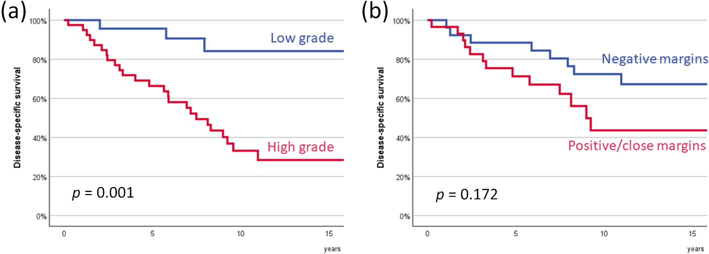
We analysed the disease-specific survival by histological grade, considering all adenoid cystic carcinoma as high-grade tumours. Patients with high-grade tumours (n = 40) had significantly lower disease-specific survival than patients with low-grade tumours (n = 24), as shown in Figure 3a (p = 0.001).
Specific survival according to histological grade (a) and resection margin status (b).

Figure 3 Long description
The image shows two line graphs. The first graph (a) displays disease-specific survival by histological grade. The x-axis is labeled 'years' and the y-axis is labeled 'disease-specific survival'. The blue line represents low-grade tumors and the red line represents high-grade tumors, with a p-value of 0.001. The second graph (b) shows survival by resection margin status. The x-axis is labeled 'years' and the y-axis is labeled 'disease-specific survival'. The blue line indicates negative margins and the red line indicates positive or close margins, with a p-value of 0.172.
Among patients who underwent surgery (n = 57), resection margins were considered free in 28 cases (49.1 per cent), close in 11 cases (19.3 per cent) and positive in 18 cases (31.6 per cent). Table S2 in the supplementary material shows the status of resection margins according to variables such as histological type, primary tumour location and local extension. Patients with adenoid cystic carcinoma, locally advanced tumours and those located in the paranasal sinuses had the highest percentage of positive or close margins. Figure 3b shows disease-specific survival as a function of resection margin status. Patients with free margins tended to have higher disease-specific survival than patients with positive or close margins. However, these differences did not reach statistical significance (p = 0.172).
In patients with adenoid cystic carcinoma, disease-specific survival was assessed according to the presence of perineural invasion. The pathology report showed the presence of perineural invasion in 19 of the 24 surgically treated adenoid cystic carcinomas (79.2 per cent). Patients with perineural invasion had a trend toward poorer disease-specific survival, although the differences did not reach statistical significance (p = 0.060). Figure S1 in the supplementary material shows the disease-specific survival of patients with adenoid cystic carcinoma as a function of perineural invasion.
Discussion
The low incidence, the large number of histological types and the variability of location within the upper aerodigestive tract make the study of minor salivary gland tumours difficult.
Benign tumours account for between 11.7 per centReference Spiro, Koss, Hajdu and Strong7 and 36.8 per centReference Wyszyńska-Pawelec, Gontarz, Zapała and Szuta8 of all minor salivary gland tumours. All authors agree that the most common histology among benign tumours is pleomorphic adenoma, with a frequency of more than 80 per cent.Reference Spiro, Koss, Hajdu and Strong7–Reference Vaidya, Pantvaidya, Metgudmath, Kane and D’Cruz9 In our group of patients, the percentage of benign tumours was 12.3 per cent, of which 89 per cent were pleomorphic adenomas.
Among malignant tumours, adenoid cystic carcinoma was the most common histological type found by some studies,Reference Wyszyńska-Pawelec, Gontarz, Zapała and Szuta8, Reference Loh, Barker, Bruch, O’Sullivan, Brown and Goldstein10–Reference Vander Poorten, Balm, Hilgers, Tan, Keus and Hart12 while mucoepidermoid carcinoma was the most common one found in others.Reference Hay, Migliacci, Karassawa Zanoni, McGill, Patel and Ganly13 The most common location found in most studies was the oral cavity, followed by the oropharynx and the nasal cavity and paranasal sinuses.Reference Loh, Barker, Bruch, O’Sullivan, Brown and Goldstein10–Reference Hay, Migliacci, Karassawa Zanoni, McGill, Patel and Ganly13 Some authors suggest a predilection for mucoepidermoid carcinoma in organs lined with squamous mucosa, such as the oral cavity or oropharynx, while the most common tumour in organs with respiratory-type mucosa, such as the nasal cavities and paranasal sinuses or the larynx, was adenoid cystic carcinoma.Reference Hay, Migliacci, Karassawa Zanoni, McGill, Patel and Ganly13, Reference Strick, Kelly, Soames and McLean14 In a study performed from the Surveillance, Epidemiology, and End Results (SEER) database, a US population-based registry that collects information on oncology patients, Baddour et al.Reference HM, Fedewa and Chen15 analysed a total of 5334 patients with minor salivary gland tumours diagnosed between 1988 and 2009, and found that the most common site was the oral cavity (58.7 per cent), followed by the pharynx (21.2 per cent) and the nasal cavities and paranasal sinuses (15.8 per cent). The most common histological type was mucoepidermoid carcinoma (29.4 per cent), followed by adenocarcinoma (24.6 per cent) and adenoid cystic carcinoma (23.0 per cent). In our series the most common histological type was adenoid cystic carcinoma and the most common location was the oropharynx.
The treatment of choice for malignant tumours of the minor salivary glands is surgical resection, with adjuvant radiotherapy for very extensive, high-grade tumours, with lymph node involvement, with perineural or lymph vascular invasion, or with positive margins.Reference Hay, Migliacci, Karassawa Zanoni, McGill, Patel and Ganly13, Reference Strick, Kelly, Soames and McLean14 Some series report a significant decrease in survival in patients who did not receive surgical treatment.Reference Loh, Barker, Bruch, O’Sullivan, Brown and Goldstein10, Reference HM, Fedewa and Chen15 In our cohort of patients, all seven patients treated with radiotherapy alone had local tumour recurrence. It has been suggested that patients who received combined treatment of surgery and radiotherapy would have worse survival. This finding would largely be a consequence of post-operative radiotherapy being preferentially indicated in patients with worse prognosis.Reference Hay, Migliacci, Karassawa Zanoni, McGill, Patel and Ganly13
When analysing the surgical outcome in our patients, the high percentage of cases in which the resection margins were considered positive (31.6 per cent) or close (19.3 per cent) stands out. The frequency of positive or close margins was higher in adenoid cystic carcinoma, locally advanced tumours and in those located in the paranasal sinuses. Other authors have reported a high percentage of positive or close resection margins, ranging from 40 per centReference Lee, Shin, Rho, Chung, Kim and Choi11 to 51 per cent.Reference Hay, Migliacci, Karassawa Zanoni, McGill, Patel and Ganly13 In the series reviewed by Strick et al.,Reference Strick, Kelly, Soames and McLean14 no patient with a tumour located in the paranasal sinuses had a resection with tumour-free resection margins. The local extension of the tumour, the anatomical complexity and the fact that the most common histological type is adenoid cystic carcinoma justify the difficulty in achieving free resection margins in tumours of this location.
A controversial aspect in the management of minor salivary gland malignant tumours is the management of the lymph nodes in patients without clinical evidence of regional disease. For patients with mucoepidermoid carcinoma, the convenience of elective neck dissection has been suggested in cN0 patients with locally advanced tumours (cT3-4), histologically high grade, pathological evidence of perineural or lymphovascular invasion, or when the tumour is located in the oropharynx.Reference Ellis, Graboyes, Day and Neskey16, Reference Reny, Ranasinghe, Magana, Kaufman, Chalian and BW17 In the case of patients with adenoid cystic carcinoma, some authors believe that the frequency of occult lymph node metastases in locations such as the oropharynx would justify performing an elective neck dissection.Reference Iyer, Kim, Nixon, Palmer, Kraus and Shaha18
Variables that have been associated with worse disease control and decreased survival in patients with malignant minor salivary gland tumours include advanced local extension,Reference Vaidya, Pantvaidya, Metgudmath, Kane and D’Cruz9–Reference Hay, Migliacci, Karassawa Zanoni, McGill, Patel and Ganly13, Reference Carrillo, Maldonado, Carrillo, Ramirez-Ortega, Pizano and Melo19–Reference Kakarala and Bhattacharyya21 the presence of lymph node metastases,Reference Carrillo, Maldonado, Carrillo, Ramirez-Ortega, Pizano and Melo19–Reference Kakarala and Bhattacharyya21 tumour location in the nasal cavity and paranasal sinuses,Reference Loh, Barker, Bruch, O’Sullivan, Brown and Goldstein10–Reference Strick, Kelly, Soames and McLean14 tumour location in the larynxReference HM, Fedewa and Chen15 or positive resection margins.Reference Hay, Migliacci, Karassawa Zanoni, McGill, Patel and Ganly13, Reference Iyer, Kim, Nixon, Palmer, Kraus and Shaha18–Reference Mücke, Robitzky, Kesting, Wagenpfeil, Holhweg-Majert and Wolff20
There is general agreement that a poorer prognosis is associated with tumours of high histological grade.Reference Vaidya, Pantvaidya, Metgudmath, Kane and D’Cruz9, Reference Loh, Barker, Bruch, O’Sullivan, Brown and Goldstein10, Reference Vander Poorten, Balm, Hilgers, Tan, Keus and Hart12, Reference Hay, Migliacci, Karassawa Zanoni, McGill, Patel and Ganly13, Reference Carrillo, Maldonado, Carrillo, Ramirez-Ortega, Pizano and Melo19 When analysing survival and disease control according to the histological tumour type, some authors have described a worse prognosis for patients with adenoid cystic carcinoma,Reference Vaidya, Pantvaidya, Metgudmath, Kane and D’Cruz9, Reference Strick, Kelly, Soames and McLean14 while others have found no significant differences according to tumour histology.Reference Loh, Barker, Bruch, O’Sullivan, Brown and Goldstein10–Reference Vander Poorten, Balm, Hilgers, Tan, Keus and Hart12, Reference Carrillo, Maldonado, Carrillo, Ramirez-Ortega, Pizano and Melo19, Reference Kakarala and Bhattacharyya21 The results of the population-based study using SEER data by Baddour et al.Reference HM, Fedewa and Chen15 showed that the overall survival of patients with mucoepidermoid carcinoma was higher than that of patients with an adenoid cystic carcinoma, whereas patients with an adenocarcinoma had an intermediate survival. Ten-year overall survival was 88.6 per cent for patients with mucoepidermoid carcinoma, 75.5 per cent for patients with adenocarcinoma and 62.1 per cent for patients with adenoid cystic carcinoma.
The 5-year and 10-year disease-specific survival rates of our patients were 77.0 and 50.8 per cent, respectively. According to our results, patients with advanced stage tumours located in the paranasal sinuses or the larynx and with high histological grade had the worst prognosis. Patients with positive or close margins had lower disease-specific survival, but the differences did not reach statistical significance. We found no differences in prognosis according to tumour histology. Some studies have found that female patients have a better survival,Reference Loh, Barker, Bruch, O’Sullivan, Brown and Goldstein10, Reference Hay, Migliacci, Karassawa Zanoni, McGill, Patel and Ganly13, Reference Lopes, Santos and Kowalski22, Reference Dubal, Unsal, Chung, Patel, Park and Baredes23 while other studies found no differences in survival according to sex.Reference Vander Poorten, Balm, Hilgers, Tan, Keus and Hart12, Reference Carrillo, Maldonado, Carrillo, Ramirez-Ortega, Pizano and Melo19 In our cohort of patients, there were no prognostic differences according to sex.
In contrast to head and neck squamous cell carcinoma patients, in whom most recurrences occur within the first two years of follow up and tumour-associated mortality usually occurs within the five years of follow up, a significant percentage of tumour-associated mortality in patients with minor salivary gland malignant tumours occurs at a later time. Most of these cases of delayed mortality are associated with the late appearance of distant metastases in patients with adenoid cystic carcinoma, as well as with the prolonged survival of these patients after the appearance of the distant metastases. This behaviour of minor salivary gland malignant tumours justifies the prolongation of the follow up in patients with this type of tumour.Reference Strick, Kelly, Soames and McLean14
The main limitation of the present study is its retrospective nature, analysing the experience obtained of a limited number of cases treated in a single institution over a long period of time. On the other hand, the accuracy of the histology needs to be reviewed and comply with the most recent version of the blue book (fifth edition) but because of its retrospective nature it was not technically possible to be included in this study.
• Minor salivary gland tumours are rare and predominantly malignant
• They show marked histological heterogeneity and variable anatomical distribution within the upper aerodigestive tract
• Prognosis has been associated with tumour stage, histological grade and anatomical site
• Adenoid cystic carcinoma and mucoepidermoid carcinoma are the most frequent histological subtypes
• This study provides long-term outcomes from a single-centre cohort with extended follow up, confirms the prognostic impact of tumour stage, location and histological grade, highlights the high rate of distant metastases, particularly in adenoid cystic carcinoma, and emphasises the need for prolonged follow up because of late tumour-related mortality
Conclusion
The percentage of benign minor salivary gland tumours treated in our centre in recent years was 12.3 per cent. The most common benign tumour was pleomorphic adenoma. Among the malignant tumours, which represented 87.7 per cent of the total, the most common location was the oropharynx and the most common histological type was adenoid cystic carcinoma. The 5- and 10-year disease-specific survival rates for patients with a malignant tumour were 77.0 and 50.8 per cent, respectively. Variables associated with disease-specific survival were tumour staging, tumour location and histological grade.
Supplementary material
The supplementary material for this article can be found at https://doi.org/10.1017/S0022215126104915
Acknowledgements
Fondo Europeo de Desarrollo Regional, A Way to Build Europe. Plan estatal de Investigación, Desarrollo e Innovación, Instituto de Salud Carlos III. Grant number: FIS PI24/01605 to XL.
Competing interests
None declared