Introduction
The larval stage of Echinococcus granulosus, the causative agent of cystic hydatid disease, is of major public health concern, with a significant economic impact on livestock and humans in many countries, including India. Globally, the existence of at least ten genotypes of the parasite have been suggested (Bowles et al., Reference Bowles, Blair and McManus1995; Scott et al., Reference Scott, Stefaniak, Pawlowski and McManus1997; Lavikainen et al., Reference Lavikainen, Lehtinen, Meri, Hirvela-Koski and Meri2003). Of these E. ortleppi (cattle strain) and E. canadensis (cervid strain) have been granted species status (Nakao et al., Reference Nakao, McManus, Schantz, Craig and Ito2007). Within the intermediate host a unilocular cyst filled with hydatid cyst fluid (HCF) is formed in the visceral organs. This fluid consists of a complex mixture of proteins of host and parasitic origin. The secreted proteins deposited in the HCF have been shown to have biological significance.
One of the most abundant parasitic antigens of HCF is the oligomeric lipoprotein antigen B (AgB), which is involved in immune invasion and is often used for immunodiagnosis (Lightowlers & Gottstein, Reference Lightowlers, Gottstein, Thompson and Lymbery1995; Fernández et al., Reference Fernández, Ferreira, Fernández, Zaha and Nieto1996; Virgino et al., Reference Virgino, Hernandez, Rott, Monteiro, Zandonai, Nieto, Zaha and Ferreira2003). Molecular studies have indicated that AgB is encoded by a gene family with five major gene clusters AgB1 (Shepherd et al., Reference Shepherd, Aitken and McManus1991), AgB2 (Fernández et al., Reference Fernández, Ferreira, Fernández, Zaha and Nieto1996), AgB3 (Chemale et al., Reference Chemale, Haag, Ferreira and Zaha2001), AgB4 (Arend et al., Reference Arend, Zaha, Ayala and Haag2004) and AgB5 (Haag et al., Reference Haag, Alves-Junior, Zaha and Ayala2004).
The molecular variation of AgB genes of the Indian bubaline isolates has not been studied. The present study has important implications in analysing the diagnostic efficacy of AgB and its family and is the first report on the AgB sequence of Indian bubaline isolates, periodicity of α and β structures and stretches of agretope.
Materials and methods
A total of 30 buffalo cysts were collected. The protoscoleces were rinsed three times in phosphate buffered saline (PBS) (pH 7.2) and stored in 70% alcohol (v/v) at − 20°C until further use. Total genomic DNA from protoscoleces was extracted by EZ-10 spin column DNA isolation kit (Bio Basic Inc., Canada).
Three separate sets of sense and antisense primers were used for amplification of different clades of AgB, as described earlier (Haag et al., Reference Haag, Araújo, Gottstein, Siles-Lucas, Thompson and Zaha1999, Reference Haag, Alves-Junior, Zaha and Ayala2004) (table 1).
Design of primers used for amplification of antigen B family.

To amplify the desired fragment, PCR was performed at 94°C for 30 s, 53–55°C for 30 s and 72°C for 30 s. Final extension was done at 72°C for 10 min. All amplicons were purified using QIAquick PCR purification kit (Qiagen, Germany). The insert was further cloned in TA vector for sequencing. Nucleotide sequence information was further interpreted to give predicted amino acid sequence information. Using the software DNASTAR (DNASTAR Inc., Madison, Wisconsin, USA) we tested α and β structure periodicity, α- and β-amphipathic regions and antigenic index. Predictions of MHC class II binding regions of more than 50% binding propensity were assessed using an online version of ProPred (Singh & Raghava, Reference Singh and Raghava2001).
Results and discussion
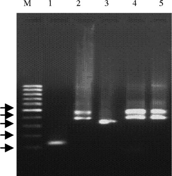
After amplification with the primer of EgAgB1 the total product size yield was 129 bp (fig. 1, lane 1); the sequence was 99% homologous to AgB1 of E. canadensis (accession number: AY 265907). Further, an amplified product of size 445 bp was completely homologous to AgB3 of E. ortlepii (DQ 137842). The open reading frame (ORF) of the amplicon showed 100% and 99% homology to E. ortleppi (DQ 137842) and E. canadensis (DQ 174241), respectively.
Amplification of antigen B family. Lanes: M, molecular weight marker (100 bp ladder) (arrows from the bottom: 100, 200, 300, 400 and 500 bp); 1, EgAgB1 (129 bp); 2, 4 and 5, EgAgB1 (445 bp) and EgAgB3 (385 bp); 3, EgAgB4 (333 bp).

Another amplicon with a product size of 384 bp, supposed to amplify AgB1, was homologous to AgB1 M1 allele (AY 773091.1), AgB1 G1 genotype (DQ 137827), AgB1 G2 genotype (DQ 137833), AgB1 M2 allele (AY 773092) and 99% homologous to AgB1 G1 genotype (DQ 137828). The predicted amino acid sequence was homologous to the AgB1 M1 allele (AY773091), AgB1 M2 allele (AY 773092), AgB1 G1 genotype (DQ 137828, DQ 137827, DQ 137143), AgB1 G2 genotype (DQ137835, DQ 137833), AgB8/1 (AF 143813), AgB1 M6 allele (AY 871018), AgB1 M5 allele (AY 871017) and Italian cattle liver cyst (Z2 6482). The same amino acid sequence information was 99% homologous to AgB1 G2 genotype (DQ 137834).
Using the third set of primers, the product size of amplicon was determined to be 333 bp. This particular sequence information was completely homologous to AgB4 of bovine origin (AY 357112) and 99% homologous to AgB4 of bovine origin (AY 357116). The length of the open reading frame was 180 bp and was completely homologous to AgB4 (AY 357116, AY 357112, AY 4614001) and 99% homologous to AgB4 (AY 357113).
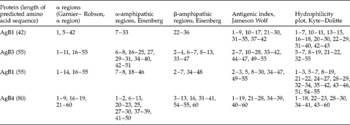
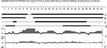
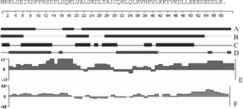
An analysis of a length of 42 amino acid residues predicted for AgB1 revealed only one stretch of α- and β-amphipathic regions, whereas for another sequence of AgB1 (with 55 amino acid residues) α- and β-amphipathic regions were present in two different stretches. In AgB3 and AgB4, α- and β-amphipathic structures were present in 4–6 different stretches (table 2). These particular regions denote the presence of T-cell antigenic sites. Delisi & Berzofsky (Reference Delisi and Berzofsky1985) have proposed that a stable amphipathic structure is required for a T-cell antigenic site. Moreover, Kaiser, & Kézdy (1984) suggested that small and intermediate-sized peptides with amphipathic structure interact with membranes. This hypothesis, suggesting that antigenic sites are strongly amphipathic, is consistent with numerous observations on other small membrane-associated peptide systems and suggests an important role for amphipathicity in molecular models of T-cell activation.
Comparison of α- and β helices of antigen B family, based on predicted amino acid sequences.

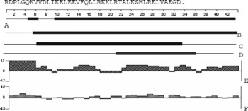
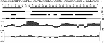
The two antigenic properties, i.e. presentation and recognition, are thought to contribute to the process of amphipathicity and α helicity (Spouge et al., Reference Spouge, Guy, Cornette, Margalit, Cease, Berzofsky and DeLisi1987). This may validate the importance of analysing the α regions for the three types of antigen considered during the study. The extended (i.e. β) peptide conformation is common in native proteins and can also be amphipathic, but β-amphipathicity has not yet been implicated in T-cell antigenicity. However, β-propensity connotes a tendency to β-conformation. During the present study a single stretch of β-amphipathicity was present in the AgB1 having 42 amino acid residues, whereas for the other antigens β-amphipathic regions were present in two to four different stretches (table 2). Further analysis of these data revealed that the antigenic index calculated by the Jameson Wolf method correlated with the Kyte–Dolitte hydrophilicity plot (figs 2–5). This kind of prediction may be correlated because hydrophobic residues are not accessible to the cells.
Analysis of predicted amino acid sequence of 42 amino acids of antigen B1 showing stretches of α-regions (A, B), α-amphipathic (C) and β-amphipathic regions (D), antigenic index (E) and hydrophilicity index (F).

Analysis of predicted amino acid sequence of 55 amino acids of antigen B3 showing stretches of α-regions (A, B), α-amphipathic (C) and β-amphipathic regions (D), antigenic index (E) and hydrophilicity index (F).

Analysis of pred icted amino acid sequence of 55 amino acids of antigen B1 showing stretches of α-regions (A, B), α-amphipathic (C) and β-amphipathic regions (D), antigenic index (E) and hydrophilicity index (F).

Analysis of predicted amino acid sequence of 60 amino acids of antigen B4 showing stretches of α-regions (A, B), α-amphipathic (C) and β-amphipathic regions (D), antigenic index (E) and hydrophilicity index (F).

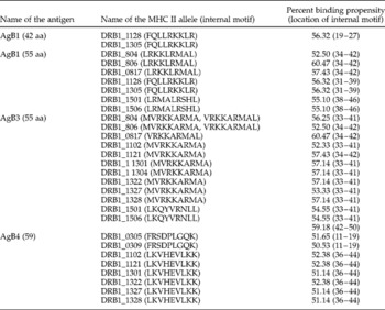
AgB1 with 42 amino acid sequences possessed more than 50% binding propensity for two common MHC molecules expressed by two MHC II alleles. AgB1 with 55 amino acid residues had two internal motifs which had more than 50% binding propensity for MHC II molecules expressed by seven different alleles. A total of four different internal motifs of AgB3 were found to have more than 50% binding propensity for a total of 12 MHC II molecules expressed by 12 different alleles. In AgB4, two internal motifs were found to have more than 50% binding propensity for eight different MHC II molecules. The details of the MHC II alleles along with stretches of amino acid sequences and positions have been depicted in table 3.
Internal motifs with more than 50% MHC II binding propensity.

Peptides that bind to class II MHC molecules comprise 7–10 amino acids that provide the major contact points (Goldsby et al., Reference Goldsby, Kindt and Osborne2000). After analysis of amino acid sequences of AgB1, AgB2, AgB3 and AgB4 it was found that the internal motifs comprised nine amino acids. Generally these sequences should have hydrophobic residues at the amino terminus. For the antigens of interest, the amino acid sequences started with hydrophobic residues such as phenylalanine (F), leucine (L), methionine (M) and valine (V). Two to three additional hydrophobic residues were also found in the middle of each internal motif. Generally three additional hydrophobic regions are thought to be present in the middle of the internal motif (Goldsby et al., Reference Goldsby, Kindt and Osborne2000) and have been compared with HLA-DR. The agretopes have been analysed in terms of their residues and their physicochemical properties. From this analysis it may be concluded that AgB and its family of bubaline isolates have internal motifs that have more than 50% binding propensity to 15 different MHC II molecules. In general, secreted and membrane-bound proteins (S/M) of helminths contribute to a wide range of parasitic properties, including penetration and establishment in host tissues, modulation of the host immune response and incorporation of metabolites from the host. Due to their potential exposure to the host immune system, S/M components are candidates for diagnostic tests, as well as being the targets for new drugs and vaccines (Rosenzvit et al., Reference Rosenzvit, Camicia, Kamenetzky, Muzulin and Gutierrez2006). However, the role of AgB in E. granulosus biology is yet to be determined. In conclusion, we believe the information presented in this study will be helpful in determining the prediction of structure and biological function of the five gene loci of the AgB family with respect to MHC II binding propensity.