


Computed tomographic and magnetic resonance imaging (MRI) studies have shown structural abnormalities in patients with schizophrenia compared with healthy controls (Reference Liddle, Hirsch and WeinbergerLiddle, 1995). Similar structural brain abnormalities have also been found in affective disorders (Reference CummingsCummings, 1993). Most of these studies have been on chronic patients. It is therefore difficult to say which of the MRI findings result from treatment (medication) or chronicity-related factors, and what their value would be in differential diagnostics. In the present study, we examined the brain volumes of patients with first-episode schizophrenia, psychotic depression and severe depression and compared them with those of healthy controls. We placed special emphasis on diagnostic procedures to distinguish as clearly as possible between schizophrenia and severe depressive disorders. Our aim was to study how regional brain volumes and their interrelationships for the patients with first-episode schizophrenia, psychotic depression and severe depression differ from those of healthy controls and from each other.
Structural central nervous system abnormalities in schizophrenia and affective disorders
Grey matter volume in patients with schizophrenia, in the neocortex generally and in various subcortical structures, is fairly consistently reduced, whereas the third and lateral ventricles, as well as the cortical sulci, are enlarged (Reference Pfefferbaum and MarshPfefferbaum & Marsh, 1995; Reference CannonCannon, 1996; Reference Buchanan and CarpenterBuchanan & Carpenter, 1997; Reference Lawrie and AbukmeilLawrie & Abukmeil, 1998). In a recent meta-analysis, Wright et al (Reference Wright, Rabe-Hesketh and Woodruff2000) concluded that regional structural differences in patients with schizophrenia include bilaterally reduced volume of medial temporal lobe structures, consistent with a pathological process in schizophrenia that involves distributed volume changes within the brain.
In three meta-analyses, Elkis et al (Reference Elkis, Friedman and Wise1995) found that the ventricles and sulci were enlarged in patients with mood disorder compared with controls, and that ventricular enlargement was greater in patients with schizophrenia compared with those with mood disorders, although the effect size was small.
Crow (Reference Crow1990; Reference Crow, Done and SackerCrow et al, 1996) suggested that brain changes in schizophrenia can be seen to include an arrest in the development of cerebral asymmetry. The normal asymmetry of the temporal horn (Reference Crow, Ball and BloomCrow et al, 1989a ; Reference Bogerts, Ashtari and DegreefBogerts et al, 1990), the Sylvian fissure (Reference Falkai, Bogerts and GreveFalkai et al, 1992; Reference Crow, Brown and BrutonCrow et al, 1992), and volume in the occipital regions (Reference Crow, Colter and FrithCrow et al, 1989b ; Reference Daniel, Myslobodsky and IngrahamDaniel et al, 1989; Reference Bilder, Wu and BogertsBilder et al, 1994) have been found to be absent in patients with schizophrenia in general, and in patients with first-episode schizophrenia specifically (Reference Bogerts, Ashtari and DegreefBogerts et al, 1990; Reference Bilder, Wu and BogertsBilder et al, 1994; Reference DeLisi, Sakuma and KushnerDeLisi et al, 1997).
Woodruff et al (Reference Woodruff, Wright and Shuriquie1997) have reported that, in comparison with unaffected controls, male patients with schizophrenia demonstrated reduced correlations between several brain regions. The most salient abnormality in patients was the dissociation between prefrontal and superior temporal gyrus volumes, suggesting a lack of mutual trophic influences during frontal and temporal lobe development. These findings suggest the possibility that not only regional volumetric differences but also the interrelationships between regional structures can differentiate schizophrenia from other psychiatric disorders and healthy people.
METHOD
Subjects
The study protocol was approved by the Ethical Committee of the Turku University/University Hospital, Turku, Finland. Written informed consent was a prerequisite for participation.
The patients were selected from a larger group of first-contact patients admitted to out-patient or in-patient facilities of the Turku University Central Hospital and the Turku City Mental Health Centre, over a 41-month period (1 November 1994 to 31 March 1998). Inclusion criteria for the patients were: schizophrenia-spectrum psychotic disorder, bipolar disorder or severe major depression according to the treating clinician; first-ever psychiatric treatment in the public sector because of this disorder; age 16-64 years; and residence in the Turku City catchment area. The sample included 116 patients, 55 (47%) men and 61 (53%) women. Nineteen subjects (16%) were out-patients and 97 (84%) in-patients, and none of the subjects had been admitted to hospital before.
Patients meeting the inclusion criteria were interviewed by one of the research psychiatrists (T.T., H.K., H.L., K-M.L. and E.W.). Diagnostic interviews consisted of three parts: (a) taking of the patient's medical history using a structured form; (b) diagnostic evaluation using a clinical interview and a simultaneously recorded structured Schedules for Clinical Assessment in Neuropsychiatry (SCAN) interview (Reference Wing, Babor and BrughaWing et al, 1990); and (c) assessment of the severity of psychiatric symptoms by the Positive and Negative Syndrome Scale (PANSS; Reference Kay, Opler and FiszbeinKay et al, 1987), the Calgary Depression Scale (CDS; Reference Addington, Addington and Maticka-TyndaleAddington et al, 1993), the Hamilton Rating Scale for Depression (HRSD; Reference HamiltonHamilton, 1967), the Brief Psychiatric Rating Scale (BPRS; Reference Overall and GorhamOverall & Gorham, 1962) and the Clinician Administered Rating Scale for Mania (CARS-M; Reference Altman, Hedeker and JanicakAltman et al, 1994). The patients also underwent somatic investigations, including brain MRI and blood test scanning for somatic disorders with psychiatric symptoms.
The diagnostic evaluation was completed and the best-estimate research diagnoses according to DSM-IV (American Psychiatric Association, 1994) were made at consensus meetings, without any knowledge of the results of the MRI scans. At these meetings, all the patients' medical records and hospital charts were available, together with the findings of the previous diagnostic interview and somatic investigations. Patients' history and symptoms were discussed by the research psychiatrists responsible for the diagnostic evaluation, a senior researcher (R.K.R.S.), and at least one research psychiatrist not personally familiar with the patient. Particular emphasis was placed on making a distinction between schizophrenia and psychotic depression. On the basis of best-estimate research, the patients were divided into four main groups: schizophrenia (n=29); bipolar disorder (n=13); major depressive disorders (n=59); and others (n=15). The collection of patients and their investigations have been described in detail previously (Taiminen et al, Reference Taiminen, Jääskeläinen and Ilonen2000, Reference Taiminen, Ranta and Karlsson2001).
For this study, only patients with the DSM-IV criteria of schizophrenia (diagnoses 295.10, 295.20, 295.30, 295.60, 295.90), severe (with psychotic features) major depressive disorder (296.24, 296.34) and severe (without psychotic features) major depressive disorder (296.23, 296.33) with successful MRI scans were included. The study sample consisted of 11 patients with schizophrenia, 20 patients with depression with psychotic features and 17 patients with depression but not psychosis. In addition, 19 healthy volunteers were included in the analyses. At clinical interview, the healthy controls had no psychiatric disorder or neurological disease or symptoms, and their first-degree relatives had no major psychiatric disorder.
MRI scanning
Magnetic resonance imaging scans of the brain were acquired with a Siemens 1.5 T scanner (Siemens, Magnetom, Germany) in Turku University Central Hospital. The scans were taken on average within 1.7 months of the clinical examinations. A conventional dual spin-echo sequence (echo time of 90 ms, repetition time 3120) was used to obtain on average 25 axial slices with a thickness of 5.4 mm and no inter-slice gap. The image matrix size was 256 × 256 (field of view of 230) and an in-plane resolution of 0.9 × 0.9 mm.
Tracing protocol
The image analyses were performed using the NRIA (New Regional Image Analysis) package, an earlier version of BBLImage. After deleting pixels corresponding to the skull and meninges using a conservative automated procedure followed by manual editing, the remaining pixels were classified into three tissue types (grey matter, white matter and cerebrospinal fluid (CSF)) using an adaptive Bayesian algorithm for three-dimensional tissue segmentation (Yan et al, 1995). To control for head tilt during scanning, the images were re-sliced according to the anterior commissure—posterior commissure plane. Detailed description of the regions of interest (ROIs) can be found in Cannon et al (Reference Cannon, van Erp and Huttunen1998). In short, the frontal lobe ROI was started on the inferior slices, where the medial border was the interhemispheric fissure and the lateral border the cortical perimeter. On the slice superior to the mamillary bodies, the posterior border was defined by extending a horizontal line from the most anterior extent of the Sylvian fissure to the interhemispheric fissure. This posterior border was continued in this fashion until the slice immediately preceding the splenium of the corpus callosum. Superior to this slice, the posterior border was defined by a horizontal line touching the most anterior part of the caudate nucleus. The temporal lobe ROI was started at the level of the midbrain, where the posteromedial border of the temporal lobe ROIs was delineated by the pons and cerebellum. The temporal lobe was separated from the frontal regions by tracing along the Sylvian fissure. More superior, the posterior boundary was defined by drawing a line extending from the contralateral cerebral peduncle to the anterior tip of the cerebellum. On the slices above the mamillary bodies, the Sylvian fissure and the diencephalon structures served as the medial borders. The posterior edge was formed by a horizontal line from the most posterior tip of the posterior fossa to the lateral perimeter. The temporal lobe ROI was discontinued on the first slice showing the splenium of the corpus callosum. The posterior regions were defined by subtracting the frontal and temporal lobe volume from the total brain volumes. The left and right hemisphere ROIs included all brain tissue except pons, medulla, cerebral peduncles and cerebellum. The ventricular ROIs included the lateral and third ventricles but not the fourth ventricle or cerebral aqueduct. The anatomical tracings were performed as described by a single rater with no knowledge of the diagnosis. Interrater reliabilities of the defined measurements based on 10 randomly selected images were excellent (intraclass correlations, 0.93).
Statistics
Statistical analyses were performed with the SAS statistical software package (SAS Institute, 1992). Differences in distributions between subject groups were analysed by chi-squared or Mantel—Haenszel chisquared test, and those in continuously scaled variables by analysis of variance (ANOVA). The data of tissue volumes were analysed with repeated-measures ANOVA and covariance models. For analyses of grey matter, white matter and sulcal CSF, hemisphere and region (frontal, temporal and posterior) served as within-subject independent variables; for analyses of ventricular CSF, hemisphere was a within-subject variable. In all models, overall intra-cranial volume and age served as continuously scaled covariates, gender as a categorised covariate and group (diagnosis) as a predictor. Multivariate analyses were followed by pair-wise group contrasts using independent sample t statistics for the sub-group (schizophrenia, psychotic depression, non-psychotic severe depression and healthy controls) comparisons. The differences in tissue volumes between patients and healthy controls are described in Figs 1,2,3,4 as Z scores, where the distances of values for patients from those for controls were scaled by s.d. P values lower than 0.05 were considered statistically significant and P values of 0.10-0.05 as marginally significant.
Regional grey matter volumes in diagnostic groups in relation to healthy controls (L, left; R, right; F, frontal; T, temporal; P, posterior; G, grey matter). [UNK], schizophrenia; [UNK], psychotic depression; [UNK], non-psychotic depression. *denotes significant values.

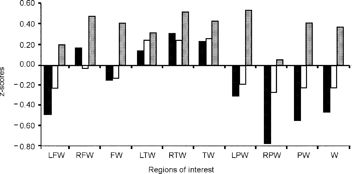
Regional white matter volumes in diagnostic groups in relation to healthy controls (L, left; R, right; F, frontal; T, temporal; P, posterior; W, white matter). [UNK], schizophrenia; [UNK], psychotic depression; [UNK], non-psychotic depression.
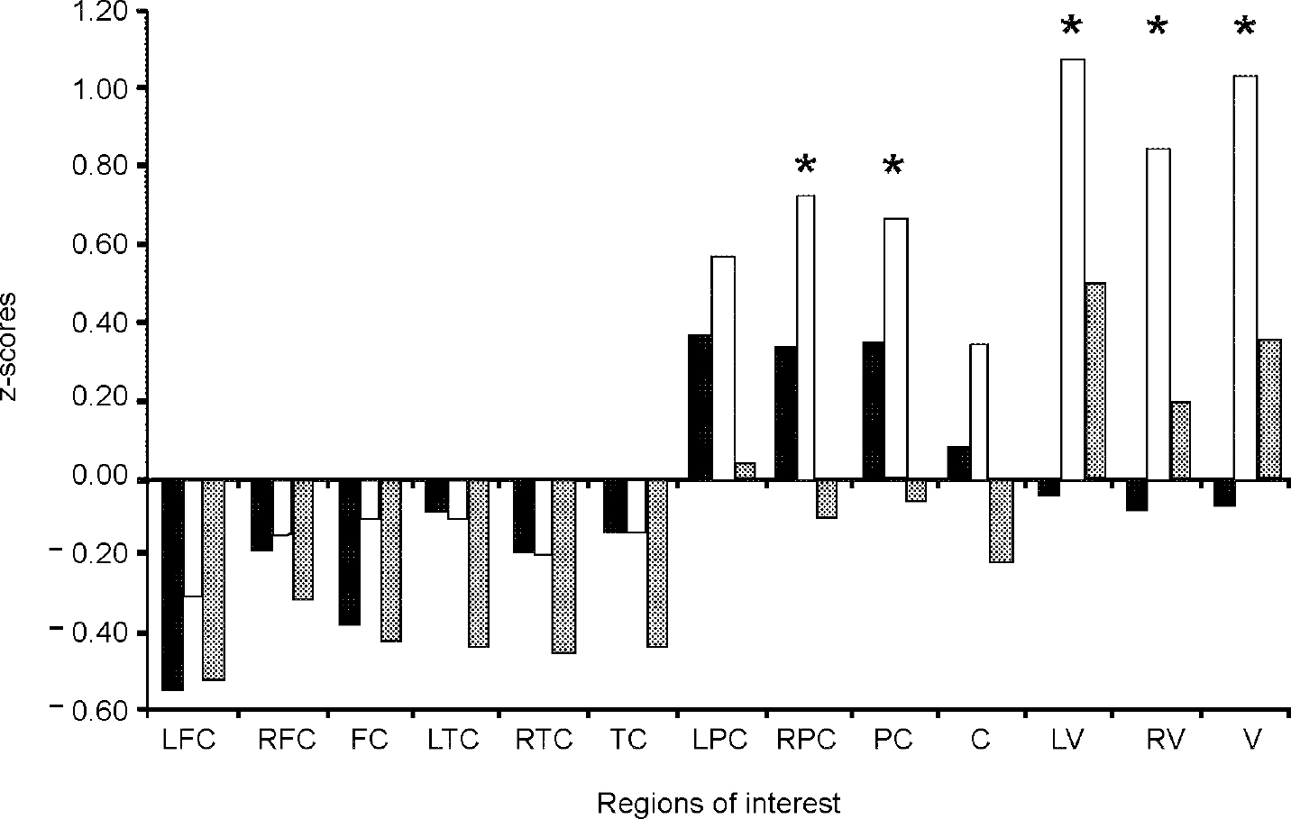
Regional sulcal and ventricular cerebrospinal fluid (CSF) volumes in diagnostic groups in relation to healthy controls (L, left; R, right; F, frontal; T, temporal; P, posterior; C, sulcal CSF; V, ventricle CSF). [UNK], schizophrenia; [UNK], psychotic depression; [UNK], non-psychotic depression.*denotes significant values.
Contralateral regional ratios of grey and white matter, as well as sulcal and ventricular volumes, in diagnostic groups in relation to healthy controls (F, frontal; T, temporal; P, posterior; G, grey matter; W, white matter; C, sulcal cerebrospinal fluid; V, ventricle cerebrospinal fluid). [UNK], schizophrenia; [UNK], psychotic depression; [UNK], non-psychotic depression.*denotes significant values.

RESULTS
There were no significant differences in gender, age, handness or use of alcohol (Table 1). No patient or control used illegal drugs.
Background of the study subjects

| Controls n=19 | Schizophrenia n=11 | Psychotic depression n=20 | Non-psychotic depression n=17 | P | |
|---|---|---|---|---|---|
| Gender | |||||
| Male | 12 | 3 | 8 | 8 | 0.2511 |
| Female | 7 | 8 | 12 | 9 | |
| Age (years) | |||||
| Mean | 30.5 | 36.6 | 34.0 | 38.4 | 0.1782 |
| s.d. | 8.4 | 14.7 | 9.8 | 12.0 | |
| Handness | |||||
| Right | 17 | 11 | 17 | 14 | 0.6423 |
| Alcohol | |||||
| > 90 mg/week | 4 | 2 | 5 | 5 | 0.7353 |
Regional volumes
Regional grey matter, white matter and CSF volumes are shown in Table 2. In repeated-measures ANOVA, intra-cranial volume had a significant effect (P < 0.05) on all tissue volumes, and age on volumes of grey matter and sulcal and ventricular CSF volumes. Diagnosis and region had a marginal interaction (P=0.091) with grey matter volume and a significant interaction (P=0.017) with sulcal CSF. Diagnosis, region and hemisphere had a significant interaction (P=0.008) with white matter volume.
Mean and s.d. of regional grey matter, white matter and cerebrospinal fluid volumes according to diagnosis

| Controls (n=19) | Schizophrenia (n=11) | Psychotic depression (n=20) | Non-psychotic depression (n=17) | |||||
|---|---|---|---|---|---|---|---|---|
| Mean | s.d. | Mean | s.d. | Mean | s.d. | Mean | s.d. | |
| Frontal lobe | ||||||||
| Grey | 123.04 | 17.67 | 114.62 | 11.36 | 115.60 | 14.26 | 119.93 | 20.03 |
| Left | 61.55 | 8.59 | 55.50 | 6.96 | 57.69 | 7.75 | 59.18 | 9.81 |
| Right | 61.49 | 9.25 | 59.12 | 6.55 | 57.91 | 6.96 | 60.75 | 10.34 |
| White | 72.82 | 10.39 | 71.84 | 9.52 | 69.34 | 12.44 | 79.09 | 13.96 |
| Left | 36.14 | 5.10 | 34.53 | 5.46 | 34.12 | 6.87 | 38.96 | 7.34 |
| Right | 36.68 | 5.78 | 37.31 | 4.67 | 35.22 | 6.03 | 40.13 | 6.91 |
| Sulci | 22.07 | 8.44 | 19.26 | 6.43 | 19.39 | 6.70 | 20.10 | 6.71 |
| Left | 11.08 | 4.20 | 8.92 | 2.90 | 9.44 | 3.23 | 9.63 | 3.29 |
| Right | 10.99 | 4.38 | 10.33 | 3.69 | 9.94 | 3.58 | 10.46 | 3.64 |
| Temporal lobe | ||||||||
| Grey | 133.84 | 16.07 | 139.71 | 12.90 | 130.63 | 17.64 | 135.26 | 13.78 |
| Left | 66.16 | 8.48 | 69.30 | 7.24 | 63.81 | 8.46 | 66.22 | 6.98 |
| Right | 67.67 | 8.08 | 70.41 | 5.89 | 66.82 | 9.72 | 69.04 | 7.53 |
| White | 59.52 | 9.01 | 62.18 | 8.14 | 60.64 | 12.07 | 66.51 | 10.09 |
| Left | 29.80 | 5.01 | 30.75 | 4.74 | 30.38 | 6.22 | 33.10 | 5.18 |
| Right | 29.72 | 4.20 | 31.42 | 4.10 | 30.26 | 6.12 | 33.40 | 5.29 |
| Sulci | 13.73 | 7.57 | 11.75 | 3.88 | 11.86 | 5.66 | 10.81 | 2.48 |
| Left | 6.59 | 3.91 | 5.65 | 1.94 | 5.79 | 2.68 | 5.14 | 1.07 |
| Right | 7.14 | 3.97 | 6.10 | 2.03 | 6.06 | 3.26 | 5.67 | 2.00 |
| Posterior region | ||||||||
| Grey | 347.84 | 33.60 | 367.82 | 37.35 | 341.96 | 43.77 | 355.63 | 44.22 |
| Left | 175.26 | 17.31 | 185.55 | 17.83 | 172.64 | 22.39 | 179.52 | 24.48 |
| Right | 172.58 | 16.97 | 182.26 | 21.40 | 169.31 | 22.03 | 176.11 | 20.27 |
| White | 243.14 | 22.60 | 229.92 | 26.10 | 229.55 | 44.80 | 257.40 | 52.75 |
| Left | 120.75 | 11.92 | 116.31 | 15.28 | 114.32 | 22.38 | 130.55 | 26.53 |
| Right | 122.38 | 11.41 | 113.60 | 11.63 | 115.22 | 22.71 | 126.84 | 26.55 |
| Sulci | 44.26 | 13.14 | 51.94 | 16.48 | 51.42 | 20.47 | 49.37 | 13.46 |
| Left | 22.31 | 6.98 | 26.39 | 7.71 | 25.40 | 9.99 | 25.07 | 7.29 |
| Right | 21.94 | 6.38 | 25.54 | 9.07 | 26.02 | 10.74 | 24.30 | 6.29 |
| Ventricles | 12.06 | 4.93 | 15.02 | 5.90 | 16.59 | 10.05 | 16.06 | 9.00 |
| Left | 6.12 | 2.62 | 7.72 | 3.13 | 8.52 | 5.84 | 8.46 | 5.10 |
| Right | 5.93 | 2.66 | 7.29 | 3.06 | 8.07 | 4.56 | 7.59 | 4.08 |
When age, gender and intra-cranial volume were taken into account in ANOVA, there was a significant group effect in left frontal grey matter volumes (P=0.043), and a marginal group effect in right posterior sulcal CSF volumes (P=0.088).
In post hoc pair comparisons, patients with schizophrenia had a smaller left frontal grey matter volume than controls, patients with depression or psychotic depression. Moreover, their total frontal grey matter volume was significantly smaller than that of controls, and their posterior grey matter volume was larger than that of controls or patients with depression (Table 2 & Fig. 1). Patients with depression had marginally larger right frontal white matter volumes than controls. They also had larger white matter volumes than patients with schizophrenia or psychotic depression in several regions (Table 2 & Fig. 2).
Patients with schizophrenia had a marginally smaller left frontal sulcal CSF volume than controls. Patients with depression also had marginally smaller left frontal sulcal CSF and temporal sulcal CSF volumes than controls. Because of the large s.d., great differences of means remained marginally significant. Patients with psychotic depression had larger ventricular CSF volumes and larger posterior sulcal CSF volumes than controls. They also had larger ventricular CSF volumes than patients with schizophrenia and a larger posterior sulcal CSF volume and total sulcal CSF volume than patients with depression (Table 2 & Fig. 3).
Regional ratios
Contralateral ratios
In inter-hemispheric comparisons, left regional volumes were divided by corresponding (contralateral) right volumes. This was called the contralateral ratio. In repeated-measures ANOVA, age had a significant effect (P < 0.05) on the contralateral ratio of volumes of grey and white matter, whereas diagnosis had a marginally significant effect (P=0.079) on the contralateral ratio of the volume of grey matter. Diagnosis and region had a marginally significant interaction (P=0.066) with white matter volume.
When the effects of age and gender were taken into account in ANOVA, there was a significant group (diagnosis) effect in the contralateral ratio of frontal grey matter (F=4.09, d.f.=3, P=0.011) and of posterior white matter (F=4.14, d.f.=3, P=0.010). In post hoc pair comparisons, when the effects of age and gender were taken into account, the contralateral ratio in patients with schizophrenia was significantly lower (i.e. left region was smaller in relation to right region) in frontal grey matter (-8.0%; F=10.2, d.f.=1, P=0.002), white matter (-9.2%; F=6.15, d.f.=1, P=0.013) and sulcal CSF volumes (-15.4; F=5.30, d.f.=1, P=0.025), and higher in posterior white matter volume (4.5%; F=4.31, d.f.=1, P=0.042) than in controls. Patients with depression had a marginally lower contralateral ratio in frontal grey matter (-4.1%; F=3.93, d.f.=1, P=0.052) and in frontal sulcal CSF (-10.8%; F=3.70, d.f.=1, P=0.059), as well as a significantly higher contralateral ratio in posterior white matter (5.5%; F=9.07, d.f.=1, P=0.004) than controls. Frontal grey matter contralateral ratio in patients with schizophrenia was lower (-7.4%; F=7.59, d.f.=1, P=0.008) than in patients with psychotic depression, whereas in patients with depression posterior white matter contralateral ratio was higher (4.8%; F=7.17, d.f.=1, P=0.010) than in patients with psychotic depression (Fig. 4).
Correlation coefficients were calculated between contralateral regional structural volumes. In general, the correlation between left and right corresponding regional volumes was high (r>0.800) and statistically significant (P <0.05). There was one exception: the correlation within frontal grey matter (r=0.413, P=0.207) was low in patients with schizophrenia, suggesting that, in schizophrenia, the contralateral inter-connection between frontal structures is rather weak.
Intra-hemispheric ratios
In intra-hemisphere comparison, frontal regional volumes were divided by ipsilateral (same side) corresponding posterior and temporal volumes, and temporal volumes by ipsilateral posterior volumes. This was called the intra-hemispheric ratio. When the effects of age and gender were taken into account, there was a significant group (diagnosis) effect in left grey matter frontal posterior intra-hemispheric ratio (F=3.94, d.f.=3, P=0.013), in left grey matter frontal temporal ratio (F=3.56, d.f.=3, P=0.020) and in left sulcal CSF frontal posterior ratio (F=5.11, d.f.=3, P=0.003).
In post hoc pair comparisons, when the effects of age and gender were taken into account, the left grey matter frontal posterior intra-hemispheric ratio (i.e. the rate of frontal grey matter divided by posterior grey matter) was lower in patients with schizophrenia than in controls (-14.6%; F=11.32, d.f.=1, P=0.001), patients with psychotic depression (-12.4; F=6.71, d.f.=1, P=0.012) and patients with depression (-13.2%; F=7.36, d.f.=1, P=0.009). The corresponding statistics for the left grey matter frontal temporal ratio were: patients with schizophrenia (-13.7%; F=9.21, d.f.=1, P=0.004), v. psychotic depression (-12.5%; F=7.70, d.f.=1, P=0.007) and v. patients with depression (-12.8%; F=7.70, d.f.=1, P=0.007). Compared with controls, the left sulcal CSF frontal posterior ratio was smaller in schizophrenia (-28.0%; F=10.08, d.f.=1, P=0.002), psychotic depression (-23.6%; F=11.21, d.f.=1, P=0.001) and depression (-19.1%; F=6.68, d.f.=1, P=0.012). Also the right sulcal CSF frontal posterior ratio was significantly lower in patients with psychotic depression than in controls (-20.3%; F=4.94; d.f.=1, P=0.030). No temporal posterior intra-hemispheric ratio values differed significantly between patients and controls.
DISCUSSION
Left frontal grey matter reduced in first-episode schizophrenia
The major finding was that the patients with first-episode schizophrenia had reduced left frontal grey matter compared with healthy controls and patients with severe psychotic or non-psychotic depression. This finding was true both in comparison of regional volumes between the groups and in inter- and intra-hemispheric comparisons, whereas in schizophrenia, intercorrelation between left and right frontal grey matter volumes remained lower than in other groups. Additionally, taking into account that the left/right ratio of all frontal tissues was lower in patients with schizophrenia than in healthy controls, we can conclude that the whole left frontal lobe, and especially its grey matter, was reduced specifically in patients with schizophrenia. Woods et al (Reference Woods, Yurgelun-Todd and Goldstein1996) found that in patients with schizophrenia, only frontal intracranial volume was reduced, suggesting a pathological process in the frontal region before the completion of brain growth. Thus, it is possible that the left frontal lobe reduction of patients with schizophrenia also found in the present study was of early origin and because of underdevelopment or early damage.
The findings of a meta-analysis (Reference Wright, Rabe-Hesketh and WoodruffWright et al, 2000) indicated that, in general, the frontal lobe reduction in schizophrenia is not extensive and is equally great on both sides. In the present study, frontal lobe reduction was rather extensive in the left frontal lobe. It is possible that changes in the brain volumes of patients with first-episode schizophrenia are more localised than those of chronic patients with a long duration of illness and drug treatment. J.H., T.C., T.v.E. and colleagues analysed regional brain volumes in never-medicated, first-episode patients and found reduced grey matter volumes in the left frontal and temporal lobe, as well as in the right posterior region, whereas differences in whole-brain grey volumes were non-significant (further details available from the author upon request). Prolonged treatments, social adversities and isolation may cause non-specific changes also in patients' cerebral structures and may explain the more extensive findings in the patients with chronic schizophrenia.
Interregional dissociation
We found a reduced inter-correlation between contralateral frontal grey matter volumes in the patients with schizophrenia. Additionally, in these patients, both the contralateral (left v. right) ratios of the frontal grey and white matter volumes and the intra-hemispheric ratios between left frontal and temporal grey matter volumes were smaller than those in healthy controls or in the patients with depression. This result is in accordance with the findings reported by Woodruff et al (Reference Woodruff, Wright and Shuriquie1997) that there is a dissociation between frontal and temporal brain regions. According to the findings of the present sample, this dissociation seems to be more pronounced between left frontal lobe and other brain regions.
We did not measure asymmetry of the central nervous system directly. The reduced volume in the left frontal lobe of patients with schizophrenia may mean, however, that the structures of their left temporal lobe have had more space to ‘move’ more anteriorally than those of healthy controls or other patients. This is in accordance with the finding that the auditory cortex of the temporal lobe was more anterior in the left than in the right hemisphere (Reference Tiihonen, Katila and PekkonenTiihonen et al, 1998). The lost asymmetry of the brain in schizophrenia, as suggested by Crow (Reference Crow1990; Reference Crow, Done and SackerCrow et al, 1996), may therefore be a result of under-development of the left frontal lobe.
Enlarged ventricles in psychotic depression but not in schizophrenia
In the present study, patients with psychotic depression had larger ventricular CSF volumes than healthy controls. They also had larger posterior sulcal CSF volumes than controls and patients with non-psychotic depression. These findings are in accordance with several other studies. Most consistently, patients with depression have shown enlarged lateral and third ventricles, as well as sulcal enlargement (Reference Nasrallah, Coffman and OlsenNasrallah et al, 1989; Reference Elkis, Friedman and WiseElkis et al, 1995).
Contrary to several other studies which have shown that ventricular CSF volumes are larger in patients with schizophrenia than in healthy people (Reference WeinbergerWeinberger, 1987; Reference Elkis, Friedman and WiseElkis et al, 1995; Reference Liddle, Hirsch and WeinbergerLiddle, 1995; Reference Lawrie and AbukmeilLawrie & Abukmeil, 1998; Reference Wright, Rabe-Hesketh and WoodruffWright et al, 2000), even in patients with first-episode schizophrenia (Reference Lim, Tew and KushnerLim et al, 1996; Reference Gur, Turetsky and BilkerGur et al, 1999), in the present study, patients with schizophrenia did not have larger ventricular volumes. In fact, adjusted ventricular volumes were slightly smaller when intra-cranial volume was taken into account. We studied first-episode patients in the early phase of their illness; more than 10% of the patients were treated in out-patient care and the time lapse between examination and MRI scans was fairly short (on average, 1.7 months). It is thus possible that in the early stage of the onset of schizophrenia, grey matter reduction and changes in ventricular volumes are less prominent. In accordance with the present study, in their analysis J.H., T.C., T.v.E. and colleagues found no significant differences in ventricular volumes between never-medicated patients with first-episode schizophrenia and healthy controls (further details available from the author upon request).
Significance of differential diagnosis
One possible explanation for the structural differences between schizophrenia and psychotic depression lies in diagnostic procedure. In the present study, we concentrated on making differential diagnoses between schizophrenia and psychotic depression, which are not always easy to distinguish from each other. According to the DSM—IV, a patient with psychotic depression may have psychotic symptoms that are also often seen in schizophrenia, and therefore, the presence of depressive state is important in differentiating between psychotic depression and schizophrenia. On the other hand, patients with schizophrenia often also have depressive symptoms, which makes differential diagnosis difficult (Reference Martin, Cloninger and GuzeMartin et al, 1985). We emphasised the presence of incoherence or clear disturbances in thinking expressed in speech and a clear decline in functioning as important criteria for schizophrenia.
An alternative explanation relates to the mean age of patients with schizophrenia in the present study: even though they were first-episode patients, mean age was rather high. A number of studies suggest that birth complications are associated with schizophrenia of severe type, male gender and early onset (Reference McGrath, Murray, Hirsh and WeinbergerMcGrath & Murray, 1995), whereas, in the Copenhagen High Risk Study, large ventricles are associated with delivery complications in high-risk subjects (Reference Cannon, Mednick and ParnasCannon et al, 1993; Reference ParnasParnas, 1999). It is therefore possible that the patients with schizophrenia in the present study represent a selected group of individuals with fewer subcortical CNS defects, including enlargement of ventricles.
Independently of the diagnostic procedure, the patients in the present study were examined with a comprehensive neuropsychological test battery. These examinations showed that the patients with schizophrenia and psychotic depression performed more poorly than controls on several tests of the Wechsler Adult Intelligence Test — Revised and Wechsler Memory Scale, but that the differences between these patients were not prominent (Ilonen et al, Reference Ilonen, Taiminen and Lauerma2000a ,Reference Ilonen, Taiminen and Karlsson b ). However, a high score on the schizophrenia index (SCZI) developed by Exner (Reference Exner1993) was found in 70% of patients with schizophrenia, but in only 7% of patients with psychotic depression (Reference Ilonen, Taiminen and KarlssonIlonen et al, 1999). The SCZI is related to disordered thinking and inaccurate perception. This finding supports the view that the pathognomonic thought disorders of schizophrenia could be related to frontal lobe (grey matter) deficiencies.
Thus, it is also possible that, because of the strict diagnostic procedure, the patients with schizophrenia in this study represent a less heterogeneous group of schizophrenias, with reduced grey matter mainly in the left frontal lobe, possibly because of genetic predisposition and associated with disturbances in cognitive performance (Reference Zipursky, Lambe and KapurZipursky et al, 1998; Reference Gur, Turetsky and BilkerGur et al, 1999), whereas the patients with psychotic depression seem to have enlarged CNS ventricles as well as reduced white matter volumes, possibly because of environmental damage. Indeed, the patients with psychotic depression had suffered more CNS damage than the patients in other diagnostic groups (Reference Salokangas, Ilonen and TaiminenSalokangas et al, 1998). This explanation is also in accordance with the findings of Cannon et al (Reference Cannon, van Erp and Huttunen1998). They proposed that cortical grey matter volume reduction in schizophrenia reflects a genetic predisposition, whereas ventricular enlargement reflects a primary non-shared causative effect, or is secondary to the illness or its treatment.
White matter reduction correlates with psychotic disorder
In the present study, the white matter volume differences between healthy controls and the patients were rather extensive but, because of large standard deviations, not significant. By contrast, the white matter volume differences between patients with non-psychotic (depression) and psychotic disorders (schizophrenia and psychotic depression) were more extensive, and the interaction of diagnosis, region and hemisphere with white matter volume was clearly significant. In general, patients with psychosis had smaller white matter volumes than patients without psychosis. Thus, we propose that white matter volume may be an important factor in the differential diagnosis of psychotic and non-psychotic disorders in general, and between psychotic and non-psychotic depression specifically.
CLINICAL IMPLICATIONS
-
▪ Magnetic resonance imaging scanning may be useful in clinical differential diagnosis of severe mental disorders even in their early phase.
-
▪ Left frontal grey matter may be specifically reduced in first-episode schizophrenia, whereas enlarged cerebral ventricles seem not to be specific to schizophrenia in its early stage.
-
▪ Enlarged cerebral ventricles and sulcal cerebrospinal fluid volumes are prevalent in psychotic depression, whereas well-preserved or expanded white matter volumes are typical of non-psychotic depression.
LIMITATIONS
-
▪ The number of patients successfully scanned remains small, especially in the case of male patients with schizophrenia.
-
▪ The different gender and age distributions, even though not statistically significant, between controls and patient groups also limit the possibilities for generalisation.
-
▪ This is a cross-sectional study and it cannot show the long-term structural central nervous system changes in each diagnostic category.




eLetters
No eLetters have been published for this article.