Introduction
Langerhans cell histiocytosis is a spectrum of diseases defined by the infiltration of pathological Langerhans cells.Reference Arico, Girschikofsky, Genereau, Klersy, McClain and Grois1 The clinical pattern may encompass localised forms, termed ‘eosinophilic granulomas’, and multi-system forms, including Hand–Schüller–Christian disease and Letterer–Siwe disease. Skull lesions are frequent, and otological manifestations occur in 15 to 61 per cent of cases.Reference McCaffrey and McDonald2–Reference Surico, Muggeo, Muggeo, Conti, Novielli and Romano4 Few cases of intra-cranial extension secondary to temporal bone erosion have been reported. Furthermore, the disease has never been reported to violate the dura.Reference Rawlings and Wilkins5
We report the first case of Langerhans cell histiocytosis of the temporal bone presenting with cerebrospinal fluid (CSF) fistula, and we review the relevant literature.
Case report
A 38-year-old Caucasian woman presented to the otolaryngologists with left otitis media with effusion.
A ventilation tube was installed, resulting in drainage of abundant clear fluid and the onset of vertigo; thus, a CSF fistula was diagnosed.
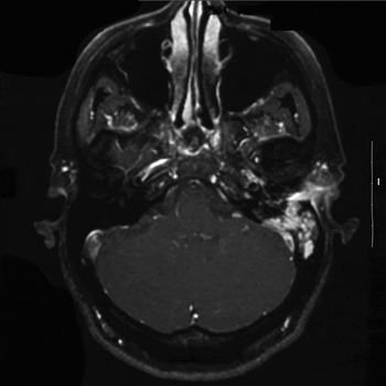
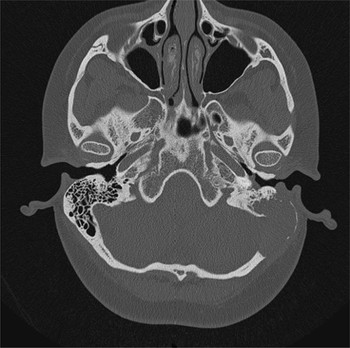
Computed tomography (CT) and magnetic resonance imaging (MRI) scans showed a significant destructive bone lesion involving the left posterior external auditory canal, middle and posterior cranial fossa, and lateral semicircular canal (Figures 1 and 2). The audiogram was normal. The differential diagnosis at that time included Langerhans cell histiocytosis, fibrous dysplasia, skull base malformation and endolymphatic sac carcinoma.
Axial, fat suppression, T1-weighted magnetic resonance imaging scan with gadolinium contrast, showing an enhancing lesion in the left mastoid. Note the involvement of the dura and sigmoid sinus. L = left

Axial computed tomography scan of mastoid, showing destruction of the posterior wall of the left external auditory canal and extension to the left sigmoid sinus and anterior cerebellar cisterna.

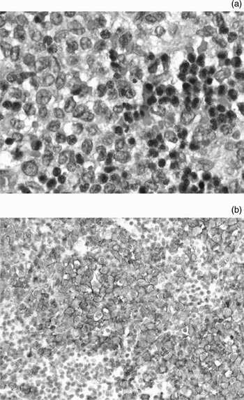
Surgical biopsies were taken via a mastoidectomy approach. Histological analysis revealed an eosinophil-rich cellular proliferation, and immunohistochemical staining was positive for cluster of differentiation 1a glycoprotein and S-100 protein (Figure 3). This confirmed the diagnosis of Langerhans cell histiocytosis.
(a) Photomicrograph of surgical biopsy material, showing eosinophils and histiocytes (H&E; ×400). (b) Immunohistochemical staining showing positivity for cluster of differentiation 1a glycoprotein (×200).

The patient was recalled to the operating theatre. The resection was completed, and the posterior fossa and tegmen were reconstructed with a titanium plate. The mastoid cavity was obliterated with temporalis fascia and abdominal fat in a watertight fashion.
During the initial investigation, panoramic dental radiography of the mandible had shown bone rarefaction in the left parasymphyseal area. This lesion was confirmed with bone scintigraphy, and the patient underwent curettage.
During the following years, the disease recurred in the patient's left mastoid and mandible, and new lesions appeared in the maxilla and lung. The patient later developed severe hearing loss on the affected side. She was treated with vinblastine.
Three years after surgery, follow-up MRI scanning showed no disease, and the patient had suffered no recurrence of her CSF leak.
Discussion
Lichtenstein and Jaffe introduced the term eosinophilic granuloma in 1940.Reference Lichtenstein and Jaffe6 In 1953, Lichtenstein linked this disease with Hand–Schüller–Christian disease and Letterer–Siwe disease, and proposed to combine these three entities under the name ‘histiocytosis X’, on the basis of their similar histopathological findings.Reference Lichtenstein7 In 1973, Nezelof described a cytoplasmic structure visible on electron microscopy in all cases of histiocytosis X, which he termed an ‘X-body’.Reference Nezelof, Basset and Rousseau8 He also noted that these bodies were similar to those described by Birbeck in Langerhans cells. He proposed that histiocytosis X was in fact a proliferation of Langerhans cells, and that the term should be changed to ‘Langerhans cell histiocytosis’. This term was officially adopted in 1986 during the Workshop on Childhood Histiocytoses.Reference Chu, Favara, Ladisch, Nezelof and Prichard9
Langerhans cell histiocytosis primarily affects children, but can manifest as late as the eighth decade.Reference Arico, Girschikofsky, Genereau, Klersy, McClain and Grois1 In the paediatric population, its incidence ranges from 2 to 5 cases per million per year.Reference Raney and D'Angio10, Reference Devaney, Putzi, Ferlito and Rinaldo11 Most cases are diagnosed before the age of 20 years.Reference Cunningham, Curtin, Jaffe and Stool3, Reference Cinberg12 The disease affects Caucasians more than Afro-Caribbeans, and males slightly more than females.Reference Arico, Girschikofsky, Genereau, Klersy, McClain and Grois1, Reference McCaffrey and McDonald2, Reference Raney and D'Angio10, Reference Cinberg12, Reference Arseni, Danaila and Constantinescu13 Langerhans cell histiocytosis is more often seen in flat bones, with the skull, ribs and pelvis being the most frequently involved.Reference McCaffrey and McDonald2, Reference Devaney, Putzi, Ferlito and Rinaldo11, Reference Brisman, Feldstein, Tarbell, Cohen, Cargan and Haddad14, Reference Groopman and Golde15 Otological involvement occurs in 15 to 61 per cent of cases, and is the first manifestation in up to 25 per cent.Reference McCaffrey and McDonald2–Reference Surico, Muggeo, Muggeo, Conti, Novielli and Romano4 The otological form seems to be more prevalent in multi-system Langerhans cell histiocytosis.Reference Cunningham, Curtin, Jaffe and Stool3 Middle and posterior fossa involvement have been documented in a few cases, but violation of the dura has not previously been reported.Reference Rawlings and Wilkins5
The aetiology of Langerhans cell histiocytosis is unknown. It is still debated whether the proliferation of Langerhans cells is of neoplastic or reactive (e.g. due to viral infection) origin.Reference Favara16–Reference Egeler and D'Angio18 Many classifications have been proposed over the years. With regard to treatment and prognosis, patients are best categorised as having either restricted or extensive disease.Reference Egeler and D'Angio18 Restricted disease consists of monostotic and polyostotic eosinophilic granulomas of the bone or skin. Extensive forms include Hand–Schüller–Christian disease and Letterer–Siwe disease. The former is defined by a chronic or subchronic course, with the classic triad of diabetes insipidus, exophthalmos and bone lesion present in only 10 per cent of patients.Reference McCaffrey and McDonald2 The latter is an acute or subacute, disseminated form which mostly affects children younger than two years and carries a much poorer prognosis.Reference McCaffrey and McDonald2, Reference Groopman and Golde15
Temporal bone involvement occurs in up to 63.2 per cent of multi-system Langerhans cell histiocytosis cases.Reference Cunningham, Curtin, Jaffe and Stool3 These patients most commonly present with purulent aural discharge.Reference McCaffrey and McDonald2–Reference Surico, Muggeo, Muggeo, Conti, Novielli and Romano4, Reference Cinberg12 Post-auricular swelling, vertigo, hearing loss and otalgia are also frequent. Facial palsy is a rare finding (less than 5 per cent).Reference Cinberg12 Physical examination shows otitis media or an external auditory canal polyp and granulation tissue. An assessment of cranial nerves, orbits and cerebellar function should be performed in all cases.Reference Broadbent, Gadner, Komp and Ladisch19
Head and neck lesions should first be investigated radiologically.Reference Cinberg12 Bone lesions typically show a ‘punched-out’ appearance, with no reactive sclerosis, although a local periosteal reaction can occur.Reference Peuchot, Eftekhari, Van Tassel and Cangir20–Reference Sweet, Kornblut and Hyams22 In the mastoid, CT scanning reveals a coalescence of cells with extension to the surrounding structures (bony canal walls, tegmentum and posterior fossa) which can appear similar to cholesteatoma. Soft tissue lesions are best characterised with MRI.
Histopathological findings include eosinophils, T-cells and ‘foamy’ macrophages on light microscopy.Reference Groopman and Golde15, Reference Sweet, Kornblut and Hyams22 Macroscopically, the lesion is often reddish or brown.Reference Sweet, Kornblut and Hyams22 Immunohistochemical staining includes S-100 protein, cluster of differentiation 1a glycoprotein antigen, peanut lectin, adenosine triphosphatase and α-D-mannosidase.Reference Devaney, Putzi, Ferlito and Rinaldo11, Reference Favara16, Reference Laman, Leenen, Annels, Hogendoorn and Egeler17 Electron microscopy shows classic Birbeck granules, although this technique is less often used nowadays.Reference Nezelof, Basset and Rousseau8 In 1987, the Writing Group of the Histiocyte Society published diagnostic criteria for Langerhans cell histiocytosis. A presumptive diagnosis is warranted when only the light microscopic appearance is consistent with the disease. A ‘designated diagnosis’ is defined by the presence of positive staining for two or more of the following: adenosine triphosphatase, S-100 protein, α-D-mannosidase or peanut lectin. A definitive diagnosis requires the demonstration of cluster of differentiation 1a glycoprotein antigens or Birbeck granules on electron microscopy.Reference Chu, D'Angio, Favara, Ladisch, Nesbit and Pritchard23
The treatment of patients with Langerhans cell histiocytosis varies widely according to the extent of the disease. Isolated bony lesions can often be observed, unless there is pain or deformity, because of a high rate of spontaneous remission.Reference Gadner, Grois, Arico, Broadbent, Ceci and Jakobson24 Restricted disease can be treated by curettage or intralesional corticosteroids. The latter is less useful in skull lesions. Low dose radiotherapy is also effective and can be combined with surgery.Reference Arico, Girschikofsky, Genereau, Klersy, McClain and Grois1, Reference Cunningham, Curtin, Jaffe and Stool3, Reference Rawlings and Wilkins5, Reference Laman, Leenen, Annels, Hogendoorn and Egeler17, Reference Egeler and D'Angio18 Extensive disease requires a systemic approach comprising chemotherapy often combined with corticosteroids. The principal agents used are vinblastine and etoposide.Reference Arico, Girschikofsky, Genereau, Klersy, McClain and Grois1, Reference Laman, Leenen, Annels, Hogendoorn and Egeler17, Reference Gadner, Grois, Arico, Broadbent, Ceci and Jakobson24 2-CdA can be administered in refractory cases.Reference Arico, Girschikofsky, Genereau, Klersy, McClain and Grois1
Factors indicating a poor prognosis include an age of less than two years, multiple organ involvement and organ failure (i.e. liver, lungs and haematopoietic system).Reference Cunningham, Curtin, Jaffe and Stool3, Reference Surico, Muggeo, Muggeo, Conti, Novielli and Romano4, Reference Egeler and D'Angio18, Reference Gadner, Grois, Arico, Broadbent, Ceci and Jakobson24, Reference Lahey25 The patient's initial response to therapy has also proven to be a prognostic factor.Reference Minkov, Grois, Heitger, Potschger, Westermeier and Gadner26 Otological involvement seems to predict a poorer outcome. In their series of 251 paediatric Langerhans cell histiocytosis patients, Surico et al. found that 93.8 per cent of cases with otological involvement had multi-system disease, and that they also had a younger age at diagnosis.Reference Surico, Muggeo, Muggeo, Conti, Novielli and Romano4
• Langerhans cell histiocytosis can present in three different forms: eosinophilic granuloma, Hand–Schüller–Christian disease and Letterer–Siwe disease
• Temporal bone involvement occurs in 67 per cent of multi-system forms
• Involvement of the middle and posterior fossae is rare, and violation of the dura has not previously been reported
• We report a case of Langerhans cell histiocytosis of the temporal bone which presented with cerebrospinal fluid fistula, showing that the disease can penetrate the dura
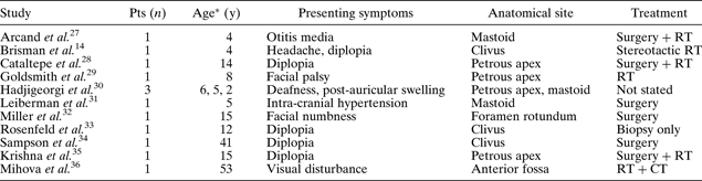
Thirteen cases of skull base Langerhans cell histiocytosis with intra-cranial involvement have previously been reported (Table I).Reference Brisman, Feldstein, Tarbell, Cohen, Cargan and Haddad14, Reference Arcand, Caouette and Dufour27–Reference Mihova, Yaneva, Sopadjieva, Andreev and Nejtcheva36 Of those, three had lesions in the mastoid, five in the petrous apex, three in the clivus, one in the floor of the anterior fossa and one near the foramen rotundum. The mean age at diagnosis was 14 years, with only two adults. Visual symptoms (mostly diplopia) were the most frequent complaints at presentation (six of 13 cases); others included intra-cranial hypertension, facial numbness, hearing loss, post-auricular swelling, refractory otitis media and facial palsy. Treatment modalities consisted of radiotherapy alone in two cases, surgery alone in three, combined surgery and radiotherapy in three, biopsy alone in one, and chemoradiotherapy in one. Treatment was not described for three cases.
Previously reported cases of Langerhans cell histiocytosis with intra-cranial involvement
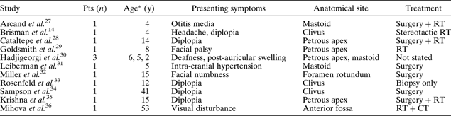
* At diagnosis. Pts = patients; y = years; RT = radiotherapy; CT = chemotherapy
Conclusion
Langerhans cell histiocytosis of the temporal bone rarely extends intra-cranially. Only 13 cases of skull base Langerhans cell histiocytosis with intra-cranial involvement have previously been reported, with only two involving adult patients. Our patient originally presented with violation of the dura and CSF otorrhoea; to our knowledge, such a manifestation of Langerhans cell histiocytosis had not previously been reported. Her fistula was successfully treated by mastoid obliteration, but the disease recurred locally and systemically, requiring chemotherapy for control. Although rare, Langerhans cell histiocytosis should be included in the differential diagnosis of any refractory otological disease.