


Introduction
Atrial fibrillation (AF) treatment with radiofrequency ablation (RFA) method has gained popularity in clinical practice in recent years. Reference Han, Ha and Sanders1–Reference Cappato, Calkins and Chen3 The treatment yields positive results, but complications such as atrioesophageal fistula (AEF) are rarely observed. Thermal injury caused by isolation of the left pulmonary vein and left atrium and subsequent ablation is the main cause of the fistula. Most commonly, AEF occurs within two to four weeks after the procedure. Reference Han, Ha and Sanders1 Patients with fistulas often present with neurological deficits, fever, chest pain, and dysphagia, whereas gastrointestinal bleeding findings are much less common. Reference Bao, Gupta, Keshavamurthy and Thomson4 The main reason for this is that the fistula anatomically allows a one-way passage from the esophagus to the atrium. Reference Stöllberger, Pulgram and Finsterer5 The incidence of fistulas is between 0.015% and 0.040% and mortality is between 40.0% and 80.0%, as reported in case series. Reference Chavez, Messerli and Dominguez6 Although patients with minor hematemesis have better survival, it has been reported that the mortality of patients with severe hematemesis exceeds 80.0%. Reference Chavez, Messerli and Dominguez6 Because of the high mortality of patients treated conservatively and undergoing endoscopy, the first treatment option is emergency surgery. Reference Han, Ha and Sanders1,Reference Kapur, Barbhaiya, Deneke and Michaud7,Reference Kim, Shim and Lee8
The aim of this report is to present the management of AEF that was detected in a patient who presented to the emergency department (ED) with gastrointestinal bleeding.
Report
A 60-year-old male patient was brought to the ED with altered mental status and red colored vomiting. The patient had palpitations and chest pain one day prior to presentation. Medical history of the patient included AF, chronic obstructive pulmonary disease, hypertension, and chronic heart failure. The patient underwent pulmonary vein and left atrium isolation using RFA 14 days before because of persistent AF. The patient was taking rivaroxaban, digoxin, ramipril, and bisoprolol.
Vital signs in the ED were as follows: blood pressure (BP), 75/45mmHg; pulse, 148/minute; oxygen saturation (SPO2), 84%; and body temperature, 37 degrees. During physical examination, the patient was determined to be unconscious; Glasgow Coma Scale was five (E1V2M2); and bilateral light reflexes were positive. There were no obvious signs of lateralization. Babinski sign was negative and pathological reflex was not detected. Rectal digital examination was normal. No apparent disorder was observed while examining other systems.
Rapid sequence intubation was performed for airway safety. Because the history of hematemesis was not clear, a nasogastric catheter was inserted and the bleeding was observed to continue actively. Laboratory findings were as follows: pH, 7.248; lactate, 9.1mmol/L; base excess, -12.4mmol/L; bicarbonate (HCO3), 13.5mmol/L; hemoglobin, 16.1g/dL; leukocyte, 13,080/µL; platelet, 159,000/µL; international nationalized ratio, 1.44; creatinine, 1.97mg/dL; and blood urea nitrogen, 36.2mg/dL.
Point-of-care ultrasound was performed in the ED which revealed patient’s inferior vena cava was collapsed, right cardiac structures were normal; pericardial effusion, B-lines, and deep vein thrombosis were not present. Thoracic computerized tomography (CT) was performed for possible fistula and brain CT was performed for neurological complications.
Although the hemoglobin level of the patient was normal, because of the presence of gastrointestinal bleeding causing vital abnormality, the ratio of transfused fresh frozen plasma (in units) to platelets (in units) to red blood cells (in units) approach (1:1:1 massive transfusion protocol) was started and tranexamic acid was given. Blood gas was measured after 30 minutes, and the analysis results were as follows: pH, 7,056; lactate, 8.3mmol/L; base excess, -15.9mmol/L; and HCO3, 11.6mmol/L.
Thoracic CT showed active contrast filling in the esophagus, and an approximately one-centimeter fistula area was detected between the left main pulmonary vein and esophagus (Figure 1).
A-B) The Axial Plane of the Mediastinal View and C) Sagittal Plane of the Thoracoabdominal Computerized Tomography.
Note: Black arrow indicates the fistula between atrium and esophagus.

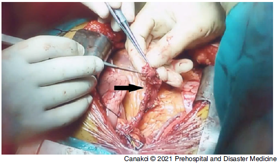
After transfusion in the ED, BP was 110/60mmHg and pulse was 90/minute. The patient was taken to an operation room urgently. A one-centimeter fistula was found between the left main pulmonary vein and esophagus (Figure 2). The esophagus was primarily repaired, but left pulmonary vein was ligatured because of severe damage. The patient was hypotensive after the operation and was taken to the intensive care unit with inotrope support. Six hours after the operation, the patient died.
From the Operation Room.
Note: Black arrow indicates damaged left pulmonary vein.

Discussion
Atrioesophageal fistula is a very rare clinical picture in the ED. Its incidence among all ablations is reported as 0.03%-0.08% in some studies, but this may be due to under-reporting and misdiagnosis. Reference Han, Ha and Sanders1,Reference Gupta, Perera and Ganesan9,Reference Nair, Shurrab and Skanes10 Because the patients reach the center where they are followed after ablation, there may be a delay in the diagnosis in centers where ablation is not performed. Furthermore, because there are mostly neurological symptoms, fever, and chest pain complaints, time may be lost during the evaluation of other clinical conditions. Emergency physicians should be aware of this important clinical picture in the differential diagnosis for patients who have recently undergone ablation. Because septic embolisms, air embolisms, mediastinitis, sepsis, and gastrointestinal bleeding are conditions that need to be treated quickly, delay in diagnosis increases mortality. Reference Kapur, Barbhaiya, Deneke and Michaud7,Reference Pappone, Vicedomini and Santinelli11 It should be remembered that some of the patients with AEF are diagnosed during autopsies. Reference Moraveji, Mesa and Siddiqui12
Serious gastrointestinal bleeding was observed in very few patients in previously reported cases. In the review by Chavez, et al, eight out of 53 patients evaluated had hematemesis and one patient had minor hematemesis; six of the remaining seven patients died. It was noted that the surviving patient was 27 years old and had no comorbidities. Reference Chavez, Messerli and Dominguez6 As this reported patient was 60 years old and had comorbidities, mortality occurred despite early diagnosis and surgical treatment.
After performing 1:1:1 blood transfusion according to massive transfusion protocols in ED management, it was believed that there was a chance for surgical treatment. Reference Holcomb, Tilley and Baraniuk13
The shunt between esophagus and atrium is commonly unilateral. Reference Stöllberger, Pulgram and Finsterer5 With this shunt, bleeding out of the atrium is much less common, whereas air leakage from the esophagus is observed more frequently, and pneumopericardium, pneumomediastinum, mediastinitis, and sepsis are more common.
Aggressive surgical treatment should be the first line of treatment in such patients. The possibility of air embolism increases with the air filling occurring during endoscopic procedures. Reference Han, Ha and Sanders1,Reference Chavez, Messerli and Dominguez6 In addition, endoscopy and stent methods may cause distraction as there may be a delay in reaching definitive therapy. This reported patient was taken directly for surgical treatment, and cause-oriented treatment was initiated quickly.
Unfortunately, the patient arrived at the ED one day after the onset of complaints, and the presence of massive bleeding and the condition of his comorbidities did not improve the prognosis despite early surgical treatment.
Limitations
The most important limitation in the case is that the patient reached the ED 24 hours after the onset of the complaints. In addition, since the procedure was performed in another center, information on how much energy was given in the ablation procedure, the use of overlapping ablation lines, esophageal temperature level, and patient education were not available.
Conclusion
It should be remembered that AEFs, together with aortoenteric fistulas, are also included in the differential diagnosis of gastrointestinal bleeding, which is frequently evaluated in EDs. Considering that radiofrequency procedures will increase in the coming years, all emergency physicians should be informed about AEF and manage it in the early period.
Conflicts of interest/funding
This research did not receive any specific grant from funding agencies in the public, commercial, or not-for-profit sectors. The authors certify that they have NO affiliations with or involvement in any organization or entity with any financial interest (such as honoraria; educational grants; participation in speakers’ bureaus; membership, employment, consultancies, stock ownership, or other equity interest; and expert testimony or patent-licensing arrangements) or non-financial interest (such as personal or professional relationships, affiliations, knowledge or beliefs) in the subject matter or materials discussed in this manuscript.